. 2015 Jun 23;112(25):7707-12.
doi: 10.1073/pnas.1503491112. Epub 2015 Jun 8.

Essential role of the cytochrome P450 CYP4F22 in the production of acylceramide, the key lipid for skin permeability barrier formation

Affiliations

Essential role of the cytochrome P450 CYP4F22 in the production of acylceramide, the key lipid for skin permeability barrier formation

Yusuke Ohno et al. Proc Natl Acad Sci U S A. .

Abstract

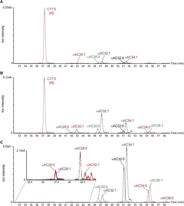
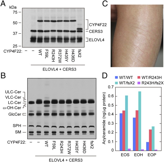
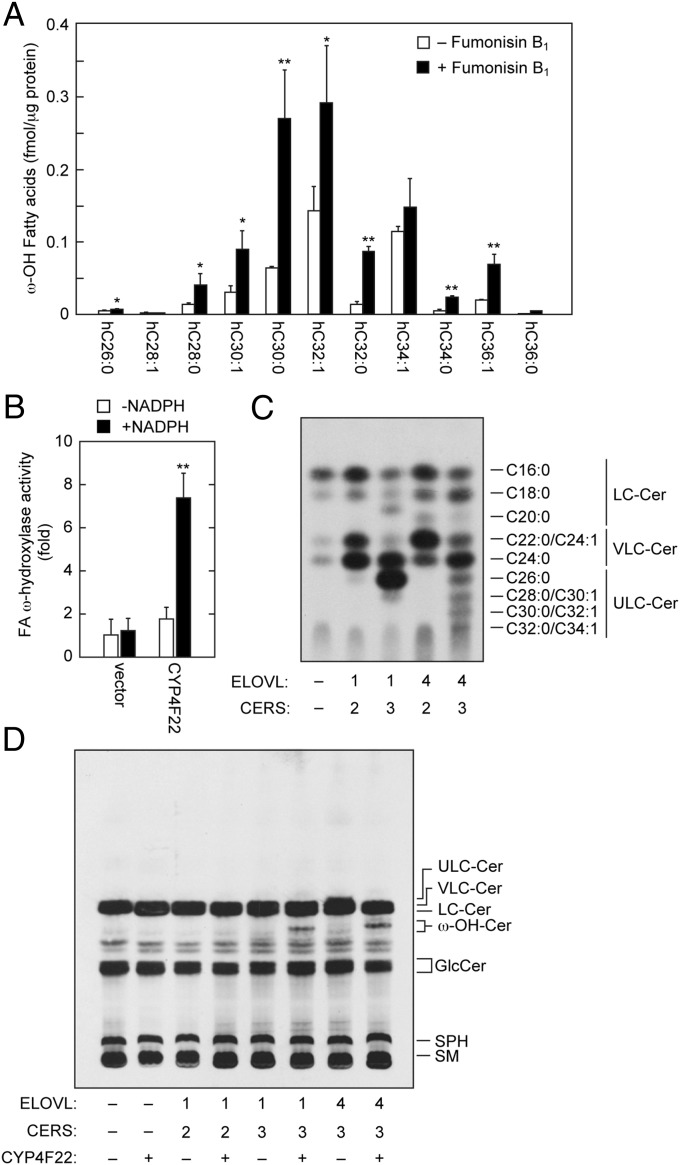
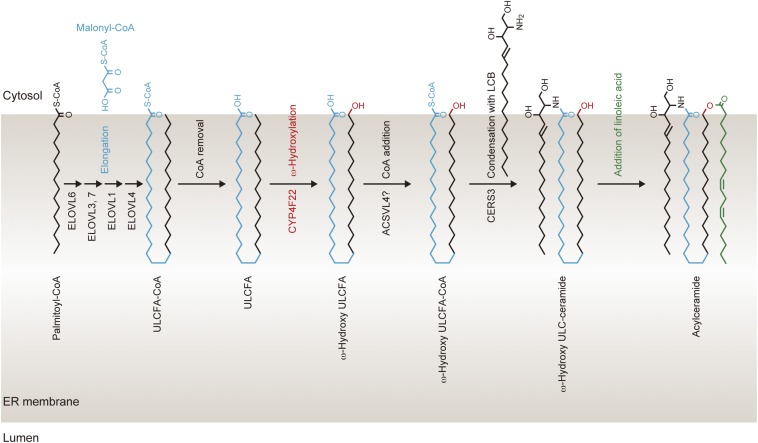
A skin permeability barrier is essential for terrestrial animals, and its impairment causes several cutaneous disorders such as ichthyosis and atopic dermatitis. Although acylceramide is an important lipid for the skin permeability barrier, details of its production have yet to be determined, leaving the molecular mechanism of skin permeability barrier formation unclear. Here we identified the cytochrome P450 gene CYP4F22 (cytochrome P450, family 4, subfamily F, polypeptide 22) as the long-sought fatty acid ω-hydroxylase gene required for acylceramide production. CYP4F22 has been identified as one of the autosomal recessive congenital ichthyosis-causative genes. Ichthyosis-mutant proteins exhibited reduced enzyme activity, indicating correlation between activity and pathology. Furthermore, lipid analysis of a patient with ichthyosis showed a drastic decrease in acylceramide production. We determined that CYP4F22 was a type I membrane protein that locates in the endoplasmic reticulum (ER), suggesting that the ω-hydroxylation occurs on the cytoplasmic side of the ER. The preferred substrate of the CYP4F22 was fatty acids with a carbon chain length of 28 or more (≥C28). In conclusion, our findings demonstrate that CYP4F22 is an ultra-long-chain fatty acid ω-hydroxylase responsible for acylceramide production and provide important insights into the molecular mechanisms of skin permeability barrier formation. Furthermore, based on the results obtained here, we proposed a detailed reaction series for acylceramide production.

Keywords: acylceramide; ceramide; lipid; skin; sphingolipid.


Conflict of interest statement

The authors declare no conflict of interest.

Figures

Fig. S1.
Fig. S1.
Structures of the epidermis, the stratum corneum, acylceramide, and protein-bound ceramide. Acylceramides are produced mainly in the stratum granulosum and partly in the stratum spinosum and are stored in lamellar bodies as glucosylated forms (acyl glucosylceramides). At the interface of the stratum granulosum and stratum corneum, the lamellar bodies fuse with the plasma membrane and release their contents into the extracellular space, where acyl glucosylceramides are converted to acylceramides. Thus, released acylceramides, FAs, and cholesterol form lipid lamellae in the stratum corneum. Some acylceramide is hydrolyzed to ω-hydroxyceramide, followed by covalent binding to corneocyte surface proteins to create corneocyte lipid envelopes. Acylceramide contains ULCFAs with carbon chain lengths of C28–C36. The FA elongase ELOVL1 produces VLCFAs, which are further elongated to ULCFAs by ELOVL4 (29). The ceramide synthase CERS3 creates an amide bond between ULCFA and LCB (17). ω-Hydroxylation of ULCFA is required for acylceramide production. However, the responsible ω-hydroxylase had not been identified previously; its identification is the subject of this research.
Fig. S2.
Fig. S2.
Structure and synthetic pathways of ceramides in mammals. (A) Structure and nomenclature of epidermal ceramides. Epidermal ceramides are classified into 12 classes depending on their differences in the LCB and FA moieties. N-type and A-type ceramides contain C16–C30 FAs (n = 1–15), whereas EO-type ceramides contain C28–C36 FAs (n = 13–21) (6, 7). (B) FA elongation and ceramide synthesis in mammals. The FA elongation pathways of saturated and monounsaturated FAs and the ceramide-synthetic pathways are illustrated. E1–E7 and C1–C6 indicate the ELOVL (ELOVL1–7) and CERS (CERS1–6) isozymes involved in each step, respectively. The differences in the letter size of E1–E7 reflect their enzyme activities in each FA elongation reaction. Cer, ceramide; MUFA, monounsaturated FA; SFA, saturated FA.
Fig. 1.
Fig. 1.
Overproduction of ELOVL4 and CERS3 causes generation of ULC-ceramides. HEK 293T cells were transfected with plasmids encoding 3xFLAG-ELOVL4 and 3xFLAG-CERS3, as indicated. Cells were labeled with [3H]sphingosine for 4 h at 37 °C. Lipids were extracted, separated by reverse-phase TLC (A) or normal-phase TLC (B), and detected by autoradiography. Cer, ceramide; GlcCer, glucosylceramide; SM, sphingomyelin; SPH, sphingosine.
Fig. S3.
Fig. S3.
MRM chromatogram of ceramides produced by combined ELOVL4 and CERS3 expression. Lipids were extracted from HEK 293T cells transfected with control vector (A) or pCE-puro 3xFLAG-ELOVL4 and pCE-puro 3xFLAG-CERS3 (B) or mouse epidermis (C) and subjected to UPLC/ESI-MS using a triple quadrupole mass spectrometer (Xevo TQ-S; Waters). Each ceramide was detected by MRM by setting the appropriate [M+H]+ and [M+H−H2O]+ values at Q1 and m/z 264.2 (corresponding to C18:0 sphingosine) at Q3. Each MRM peak was overlaid using MassLynx software. Insets show enlarged views of the indicated areas of the original chromatograms.
Fig. 2.
Fig. 2.
CYP4F22 is involved in ω-hydroxyceramide synthesis. HEK 293T cells were transfected with plasmids encoding 3xFLAG-ELOVL4, 3xFLAG-CERS3, and 3xFLAG-CYP4F subfamily members, as indicated. (A) Total lysates prepared from the transfected cells were separated by SDS/PAGE, followed by immunoblotting with anti-FLAG antibodies. (B) Cells were labeled with [3H]sphingosine for 4 h at 37 °C. Lipids were extracted, separated by normal-phase TLC, and detected by autoradiography. Cer, ceramide; GlcCer, glucosylceramide; SM, sphingomyelin; SPH, sphingosine; ω-OH, ω-hydroxy.
Fig. S4.
Fig. S4.
TLC chromatogram of ceramides. The acylceramide EOS and ω-hydroxyceramide were prepared as follows. Lipids were prepared from mouse epidermis and separated by normal-phase TLC. Silica containing EOS ceramide was scraped from the TLC plate and eluted with chloroform/methanol (1:2, vol/vol). A portion of EOS ceramide was converted to ω-hydroxyceramide by hydrolysis of the ester bond connecting ω-hydroxyceramide and linoleic acid with 0.1 M NaOH. The prepared EOS ceramide and ω-hydroxyceramide, as well as C24:0 ceramide (Avanti Polar Lipids), C16:0 ceramide (Avanti Polar Lipids), and glucosylceramide (Avanti Polar Lipids), were separated by normal-phase TLC and visualized by cupric acetate/phosphoric acid staining. Cer, ceramide; GlcCer, glucosylceramide; ω-OH Cer, ω-hydroxyceramide.
Fig. S5.
Fig. S5.
MRM chromatogram of ω-hydroxyceramide species produced by CYP4F22. Lipids were extracted from HEK 293T transfected with pCE-puro 3xFLAG-ELOVL4 and pCE-puro 3xFLAG-CERS3, together with control vector (A) or pCE-puro 3xFLAG-CYP4F22 (B). EOS from mouse epidermis was treated with an alkali to liberate ω-hydroxyceramides (C). Lipids were subjected to UPLC/ESI-MS. Each ceramide was detected by MRM by setting the appropriate [M+H]+ and [M+H−H2O]+ values at Q1 and m/z 264.2 at Q3. Each MRM peak was overlaid using MassLynx software. ωhC30:1, ω-hydroxyceramide with a chain length of C30:1. IS, internal control (C17:0 ceramide). The Inset in C shows an enlarged large view of the indicated area of the original chromatogram.
Fig. 3.
Fig. 3.
Hydroxylase activity of CYP4F22 is impaired by ichthyosis-causing mutations. (A and B) HEK 293T cells were transfected with plasmids encoding 3xFLAG-ELOVL4, 3xFLAG-CERS3, and 3xFLAG-CYP4F22 (wild type or mutant), as indicated. (A) Total cell lysates prepared from the transfected cells were separated by SDS/PAGE and subjected to immunoblotting with anti-FLAG antibodies. (B) The transfected cells were labeled with [3H]sphingosine for 4 h at 37 °C. Extracted lipids were separated by normal-phase TLC and detected by autoradiography. Cer, ceramide; GlcCer, glucosylceramide; SM, sphingomyelin; SPH, sphingosine; ω-OH, ω-hydroxy. (C) Representative clinical feature of a 2-y-old ARCI patient. Leaf-like flakes presented on the extensor side of the left lower limb before tape stripping. (D) Acylceramide (EOS, EOH, and EOP) levels in stratum corneum of a control (WT/WT), carriers (WT/R243H, the ichthyosis patient’s father, and WT/D380T fs2X (fs2X), the patient’s mother), and an ARCI patient (R243H/D380T fs2X) were measured by LC-MS.
Fig. 4.
Fig. 4.
CYP4F22 is a type I ER membrane protein. (A, D, and E) HeLa cells were transfected with plasmids encoding HA-ELOVL4, HA-CERS3, 3xFLAG-CYP4F22, and 3xFLAG-CYP4F22ΔN (CYP4F22 lacking 54 N-terminal amino acids), as indicated. (A and D) Cells were subjected to indirect immunofluorescence microscopic observation. (Scale bars, 10 μm.) (B) The hydrophilicity of CYP4F22 was analyzed by MacVector software (MacVector) using the Kyte and Doolittle algorithm (window size, 15). (C) HEK 293T cells were transfected with pCE-puro 3xFLAG-CYP4F22, pCE-puro 3xFLAG-CYP4F22 (N-term: insertion of the N-glycosylation cassette to the N terminus), pCE-puro 3xFLAG-CYP4F22 (E85/K: insertion of the cassette between Glu-85 and Lys-86), pCE-puro 3xFLAG-CYP4F22 (H155/R), pCE-puro 3xFLAG-CYP4F22 (A285/L), pCE-puro 3xFLAG-CYP4F22 (C361/R), pCE-puro 3xFLAG-CYP4F22 (D455/N), or pCE-puro 3xFLAG-CYP4F22 (R508/K). Lysates (3 μg) prepared from transfected cells were treated with or without endoglycosidase H (Endo H) and were separated by SDS/PAGE, followed by immunoblotting with anti-FLAG antibodies. (E) Total cell lysates (10 μg) were centrifuged at 100,000 × g for 30 min at 4 °C. The resulting supernatant (soluble fraction, S) and pellet (membrane fraction, M) were subjected to immunoblotting using anti-FLAG, anti-calnexin (membrane protein marker) or anti-GAPDH (soluble protein marker) antibodies. IB, immunoblotting. (F) HEK 293T cells transfected with plasmids encoding 3xFLAG-ELOVL4, 3xFLAG-CERS3, and 3xFLAG-CYP4F22 [wild type or CYP4F22ΔN (ΔN)], as indicated, were labeled with [3H]sphingosine for 4 h at 37 °C. Lipids were extracted, separated by normal-phase TLC, and detected by autoradiography. Cer, ceramide; GlcCer, glucosylceramide; SM, sphingomyelin; SPH, sphingosine; ω-OH, ω-hydroxy.
Fig. 5.
Fig. 5.
CYP4F22 hydroxylates ULCFAs. (A) Keratinocytes were differentiated for 7 d in the presence or absence of 10 μM fumonisin B1. Lipids were extracted, treated with an alkali, and derivatized to AMPP amides. Derivatized FAs were analyzed by a Xevo TQ-S LC/MS system and quantified by MassLynx software. Statistically significant differences are indicated; *P < 0.05, **P < 0.01; t test. hC26:0, hydroxy C26:0 FA. (B) Total membrane fractions (50 μg) prepared from BY4741 bearing the pAK1017 (vector) or pNS29 (His6-Myc-3xFLAG-CYP4F22) plasmids were incubated with 10 μM C30:0 FA and 1 mM NADPH as indicated for 1 h at 37 °C. Lipids were extracted, derivatized to AMPP amides, and analyzed as in A. The values represent the amount of each FA ω-hydroxylase activity relative to that of the vector/−NADPH sample. The value of the CYP4F22/+NADPH sample was statistically different from the values of all other samples (**P < 0.01; t test). hC30:0, hydroxy C30:0 FA. (C and D) HEK 293T cells were transfected with the plasmids encoding ELOVL1 or ELOVL4, CERS2 or CERS3, and CYP4F22. Transfected cells were labeled with [3H]sphingosine for 4 h at 37 °C. Extracted lipids were separated by reverse-phase TLC (C) or by normal-phase TLC (D) and detected by autoradiography. Cer, ceramide; GlcCer, glucosylceramide; SM, sphingomyelin; SPH, sphingosine; ω-OH, ω-hydroxy.
Fig. S6.
Fig. S6.
Working model for the acylceramide synthesis in the ER. Palmitoyl-CoA is elongated to ULCFA-CoA on the cytosolic side of the ER membrane. During FA elongation, ULCFA (C30–C36) portions of ULCFA-CoAs may be bent in the cytosolic leaflet of the ER membrane. After conversion of ULCFA-CoA to ULCFA, the ω-carbon of ULCFA is hydroxylated by CYP4F22. ω-Hydroxy-ULCFA then is converted to ω-hydroxy-ULCFA-CoA by acyl-CoA synthetase, followed by synthesis of ω-hydroxyceramide by CERS3. Finally, an acyltransferase catalyzes the formation of an ester bond between linoleic acid and the ω-hydroxy group of ω-hydroxyceramide, producing acylceramide.

References

    1. Uchida Y, Holleran WM. Omega-O-acylceramide, a lipid essential for mammalian survival. J Dermatol Sci. 2008;51(2):77–87. - PubMed
    1. Mizutani Y, Mitsutake S, Tsuji K, Kihara A, Igarashi Y. Ceramide biosynthesis in keratinocyte and its role in skin function. Biochimie. 2009;91(6):784–790. - PubMed
    1. Breiden B, Sandhoff K. The role of sphingolipid metabolism in cutaneous permeability barrier formation. Biochim Biophys Acta. 2014;1841(3):441–452. - PubMed
    1. Kihara A. Very long-chain fatty acids: Elongation, physiology and related disorders. J Biochem. 2012;152(5):387–395. - PubMed
    1. Sassa T, Kihara A. Metabolism of very long-chain Fatty acids: Genes and pathophysiology. Biomol Ther (Seoul) 2014;22(2):83–92. - PMC - PubMed

Publication types