. 2015 Jun 23;112(25):E3179-88.
doi: 10.1073/pnas.1422363112. Epub 2015 Jun 8.

Conserved SMP domains of the ERMES complex bind phospholipids and mediate tether assembly

Affiliations

Conserved SMP domains of the ERMES complex bind phospholipids and mediate tether assembly

Andrew P AhYoung et al. Proc Natl Acad Sci U S A. .

Abstract

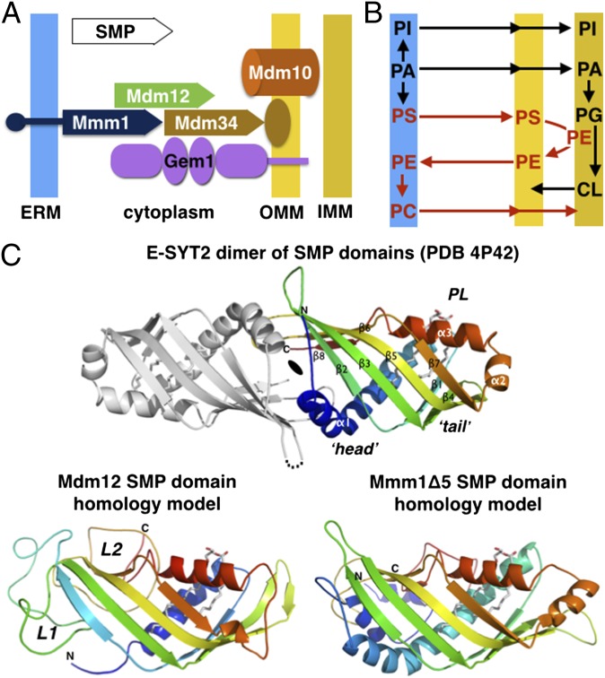
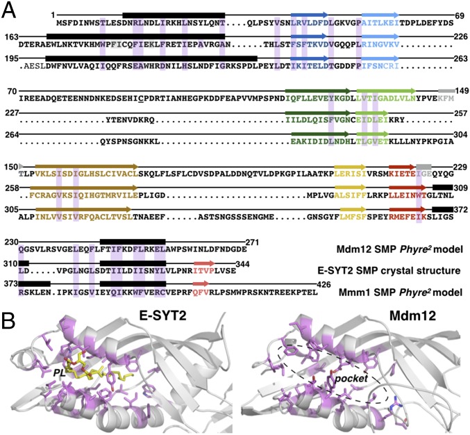
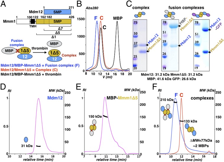
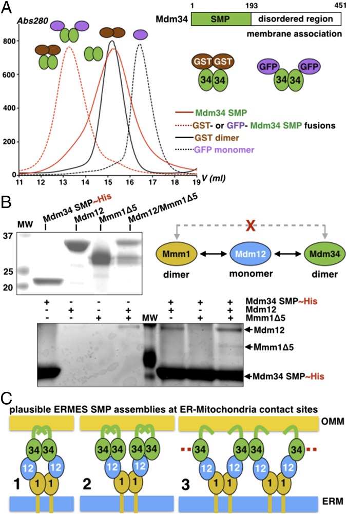
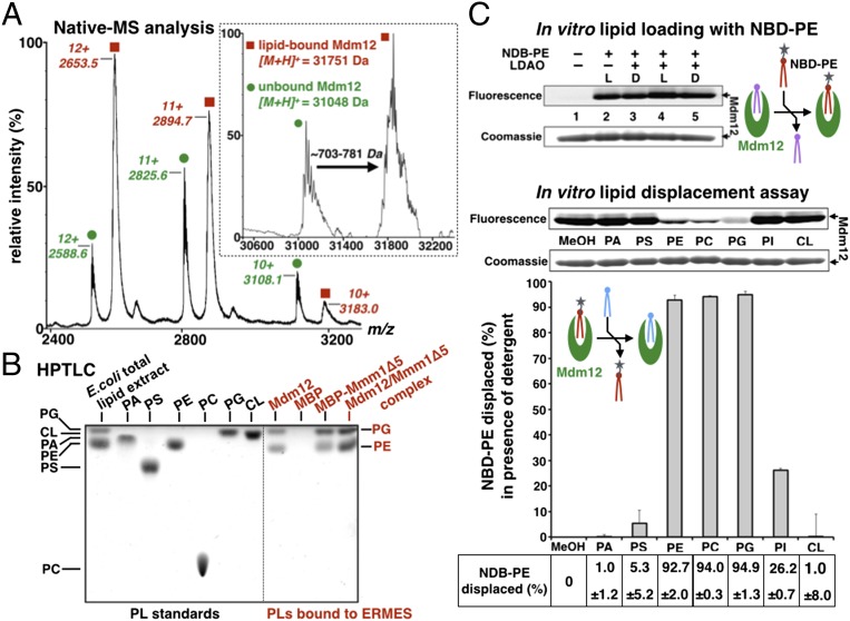
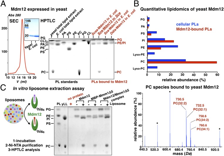
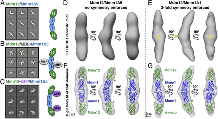
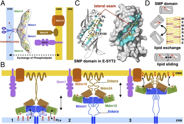
Membrane contact sites (MCS) between organelles are proposed as nexuses for the exchange of lipids, small molecules, and other signals crucial to cellular function and homeostasis. Various protein complexes, such as the endoplasmic reticulum-mitochondrial encounter structure (ERMES), function as dynamic molecular tethers between organelles. Here, we report the reconstitution and characterization of subcomplexes formed by the cytoplasm-exposed synaptotagmin-like mitochondrial lipid-binding protein (SMP) domains present in three of the five ERMES subunits--the soluble protein Mdm12, the endoplasmic reticulum (ER)-resident membrane protein Mmm1, and the mitochondrial membrane protein Mdm34. SMP domains are conserved lipid-binding domains found exclusively in proteins at MCS. We show that the SMP domains of Mdm12 and Mmm1 associate into a tight heterotetramer with equimolecular stoichiometry. Our 17-Å-resolution EM structure of the complex reveals an elongated crescent-shaped particle in which two Mdm12 subunits occupy symmetric but distal positions at the opposite ends of a central ER-anchored Mmm1 homodimer. Rigid body fitting of homology models of these SMP domains in the density maps reveals a distinctive extended tubular structure likely traversed by a hydrophobic tunnel. Furthermore, these two SMP domains bind phospholipids and display a strong preference for phosphatidylcholines, a class of phospholipids whose exchange between the ER and mitochondria is essential. Last, we show that the three SMP-containing ERMES subunits form a ternary complex in which Mdm12 bridges Mmm1 to Mdm34. Our findings highlight roles for SMP domains in ERMES assembly and phospholipid binding and suggest a structure-based mechanism for the facilitated transport of phospholipids between organelles.

Keywords: electron microscopy; interorganelle tether; membrane contact sites; membrane protein complex; phospholipid exchange.


Conflict of interest statement

The authors declare no conflict of interest.

Figures

Fig. 1.
Fig. 1.
ERMES, phospholipid metabolism, and the signature SMP domain. (A) Subunit topology and composition of the ERMES complex. The three SMP domain-containing subunits Mdm12, Mmm1, and Mdm34 together with a β-barrel outer mitochondrial membrane protein Mdm10 and the calcium-activated regulatory GTPase Gem1 constitute the ERMES in yeast. The SMP domains are depicted with a plain arrow. (B) Interconnection of the phospholipid metabolic pathways at the interface between ER and mitochondria. (C, Upper) Homology models for the SMP domains of yeast Mdm12 and Mmm1. The crystal structure of the head-to-head dimer of SMP domain of E-SYT2 (23) present in an ER-to-plasma membrane tether is shown with the observed bound phospholipid (PL). Only one SMP monomer has been colored. (Lower) Phyre2 homology models of the yeast Mdm12 and Mmm1 SMP. L1 and L2 refer to the nonconserved insertions present in Mdm12 sequences.
Fig. 2.
Fig. 2.
The predicted hydrophobic binding pockets of Mdm12 and Mmm1 SMP domains resemble that of E-SYT2. (A) Alignment of Mdm12 and Mmm1 amino acid sequences to E-SYT2 using predicted structural homology. The structures of the SMP domains of Mdm12 and Mmm1 (Phyre2 homology models) and E-SYT2 (crystal structure) were superposed to identify residues (shaded in magenta) of Mdm12 and Mmm1 equivalent in terms of position to those involved in phospholipid binding in E-SYT2. α-Helices and β-strands are depicted as black rectangles and colored arrows, respectively. (B) The binding pockets of the SMP domain of E-SYT2 and Mdm12. Equivalent residues (magenta) involved in phospholipid binding in E-SYT2 are mapped on the Mdm12 structure. The bound phospholipid (yellow) is shown in E-SYT2; the corresponding pocket in Mdm12 is hydrophobic and of similar dimensions.
Fig. 3.
Fig. 3.
Reconstitution and characterization of the ERMES Mdm12/Mmm1 heterotetramer. (A) Organization of Mdm12 and Mmm1 (Upper) and strategy used to coexpress and copurify Mdm12 associated to an MBP-Mmm1Δ construct as a fusion complex (Lower). (B) Reconstitution of the Mdm12/Mmm1Δ5 complex. Thrombin treatment of the fusion complex releases the monomeric MBP carrier protein from the Mdm12/Mmm1 complex assembled in vivo. The Mdm12/Mmm1Δ5 complex obtained after quantitative removal of the MBP fusion partner is identical to the native Mdm12/Mmm1Δ5. (C) Coomassie-stained SDS/PAGE gel of complexes Mdm12/Mmm1Δ5, Mdm12/MBP-Mmm1Δ5, and Mdm12-GFP/Mmm1Δ5 expressed and reconstituted in E. coli. These samples also were used for the EM study. (DF) Determination of the mass of Mdm12 (D), the MBP-Mmm1Δ5 (E), and the Mdm12/MBP-Mmm1Δ5 fusion and Mdm12/Mmm1Δ5 complexes (F) by SEC-MALS. Mdm12 is a monomer (31 kDa), whereas MBP-Mmm1Δ5 forms a homodimer (150 kDa). The difference in mass of 77 kDa, measured between the fusion (210 kDa) and the native (133 kDa) complexes, corresponds to two MBP proteins (40 kDa each).
Fig. 4.
Fig. 4.
The SMP domain of Mdm34 interacts with Mdm12 and the Mdm12/Mmm1 complex but not with Mmm1. (A) Organization of Mdm34. Strategy used to purify Mdm34 as a GST or a GFP fusion. SEC analysis of the GST and GFP fusion proteins of SMP Mdm34 and comparison with free monomeric GFP (26 kDa) and dimeric GST (2 × 26 = 52 kDa). The SMP domain of Mdm34 (22 kDa) is a dimer. (B) Pull-down assay analysis of the interactions between Mdm34 and Mdm12, MBP-Mmm1Δ5, or the Mdm12/Mmm1Δ5 complex. The upper gel shows the proteins used for the pull-down in the lower gel (elutions). Mdm12 bridges Mmm1 to Mdm34. (C) Three plausible modes of association among the three SMP domains of ERMES at ER to mitochondria contact sites.
Fig. 5.
Fig. 5.
Mdm12, Mmm1, and their complex bind phospholipids promiscuously. (A) Native-MS analysis of yeast Mdm12 expressed in E. coli. Yeast Mdm12 purified from E. coli copurifies with noncovalently bound ligands with a mass ranging from ∼703–781 Da. The Inset represents the deconvolution of the raw data. (B) Mdm12, Mmm1, and the Mdm12/Mmm1 complex bind phospholipids PE and PG. HPTLC of phospholipids extracted from yeast Mdm12 and Mdm12/Mmm1Δ5 complex purified from E. coli. Standards of bacterial polar lipid extract and pure phospholipids are run next to the phospholipid extracted from purified yeast Mdm12 and Mdm12/Mmm1Δ5 complex. For Mmm1Δ5, the more stable MBP-Mmm1Δ5 fusion was used. As a negative control, we did not detect any phospholipid bound to MBP. (C) In vitro lipid displacement assay showing that PC binds to Mdm12. Purified Mdm12 can be preloaded with fluorescent PE (NBD-PE) as observed by BN-PAGE. Loading of NBD-PE is enhanced by the detergent LDAO. Seven different phospholipids (at 18-fold molar excess) are incubated with Mdm12 preloaded with NBD-PE in the presence of LDAO. MeOH, methanol-only control for the preloaded protein without added competitor phospholipid. NBD-PE displacement percentages are quantified. PE, PC, and PG nearly quantitatively displace the fluorescent PE preloaded in Mdm12. For comparison, the lipid displacement assay performed in absence of detergent is shown in Fig. S7D.
Fig. 6.
Fig. 6.
Phosphatidylcholines are bona fide ligands of Mdm12 and Mmm1. (A) Yeast Mdm12 purified from yeast is monomeric and binds PC, PE, and PI. SEC profile of Mdm12 purified from yeast. In BN-PAGE of yeast purified from yeast or E. coli, the two proteins are monomeric and undistinguishable. HPTLC analyses of three different Mdm12 proteins from S. cerevisiae (Sce), S. castellii (Scas), and the amoeba D. discoideum (Ddis). All three proteins expressed in E. coli contain PE and PG. Mdm12 purified from its native source (yeast) contains PC, PE, and PI. (B) Lipid profiling by ESI-MS of yeast Mdm12 purified from yeast shows that Mdm12 preferentially binds to PCs in vivo. (Upper) Comparison between the levels of phospholipids (PLs) bound to Mdm12 purified from yeast (red bars) and overall phospholipid levels in yeast (blue bars) showing at least twofold enrichment of PC bound to Mdm12. (Lower) The ESI-MS spectrum reveals the PC species bound to Mdm12. The main PC species is PC(32:2) at m/z 730.5. Lyso-PCs do not bind. (C) In vitro liposome-binding assay to determine the bona fide phospholipid ligands of Mdm12, Mmm1, and their complex. Liposomes prepared with yeast total polar lipid extract (yLL) are incubated with purified His-tagged ERMES proteins (Mdm12, Mmm1Δ5, or their complex). After incubation with liposomes, proteins were purified on nickel-nitrilotriacetic acid (Ni-NTA), and their lipid content was analyzed by HPTLC. The phospholipid contents of proteins incubated (+) or not (−) with liposomes are shown together with a liposome-only control (no protein) showing that no liposome carryover contaminates the protein samples. A phospholipid standard is shown.
Fig. 7.
Fig. 7.
Architecture of the Mdm12/Mmm1 subcomplex of the ERMES. Class averages obtained by negative-stain EM analysis and subunit positions: (A) Mdm12/Mmm1Δ5; (B) Mdm12/MBP-Mmm1Δ5; (C) Mdm12-GFP/Mmm1Δ5. Yellow arrowheads in B and C indicate additional electron-dense structures assigned to the GFP and MBP carrier proteins used to locate the Mdm12 and Mmm1Δ5 subunits, respectively. (D and E) Negative-stain EM 3D reconstructions of the Mdm12/Mmm1 heterotetramer. RCT reconstruction of the Mdm12/Mmm1Δ5 complex (35-Å resolution) (D) and RCT reconstruction of the Mdm12/Mmm1Δ1 complex (17-Å resolution) using untilted particle images and refined with twofold symmetry (E). (F and G) Modeling of four SMP domains by rigid body fitting of the E-SYT2 SMP domain crystal structure (PDB ID code 4P42) in the 3D electron-density maps. A central SMP dimer (blue) represents the Mmm1 homodimer and is decorated by two distal SMP domains corresponding to Mdm12 (green). The asterisks and dots indicate the positions of the C terminus of Mdm12 and N terminus of Mmm1, respectively; they agree with class averages shown in AC. Scale bars are shown.
Fig. 8.
Fig. 8.
ERMES and the exchange of phospholipids at ER–mitochondria membrane contact sites. (A) Structural and functional model of the ERMES Mdm12/Mmm1 subcomplex as a phospholipid transfer system at ER-to-mitochondria contact sites. (B) Model of ERMES accounting for the SMP-facilitated exchange of phospholipids. A ternary complex composed of all three SMPs diffuses back and forth between membranes to transport PLs. The long and flexible linkers on Mmm1 and Mdm34 tether the assembly to both membranes and enable movement. The SMP domains of Mdm12 and Mmm1 (and possibly Mdm34) extract phospholipids (PC and possibly others) from the ER membrane (1), and the complex diffuses between the two membranes (2) then delivers phospholipids to the outer mitochondria surface (3). This model is compatible with bidirectional exchange. Gem1 and Mdm10 subunits are depicted only on the side; the exact stoichiometry of the ternary assembly of SMPs is currently unknown. (C) A lateral opening in the SMP domain mediates phospholipid exchange and/or sliding. Ribbon and surface representations show the E-SYT2 SMP domain bound to a phospholipid and a Triton X-100 detergent molecule (TX100). Residues lining the lateral opening along the tubular TULIP fold are colored in cyan. (D) The SMP domains present in ERMES might act as phospholipid-extracting proteins capable of partitioning phospholipids in and out of membranes. Two aligned SMP domains also could exchange phospholipids by a sliding mechanism along their lateral openings.

References

    1. Helle SC, et al. Organization and function of membrane contact sites. Biochim Biophys Acta. 2013;1833(11):2526–2541. - PubMed
    1. Elbaz Y, Schuldiner M. Staying in touch: The molecular era of organelle contact sites. Trends Biochem Sci. 2011;36(11):616–623. - PubMed
    1. Tavassoli S, et al. Plasma membrane—endoplasmic reticulum contact sites regulate phosphatidylcholine synthesis. EMBO Rep. 2013;14(5):434–440. - PMC - PubMed
    1. Tatsuta T, Scharwey M, Langer T. Mitochondrial lipid trafficking. Trends Cell Biol. 2014;24(1):44–52. - PubMed
    1. Tamura Y, Sesaki H, Endo T. Phospholipid transport via mitochondria. Traffic. 2014;15(9):933–945. - PMC - PubMed

Publication types