. 2015 Jun 30;112(26):7972-7.
doi: 10.1073/pnas.1500625112. Epub 2015 Jun 8.

High-resolution helix orientation in actin-bound myosin determined with a bifunctional spin label

Affiliations

High-resolution helix orientation in actin-bound myosin determined with a bifunctional spin label

Benjamin P Binder et al. Proc Natl Acad Sci U S A. .

Abstract

Using electron paramagnetic resonance (EPR) of a bifunctional spin label (BSL) bound stereospecifically to Dictyostelium myosin II, we determined with high resolution the orientation of individual structural elements in the catalytic domain while myosin is in complex with actin. BSL was attached to a pair of engineered cysteine side chains four residues apart on known α-helical segments, within a construct of the myosin catalytic domain that lacks other reactive cysteines. EPR spectra of BSL-myosin bound to actin in oriented muscle fibers showed sharp three-line spectra, indicating a well-defined orientation relative to the actin filament axis. Spectral analysis indicated that orientation of the spin label can be determined within <2.1° accuracy, and comparison with existing structural data in the absence of nucleotide indicates that helix orientation can also be determined with <4.2° accuracy. We used this approach to examine the crucial ADP release step in myosin's catalytic cycle and detected reversible rotations of two helices in actin-bound myosin in response to ADP binding and dissociation. One of these rotations has not been observed in myosin-only crystal structures.

Keywords: BSL; actomyosin; electron paramagnetic resonance; muscle.


Conflict of interest statement

The authors declare no conflict of interest.

Figures

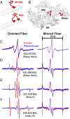
Fig. 1.
Fig. 1.
(A) Chemical structure of BSL. (B) BSL bound stereospecifically to an α-helix at positions i and i+4, as in ref. . (C) Angles θNB and ϕNB that define the orientation of the nitroxide spin label (defined by axes xN, yN, zN) relative to the applied magnetic field B, which directly determine the orientation dependence of the EPR spectrum. (D) Orienting the helically ordered muscle fiber (and thus the actin filament axis) with B permits direct measurement of the nitroxide orientation relative to actin.
Fig. 2.
Fig. 2.
(A) MTSSL and BSL on the relay helix. (B) BSL labeling sites chosen on three stable helices throughout the myosin CD. (C–F) (Left) EPR spectra of the spin-labeled constructs on oriented fiber bundles, in the absence of nucleotide (rigor), with the fiber axis aligned parallel (red) and perpendicular (blue) to the magnetic field. (Right) EPR spectra of randomly-oriented (minced fiber) preparations of all spin-labeled constructs. (C) S1dC labeled at residue 492 with MTSSL (relay helix). (D) S1dC labeled at residues 492 and 496 with BSL (relay helix, bifunctional analog to C). (E) S1dC labeled at residues 325 and 329 with BSL (helix K). (F) S1dC labeled at residues 639 and 643 with BSL (helix W).
Fig. S1.
Fig. S1.
Actin-activated ATPase data for bifunctional constructs. (Upper) Di-Cys myosin S1dC constructs with labeling sites highlighted and spin labels modeled in red. (Lower) Actin-activated ATPase data for each mutant construct, before labeling (red) and after labeling with BSL (blue). Errors are ±SEM (n = 3).
Fig. S2.
Fig. S2.
Comparison of BSL spectra from single-Cys and di-Cys constructs. Spectra were acquired on parallel-oriented fiber bundles in the absence of nucleotide. Black, spectrum of BSL attached to a di-Cys S1dC construct at positions 639 and 643 (helix W); blue, spectrum of MTSSL attached to a single-Cys S1dC construct at position 639 (helix W); orange, spectrum of BSL attached to same construct as in blue. Absence of blue or orange spectral components from the black spectrum indicates complete bifunctional labeling on the di-Cys construct.
Fig. 3.
Fig. 3.
(A) BSL-labeled myosin bound to actin in skinned muscle fibers in the absence of nucleotide (black) and the presence of 5 mM MgADP (green). (B) θNA distributions for nucleotide-free (black) and ADP-bound (green) biochemical states, obtained by spectral simulation and least-squares fitting of the data in A.
Fig. S3.
Fig. S3.
EPR spectra of fibers oriented parallel to the applied magnetic field. (Left) Data (black), best fit simulated spectrum (red) and residual (purple). (Center and Right) Angular distributions associated with each simulated spectrum, showing angular component 1 (cyan), angular component 2 (violet), and envelope (black). EPR is subject to some intrinsic ambiguity in the determination of angular centers, because the orientation dependence in EPR depends on squared cosines of angles. For simplicity, a single distribution on the interval [0,90] is reported here for each parameter. In all cases in this study, only one plausible helix orientation was found. For each construct, we observed two well-resolved spectral components, corresponding to a major, highly ordered spin label population, and a minor, more weakly ordered population. We hypothesize that the second component arises predominantly either from unbound S1dC trapped in the myofilament lattice during fiber decoration, or (in the case of the site on helix W, where the minor component is relatively well ordered) from a small population of alternate BSL conformers stabilized by the structural environment of the specific labeling site. Thus, in the main text, only the primary component is considered.
Fig. 4.
Fig. 4.
Visualization of coordinate transformations. (A) Actin (yellow) in complex with myosin S1 (blue) labeled with BSL (red). The actin long axis (gold, same as the magnetic field axis B in Fig. 1C, because fibers are aligned parallel to B) has a well-defined orientation within BSL’s nitroxide coordinate frame (white), defined by angles θNA and ϕNA (B). (C) BSL bound to a helix, with the nitroxide frame (gray) and helix axis (red) highlighted, defining angles θNH and ϕNH that describe the helix orientation relative to BSL (D). (E) Actin (yellow) and myosin (blue) in complex, as in A, showing the two vectors of interest, actin (gold) and a representative helix axis (red), defining angle θAH between them (F).
Fig. 5.
Fig. 5.
(A) Structural alignment of three Dicty myosin relay helix crystal structures, showing the nucleotide-induced C-terminal bend. (B) Relay helix from our actomyosin model (32), showing the EPR-derived change induced by ADP (black to green), with the ADP.Pi structure from A shown for reference. (C) Actin (yellow) in complex with myosin (blue), with EPR-derived mean relay helix orientations highlighted. (D and E) EPR-derived amplitudes of angular disorder.

References

    1. Lymn RW, Taylor EW. Mechanism of adenosine triphosphate hydrolysis by actomyosin. Biochemistry. 1971;10(25):4617–4624. - PubMed
    1. Preller M, Manstein DJ. Myosin structure, allostery, and mechano-chemistry. Structure. 2013;21(11):1911–1922. - PubMed
    1. Thomas DD, Prochniewicz E, Roopnarine O. Changes in actin and myosin structural dynamics due to their weak and strong interactions. Results Probl Cell Differ. 2002;36:7–19. - PMC - PubMed
    1. Spudich JA. Hypertrophic and dilated cardiomyopathy: Four decades of basic research on muscle lead to potential therapeutic approaches to these devastating genetic diseases. Biophys J. 2014;106(6):1236–1249. - PMC - PubMed
    1. Tajsharghi H, Oldfors A. Myosinopathies: Pathology and mechanisms. Acta Neuropathol. 2013;125(1):3–18. - PMC - PubMed

Publication types

LinkOut - more resources