. 2015 Jun 23;112(25):E3255-64.
doi: 10.1073/pnas.1509322112. Epub 2015 Jun 8.

Molecular transitions from papillomavirus infection to cervical precancer and cancer: Role of stromal estrogen receptor signaling

Affiliations

Molecular transitions from papillomavirus infection to cervical precancer and cancer: Role of stromal estrogen receptor signaling

Johan A den Boon et al. Proc Natl Acad Sci U S A. .

Abstract

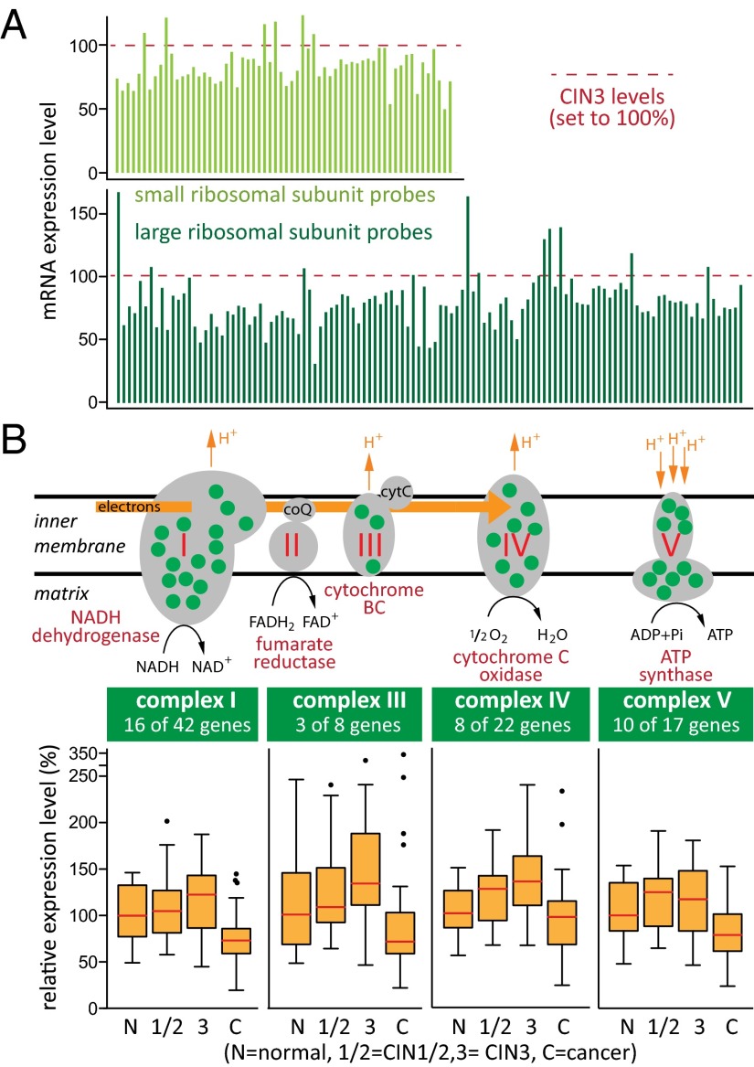
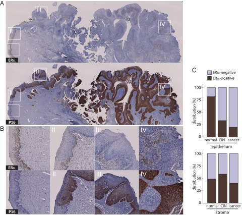
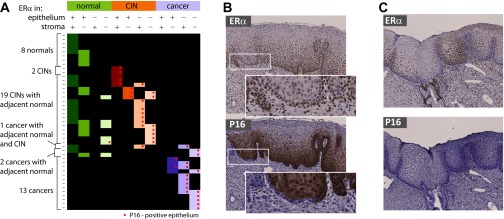
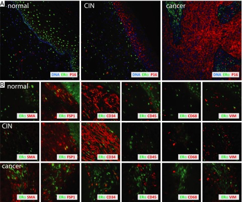
To study the multistep process of cervical cancer development, we analyzed 128 frozen cervical samples spanning normalcy, increasingly severe cervical intraepithelial neoplasia (CIN1- CIN3), and cervical cancer (CxCa) from multiple perspectives, revealing a cascade of progressive changes. Compared with normal tissue, expression of many DNA replication/repair and cell proliferation genes was increased in CIN1/CIN2 lesions and further sustained in CIN3, consistent with high-risk human papillomavirus (HPV)-induced tumor suppressor inactivation. The CIN3-to-CxCa transition showed metabolic shifts, including decreased expression of mitochondrial electron transport complex components and ribosomal protein genes. Significantly, despite clinical, epidemiological, and animal model results linking estrogen and estrogen receptor alpha (ERα) to CxCa, ERα expression declined >15-fold from normalcy to cancer, showing the strongest inverse correlation of any gene with the increasing expression of p16, a marker for HPV-linked cancers. This drop in ERα in CIN and tumor cells was confirmed at the protein level. However, ERα expression in stromal cells continued throughout CxCa development. Our further studies localized stromal ERα to FSP1+, CD34+, SMA- precursor fibrocytes adjacent to normal and precancerous CIN epithelium, and FSP1-, CD34-, SMA+ activated fibroblasts in CxCas. Moreover, rank correlations with ERα mRNA identified IL-8, CXCL12, CXCL14, their receptors, and other angiogenesis and immune cell infiltration and inflammatory factors as candidates for ERα-induced stroma-tumor signaling pathways. The results indicate that estrogen signaling in cervical cancer has dramatic differences from ERα+ breast cancers, and imply that estrogen signaling increasingly proceeds indirectly through ERα in tumor-associated stromal fibroblasts.

Keywords: HPV; cervical cancer; estrogen; stroma; tumor microenvironment.


Conflict of interest statement

The authors declare no conflict of interest.

Figures

Fig. 1.
Fig. 1.
Differential gene expression and biological processes affected during cervical cancer progression. (A) Heatmap presentations of gene expression levels measured by 2,084 Affymetrix probe sets measuring genes whose expression predominantly increases or decreases at only one disease-stage transition. (B) Blue/red plots hierarchically clustered Gene Ontology classes that are enriched for these genes and reveal prominent GO clusters (circled) annotated (Right). Full details on gene expression measurements and GO enrichment analyses are provided in Datasets S2 and S5, respectively.
Fig. 2.
Fig. 2.
Expression of genes encoding ribosomal proteins and mitochondrial electron transport chain complexes I, III, IV, and V is reduced at the transition from CIN3 to cancer. (A) Twenty-four small and 35 large ribosomal subunit proteins (∼70% of all ribosomal proteins) are expressed at statistically significant reduced levels in cancers compared with precancerous CIN3 lesions. Histograms show measurements in cancers by all 177 Affymetrix microarray probe sets for ribosomal protein-encoding genes, normalized to levels measured in CIN3s. (B) Green dots in the five complexes of the mitochondrial electron transport chain indicate proteins with reduced gene expression in cancers. Box plots (Tukey) show the average distribution of gene expression of all differentially expressed ETC complex I, III, IV, and V components (median levels are in red).
Fig. 3.
Fig. 3.
Differential expression of estrogen-responsive genes correlates strongly with cervical cancer progression. (A) Hierarchical clustering of 128 specimens based on changes in gene expression measured by 365 Affymetrix probe sets measuring expression of 217 estrogen-responsive genes shows strong correlation with disease progression. (B) Box plots (Tukey) show the distribution of Affymetrix microarray-based gene expression measurements of CDKN2A (p16), ERα, and estrogen-responsive genes GREB1, PGR1, and AR. White and red horizontal lines indicate median expression levels.
Fig. 4.
Fig. 4.
Negative correlation between ERα and p16 expression during cervical cancer progression. (A) IHC stains for ERα (Top) and p16 (Bottom) in a cervical specimen representing complete disease progression. (B) Higher magnifications (I–IV) show that ERα resides in the nuclei of dividing cells in stratified differentiating normal healthy epithelium (I), is lost from progressively p16-positive undifferentiated epithelial CIN lesions (II and III), and is entirely lacking in cancerous epithelium (IV), and that ERα becomes exclusive to interspersed stroma. (C) Epithelial and adjacent stromal distribution of ERα by disease pathology, based on 77 observations in 43 individual specimens, shows loss of ERα in epithelium but not in stroma.
Fig. S1.
Fig. S1.
ERα and p16 transitional expression phenotypes. (A) Overview of IHC observations for ERα and p16 from 43 specimens shows distinct local sites of varying levels of disease progression within single sections, accompanied by changes in epithelial distribution of ERα in epithelium and stroma. Cancer progression gravitates toward epithelial loss of ERα expression and increased expression of p16. (B) Example of a specimen with ERα-positive epithelium throughout but with p16 staining progressing from the right-hand side of the image. A single epithelial lobe at the border of p16-negative and p16-positive epithelium shows reduced ERα staining and ERα-positive/p16-negative basal epithelium (Insets). (C) Example of a specimen with distinct convoluted epithelial regions with reduced ERα staining that are nevertheless still negative for p16.
Fig. 5.
Fig. 5.
ERα costains stromal fibroblasts. (A) Low-magnification immunofluorescent ERα, p16, and DAPI in three cervical specimens representing disease progression. With increased expression of p16 in CIN epithelium and epithelial cancer compartments, ERα expression is lost and becomes restricted to stroma. (B) Higher magnifications of tissues stained with various cell-type markers show that in normal and precancerous lesions, ERα resides in the nuclei of stromal cells that stain positive for FSP1 and CD34. Only a subset of ERα-positive cancer-associated stromal cells expresses CD34 and is increasingly positive for αSMA and occasionally also for vimentin. ERα is not expressed in CD45-positive hematopoietic cells and CD68-positive macrophages.

References

    1. World Health Organization (2015) Human papillomavirus (HPV) and cervical cancer, Fact Sheet No 380. Available at www.who.int/mediacentre/factsheets/fs380/en/. Accessed May 26, 2015.
    1. Wentzensen N, Schiffman M. Filling a gap in cervical cancer screening programmes. Lancet Oncol. 2014;15(3):249–251. - PMC - PubMed
    1. Schiffman M, Wentzensen N. Human papillomavirus infection and the multistage carcinogenesis of cervical cancer. Cancer Epidemiol Biomarkers Prev. 2013;22(4):553–560. - PMC - PubMed
    1. Hanahan D, Weinberg RA. Hallmarks of cancer: The next generation. Cell. 2011;144(5):646–674. - PubMed
    1. zur Hausen H. Papillomaviruses in the causation of human cancers—A brief historical account. Virology. 2009;384(2):260–265. - PubMed

Publication types

MeSH terms

Substances

Associated data