. 2015 Jun 23;112(25):7713-8.
doi: 10.1073/pnas.1504615112. Epub 2015 Jun 8.

Retro-translocation of mitochondrial intermembrane space proteins

Affiliations

Retro-translocation of mitochondrial intermembrane space proteins

Piotr Bragoszewski et al. Proc Natl Acad Sci U S A. .

Abstract

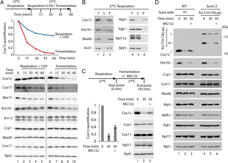
The content of mitochondrial proteome is maintained through two highly dynamic processes, the influx of newly synthesized proteins from the cytosol and the protein degradation. Mitochondrial proteins are targeted to the intermembrane space by the mitochondrial intermembrane space assembly pathway that couples their import and oxidative folding. The folding trap was proposed to be a driving mechanism for the mitochondrial accumulation of these proteins. Whether the reverse movement of unfolded proteins to the cytosol occurs across the intact outer membrane is unknown. We found that reduced, conformationally destabilized proteins are released from mitochondria in a size-limited manner. We identified the general import pore protein Tom40 as an escape gate. We propose that the mitochondrial proteome is not only regulated by the import and degradation of proteins but also by their retro-translocation to the external cytosolic location. Thus, protein release is a mechanism that contributes to the mitochondrial proteome surveillance.

Keywords: mitochondria; protein biogenesis; protein quality control; protein transport; protein turnover.


Conflict of interest statement

The authors declare no conflict of interest.

Figures

Fig. 1.
Fig. 1.
Proteins that are defective in oxidative folding can be released from the IMS to the cytosol. (A) Schematic representation and cellular levels of b2-Mia40core variants. (B) Cellular levels of b2-Mia40core expressed in WT and erv1-2int strains. (C) Import of [35S]b2-Mia40core and [35S]b2(167)-DHFR into WT mitochondria with or without IA (50 mM). (D) Cellular levels of b2-Mia40core-C3S in WT, Δrad6, and Δate1 strains. Proteins were analyzed by SDS/PAGE and immunodetection (A, B, and D) or autoradiography (C). i, intermediate; m, mature; p, precursor; WT, wild type; *, altered migration of b2-Mia40core-C3S m-form.
Fig. 2.
Fig. 2.
Monitoring the release of MIA substrate proteins in organello. (A) Protein levels in WT mitochondria and corresponding supernatants (release) upon treatment with DTT. (B) Protein levels and redox state in WT mitochondria and supernatants (release) upon treatment with DTT followed by denaturation in the presence of thiol-modifying agent AMS. (C) Levels of b2-Mia40core-C4S in mitochondria and supernatants (release) upon reduction with DTT with or without protease pretreatment. (D) Protein levels in human HEK293 mitochondria and supernatants (release) upon reduction with DTT. (AD) Proteins were analyzed by SDS/PAGE and immunodetection. Protein molecular weight (A, C, and D; kDa) or number of cysteine residues (B) is given in parentheses. i, intermediate; m, mature; ox, oxidized; red, reduced; WT, wild type.
Fig. 3.
Fig. 3.
Erv1-2 is destabilized and released from mitochondria upon heat shock. (A) Cellular protein levels of WT and erv1-2 strains grown at 19 °C and 37 °C. (B) Levels of Erv1 and Erv1-2 proteins in mitochondria and supernatants (release) upon incubation at the indicated temperatures. (A and B) Proteins were analyzed by SDS/PAGE and immunodetection. WT, wild type; *, cleaved forms of Erv1.
Fig. 4.
Fig. 4.
Protein release is size dependent and involves the TOM complex. (A) Release of Pet191FLAG and Pet191_Pet191FLAG from mitochondria upon treatment with DTT. (B) Release of Pet191 upon modification with mPEG (5,000 Da) and quantification. Signals from mitochondrial fractions not treated with DTT were set to 1. (C) Protein release from WT and Δtom5 mitochondria upon treatment with DTT and quantification. Signals from the mitochondrial fractions of WT and Δtom5 not treated with DTT were set to 1. Mitochondria fractions are shown in Fig. S5A. Mean ± SEM (n = 3). (D) Susceptibility of Tom40 and Tom40C130/C138 to thiol-modifying agents (AMS or mPEG) under native conditions. (E) Protein release from WT and Tom40C130/C138 mitochondria pretreated with mPEG upon treatment with DTT. Mitochondria fractions are shown in Fig. S5E. (F) Mix17FLAG release from Tom40C130/C138 mitochondria, with or without pretreatment with mPEG, upon treatment with DTT and quantification. The signal from the sample not treated with mPEG and incubated with DTT for 25 min was set to 1. Signals from DTT untreated samples were subtracted. Mean ± SEM (n = 3). Mitochondria fractions are shown in Fig. S5G. (AF) Proteins were analyzed by SDS/PAGE and immunodetection. WT, wild type; *, full-size Mix17FLAG; **, Mix17FLAG degradation product.
Fig. 5.
Fig. 5.
Respiration-to-fermentation shift induces protein escape to the cytosol and proteasomal clearance. (A) Cellular protein levels upon inhibition of protein synthesis or shift from respiration to fermentation. Experimental scheme and quantification of Cox12 are provided. Signals from time point 0 were set to 1. (B) Subcellular localization of MIA substrate proteins in the cells grown under respiratory conditions. (C) Protein levels upon shift from respiration to fermentation with or without MG132 treatment. Experimental scheme and quantification of Cox12 are provided. Signals from time point 0 were set to 1. Mean ± SEM (n = 7). (D) Protein levels in WT and ∆cox12 cells with plasmid expressing b2-Cox12FLAG upon shift from respiration to fermentation with or without MG132 treatment. Experiment was done based on the scheme as in C. (AD) Proteins were analyzed by SDS/PAGE and immunodetection. CHX, cycloheximide; ev, empty vector; i, intermediate; m, mature; P, mitochondrial pellet; S, postmitochondrial supernatant, T, total; WT, wild type.

References

    1. Neupert W, Herrmann JM. Translocation of proteins into mitochondria. Annu Rev Biochem. 2007;76:723–749. - PubMed
    1. Chacinska A, Koehler CM, Milenkovic D, Lithgow T, Pfanner N. Importing mitochondrial proteins: Machineries and mechanisms. Cell. 2009;138(4):628–644. - PMC - PubMed
    1. Schmidt O, Pfanner N, Meisinger C. Mitochondrial protein import: From proteomics to functional mechanisms. Nat Rev Mol Cell Biol. 2010;11(9):655–667. - PubMed
    1. Endo T, Yamano K, Kawano S. Structural insight into the mitochondrial protein import system. Biochim Biophys Acta. 2011;1808(3):955–970. - PubMed
    1. Dudek J, Rehling P, van der Laan M. Mitochondrial protein import: Common principles and physiological networks. Biochim Biophys Acta. 2013;1833(2):274–285. - PubMed

Publication types

MeSH terms

Substances