. 2015 Jun 23;112(25):7833-8.
doi: 10.1073/pnas.1423088112. Epub 2015 Jun 8.

Caffeine acts through neuronal adenosine A2A receptors to prevent mood and memory dysfunction triggered by chronic stress

Affiliations

Caffeine acts through neuronal adenosine A2A receptors to prevent mood and memory dysfunction triggered by chronic stress

Manuella P Kaster et al. Proc Natl Acad Sci U S A. .

Abstract

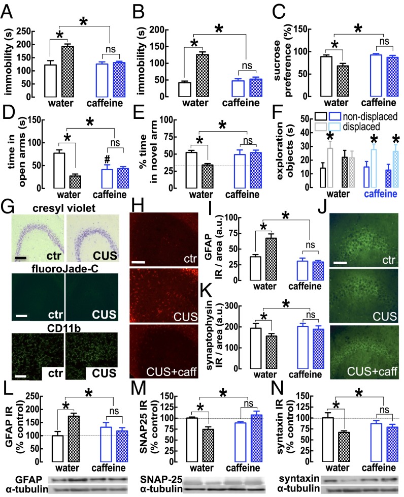
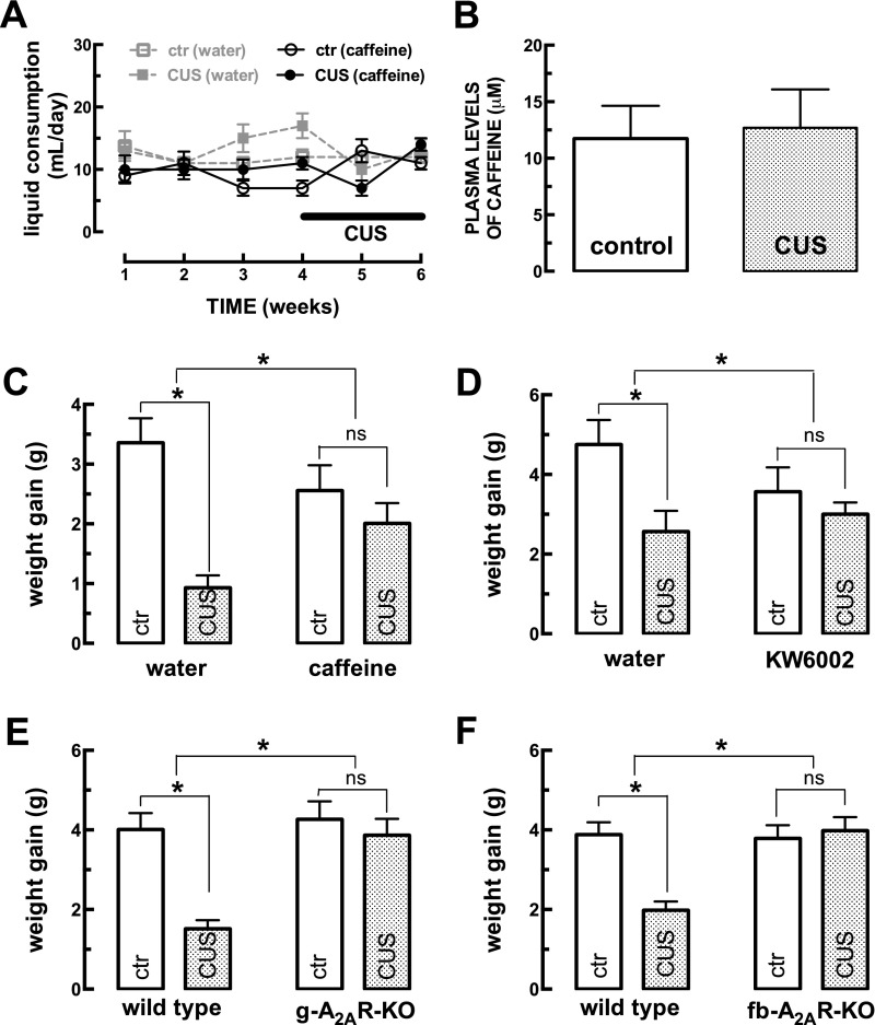
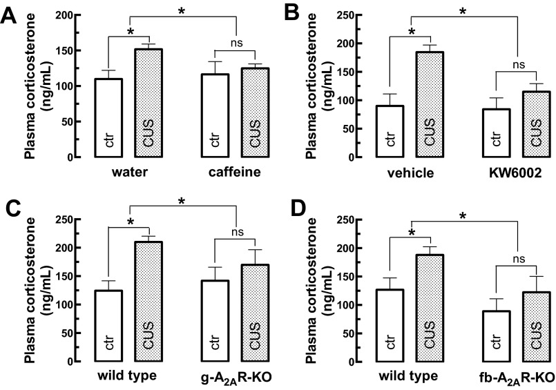
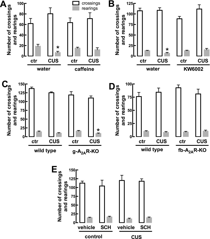
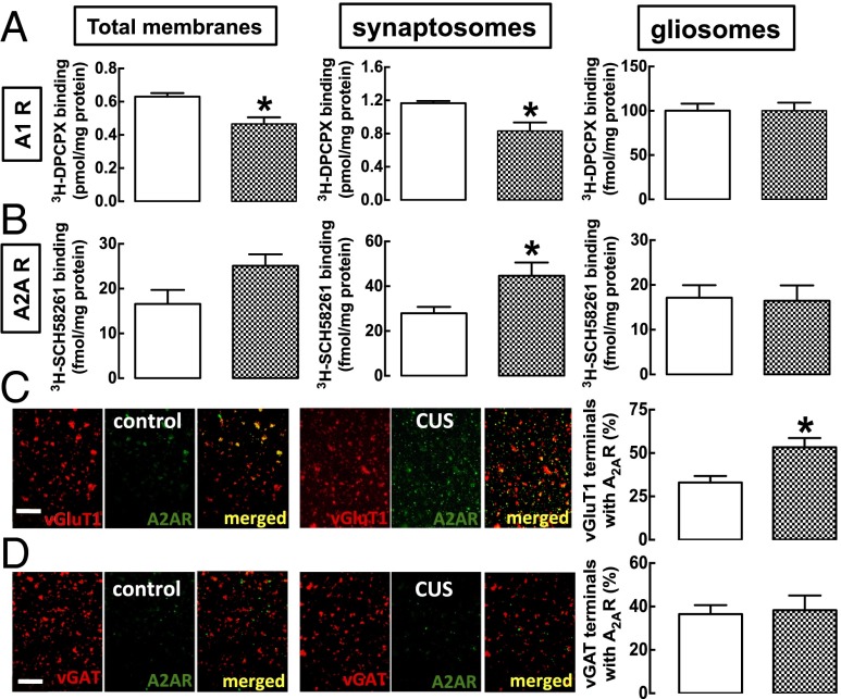
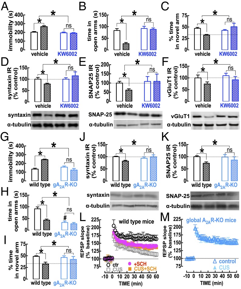
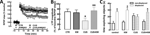
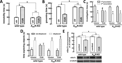
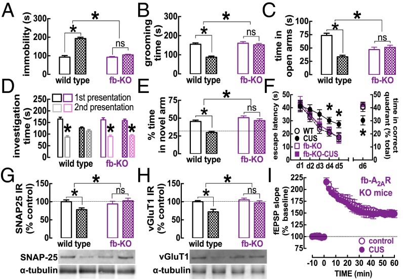
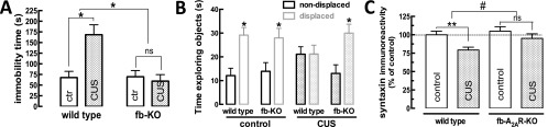
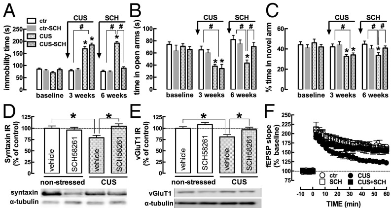
The consumption of caffeine (an adenosine receptor antagonist) correlates inversely with depression and memory deterioration, and adenosine A2A receptor (A2AR) antagonists emerge as candidate therapeutic targets because they control aberrant synaptic plasticity and afford neuroprotection. Therefore we tested the ability of A2AR to control the behavioral, electrophysiological, and neurochemical modifications caused by chronic unpredictable stress (CUS), which alters hippocampal circuits, dampens mood and memory performance, and enhances susceptibility to depression. CUS for 3 wk in adult mice induced anxiogenic and helpless-like behavior and decreased memory performance. These behavioral changes were accompanied by synaptic alterations, typified by a decrease in synaptic plasticity and a reduced density of synaptic proteins (synaptosomal-associated protein 25, syntaxin, and vesicular glutamate transporter type 1), together with an increased density of A2AR in glutamatergic terminals in the hippocampus. Except for anxiety, for which results were mixed, CUS-induced behavioral and synaptic alterations were prevented by (i) caffeine (1 g/L in the drinking water, starting 3 wk before and continued throughout CUS); (ii) the selective A2AR antagonist KW6002 (3 mg/kg, p.o.); (iii) global A2AR deletion; and (iv) selective A2AR deletion in forebrain neurons. Notably, A2AR blockade was not only prophylactic but also therapeutically efficacious, because a 3-wk treatment with the A2AR antagonist SCH58261 (0.1 mg/kg, i.p.) reversed the mood and synaptic dysfunction caused by CUS. These results herald a key role for synaptic A2AR in the control of chronic stress-induced modifications and suggest A2AR as candidate targets to alleviate the consequences of chronic stress on brain function.

Keywords: adenosine A2A receptor; caffeine; chronic stress; mood dysfunction; synaptic dysfunction.


Conflict of interest statement

The authors declare no conflict of interest.

Figures

Fig. 1.
Fig. 1.
Mice subjected to CUS display the expected features of depressed mice, which are largely prevented by the regular consumption of caffeine. Male mice (10 wk old) were subjected to a 3-wk period of CUS (Table S1) and were evaluated behaviorally 24 h after the last stressor. Compared with nonstressed control mice (ctr, open bars), CUS-mice (checkered bars) displayed helpless-like behavior as evaluated by the forced-swimming (A) and tail-suspension (B) tests, anhedonia as evaluated by a sucrose preference test (C), anxiety-like behavior as evaluated by the elevated-plus maze test (D), and impaired memory performance as evaluated by a modified Y maze test (E) and an object-displacement test (Student’s t test comparing displaced vs. nondisplaced object) (F). After mice were killed, the CA3 area of hippocampi from CUS-subjected mice did not display overt neuronal damage, as gauged by the preservation of cresyl violet staining (G, Top Row) and lack of FluoroJade C staining (G, Middle Row) or microgliosis as evaluated by CD11b immunoreactivity (G, Bottom Row) but did display increased GFAP (H and I) and decreased synaptophysin immunoreactivity (J and K). Similar findings were obtained in the hippocampal CA1 area. Western blot analysis of whole hippocampal membranes confirmed the increase in GFAP density with CUS (L) and the decrease of synaptic markers, namely SNAP25 (M) and syntaxin (N). The administration of caffeine (1 g/L via the drinking water) to mice beginning 3 wk before CUS and continuing until mice were killed did not modify behavior or histology, except for increased anxiety in the elevated-plus maze (D), but did prevent all CUS-induced behavioral and morphological alterations. (Scale bars, 100 μm.) Data are shown as mean ± SEM; n = 9–19 mice per group in the behavioral assays (AF); n = 4–7 mice per group in the morphological analysis; n = 5 or 6 mice per group in the neurochemical analysis. *P < 0.05 and #P < 0.05 using a two-way ANOVA followed by a Newman–Keuls post hoc test, except when stated otherwise. ns, not significant.
Fig. S1.
Fig. S1.
Mice subjected to CUS do not change their liquid intake and have a decreased weight gain. Male mice (10 wk old) were subjected to a 3-wk period of CUS (Table S1). (A) Liquid intake is not significantly altered upon CUS or with caffeine consumption. (B) Caffeine reaches similar plasma concentrations in control mice and in mice subjected to CUS, as measured by HPLC. (CF) Compared with control mice (ctr), CUS caused decreased weight gain, which was prevented by the regular consumption of either caffeine (1 g/L) [CUS F(1,70) = 17.95; caffeine F(1,70) = 0.16; interaction F(1,70) = 7.15] (C) or the selective A2AR antagonist KW6002 (3 mg/kg) [CUS F(1,33) = 6.64; KW6002 F(1,33) = 0.49; interaction F(1,33) = 2.27] (D) and which also was abrogated by the global deletion of A2AR (g-A2AR-KO) [CUS F(1,36) = 14.33; genotype F(1,36) = 11.69; interaction F(1,36) = 7.50] (E) or by the selective deletion of neuronal A2AR in fb-A2AR-KO mice [CUS F(1,32) = 7.92; genotype F(1,32) = 9.89; interaction F(1,32) = 11.84] (F). Data are shown as mean ± SEM. n = 19–27 mice per group in the liquid consumption experiment (A); n = 6 or 7 mice per group in the HPLC experiments (B); n = 7–19 mice per group in the weight-gain determinations (CF). *P < 0.05 using two-way ANOVA followed by a Newman–Keuls post hoc test; ns, nonsignificant.
Fig. S2.
Fig. S2.
CUS increased the plasma levels of corticosterone, and caffeine, and A2AR blockade prevented this increase. Male mice (10 wk old) were subjected to a 3-wk period of CUS (Table S1). Three days later, animals were gently immobilized, and blood was collected from the tail vein. Animals were killed at 9:00 AM on the day of blood collection. Compared with control mice (ctr), CUS increased the plasma levels of corticosterone. This increase was prevented by the regular consumption of either caffeine (1 g/L) [CUS F(1,70) = 7.95; caffeine F(1,70) = 2.34; interaction F(1,70) = 4.36] (A) or the selective A2AR antagonist KW6002 (3 mg/kg) [CUS F(1,33) = 15.30; KW6002 F(1,33) = 6.78; interaction F(1,33) = 4.99] (B) and also was abrogated by the global deletion of A2AR (g-A2AR-KO) [CUS F(1,36) = 14.91; genotype F(1,36) = 1.50; interaction F(1,36) = 6.41] (C) or by the selective deletion of neuronal A2AR in fb-A2AR-KO mice [CUS F(1,32) = 16.79; genotype F(1,32) = 20.55; interaction F(1,32) = 4.56] (D). Data are shown as mean ± SEM of 7–19 mice per group. *P < 0.05 using two-way ANOVA followed by a Newman–Keuls post hoc test; ns, nonsignificant.
Fig. S3.
Fig. S3.
CUS does not significantly affect spontaneous locomotion. Male mice (10 wk old) were subjected to a 3-wk period of CUS (Table S1) and were evaluated 24 h after the last stressor in an open-field arena. Both the number of crossings and the number of rearing events were globally similar in nonstressed control mice (ctr) and in mice subjected to CUS. Furthermore, neither the regular consumption of caffeine (1 g/L) (A) or of the selective A2AR antagonist KW6002 (3 mg/kg) (B) nor the global deletion of A2AR (g-A2AR-KO) (C) or the selective deletion of neuronal A2AR in fb-A2AR-KO mice (D) affected spontaneous locomotion. (E) Likewise, the pattern of spontaneous locomotion was similar in control mice and mice subjected to CUS that subsequently were treated for 3 wk with the A2AR antagonist SCH58261. Data are shown as mean ± SEM of 7–19 mice per group. *P < 0.05 using two-way ANOVA followed by a Newman–Keuls post hoc test; ns, nonsignificant.
Fig. 2.
Fig. 2.
CUS alters the adenosine neuromodulation system in the hippocampus. Male mice (10 wk old) were subjected to a 3-wk period of CUS (Table S1) and were killed for preparation of total, synaptosomal (i.e., from synapses), and gliosomal (i.e., from astrocytes) membranes from the hippocampus. (A) The binding density of A1R [evaluated with the A1R antagonist 3H-DPCPX (10 nM)] was decreased in total and synaptosomal membranes and was unaltered in gliosomal membranes of CUS-subjected mice (checkered bars) compared with nonstressed mice (control; open bars). (B) In contrast, there was a selective increase in the binding density of the A2AR antagonist 3H-SCH58261 in synaptosomal membranes from CUS-subjected mice, without changes in its binding density in total or gliosomal membranes. (C and D) Double-labeling immunocytochemical analysis of plated purified nerve terminals confirmed that A2AR are located mostly in glutamatergic (immunopositive for vGluT1) rather than GABAergic (immunopositive for vGAT) nerve terminals and showed that in CUS-exposed mice the number of glutamatergic terminals was enhanced selectively (C), rather than GABAergic terminals endowed with A2AR (D). Note that this immunocytochemistry approach allows only the relative colocalization of epitopes to be quantified, irrespective of their absolute staining, which varies among groups of plated nerve terminals. We previously validated the selectivity of 3H-SCH58261 and of the anti-A2AR antibodies used, which do not yield any signal in tissue from A2AR-KO mice (47). Data are shown as mean ± SEM of 5 or 6 mice per group; *P < 0.05 using an unpaired Student’s t test.
Fig. 3.
Fig. 3.
The pharmacological or genetic blockade of A2AR prevents CUS-induced behavioral, neurochemical, and electrophysiological alterations in the hippocampus. Male mice (10 wk old) were subjected to a 3-wk period of CUS (Table S1) before behavioral evaluation 24 h after the last stressor. In CUS-subjected mice (checkered bars), as compared with vehicle-treated mice (AF) (open bars) or wild-type mice (GM) (open bars and symbols), the consumption of the A2AR antagonist KW6002 (3 mg/kg, through the drinking water, starting 3 d before CUS until mice were killed) (AF), or the genetic elimination of A2AR in global A2AR-KO mice (GM), prevented the CUS-induced helpless-like behavior evaluated in the forced-swimming test (A and G), the anxiety-like behavior evaluated in the elevated-plus maze test (B and H), the impaired memory performance evaluated in a modified Y maze test (C and I), and the decreases in synaptic markers such as syntaxin (D and J), SNAP-25 (E and K), and markers of glutamatergic terminals (vGluT1) (F) in hippocampal nerve terminals. Additionally, A2AR blockade with the antagonist SCH58261 (SCH, 50 nM) prevented the CUS-induced depression of LTP [triggered by a high-frequency stimulation train at time 0 in Schaffer fibers (collateral synapses of CA1 pyramidal cells)] of hippocampal slices from wild-type mice (L) and CUS failed to modify LTP in A2AR-KO mice (M) (Student’s t test). Data are shown as mean ± SEM; n = 8–10 mice per group in the behavioral assays (AC and GI); n = 5 or 6 mice per group in the neurochemical analyses (DF, J, and K); and n = 5 or 6 mice per group in the electrophysiological analyses (L and M). *P < 0.05 using a two-way ANOVA followed by a Newman–Keuls post hoc test, except when stated otherwise; ns, nonsignificant.
Fig. S4.
Fig. S4.
The pharmacological inhibition of A2AR blunts behavioral and electrophysiological alterations caused by CUS. Male mice (10 wk old) were subjected to a 3-wk period of CUS (Table S1) and were evaluated 24 h after the last stressor in an open-field arena. Mice were killed 8 d after the end of the CUS period. (A and B) Synaptic plasticity (induced by a high-frequency stimulation train at time 0) recorded in Schaffer fibers (CA1 pyramid synapses) was decreased in hippocampal slices from mice subjected to CUS, and this decrease was prevented in mice consuming the A2AR antagonist KW6002 (KW) (3 mg⋅kg−1⋅d−1) from 3 d before the beginning of the protocol until the end of the CUS protocol. n = 5–6 mice per group. *P < 0.05 vs. control; **P < 0.05 vs. CUS; two-way ANOVA [CUS F(1,11) = 5.24; KW6002 F(1,11) = 8.71; interaction F(1,11) = 12.60] followed by a Newman–Keuls post hoc test. CTR, control. (C) In an object-displacement test, mice were exposed to two identical objects for 3 min and 90 min later were exposed for 3 min to the same objects, but with one of the objects in a different location. Control mice spent more time sniffing/exploring the displaced object than the nondisplaced object. This spatial recognition memory was blunted in mice subjected to CUS but was present in mice consuming KW6002. n = 8–10 mice per group. *P < 0.05 vs. time exploring the nondisplaced object; Student’s t test.
Fig. S5.
Fig. S5.
The genetic deletion of A2AR blunts behavioral and neurochemical alterations caused by CUS. Male mice (10 wk old) were subjected to a 3-wk period of CUS (Table S1) and were evaluated in an open-field arena 24 h after the last stressor. Mice were killed 8 d after the end of the CUS period. In contrast with the effect of CUS (patterned bars) in wild-type mice, in global A2AR-KO mice CUS failed to trigger helpless-like behavior in the tail-suspension test (A) [CUS F(1,36) = 65.33; genotype F(1,36) = 58.16; interaction F(1,36) = 50.48], anhedonia-like behavior in the splash test (B) [CUS F(1,36) = 35.61; genotype F(1,36) = 22.87; interaction F(1,36) = 25.51], or impaired social interaction (C) [first presentation, CUS F(1,36) = 4.32; genotype F(1,36) = 2.09; interaction F(1,36) = 5.04; social interaction memory, Student’s t test comparing first vs. second presentation of the foreign mouse] or impaired short-term spatial memory in the object-displacement test (D) (n = 10; *P < 0.05 vs. time exploring the nondisplaced object, Student’s t test). (E) The density of the vGluT1, a marker of glutamatergic terminals, was reduced in the hippocampus of wild-type mice subjected to CUS. This reduction was not observed in A2AR-KO mice subjected to CUS. n = 5–6 mice per group. *P < 0.05, two-way ANOVA [CUS F(1,20) = 11.42; genotype F(1,20) = 7.53; interaction F(1,20) = 5.29] followed by a Newman–Keuls post hoc test. n = 5–6 mice per group; nonsignificant (ns) differences at P > 0.05, two-way ANOVA [CUS F(1,20) = 0.19; genotype F(1,20) = 0.01; interaction F(1,20) = 0.02] followed by a Newman–Keuls post hoc test.
Fig. 4.
Fig. 4.
The selective deletion of neuronal A2AR prevents CUS-induced behavioral, neurochemical, and electrophysiological alterations in the hippocampus. Male mice (10 wk old) were subjected to a 3-wk period of CUS (Table S1) before behavioral evaluation 24 h after the last stressor. In contrast with the impact of CUS in wild-type mice (checkered bars and filled symbols; control: open bars and symbols), in fb-A2AR-KO mice (in which neuronal A2AR is eliminated selectively in the forebrain and A2AR-mediated control of glutamatergic synapses is blunted) CUS failed to trigger helpless-like behavior in the forced-swimming test (A), anhedonia-like behavior in the splash test (B), anxiety-like behavior in the elevated-plus maze test (C), impaired social interaction and social interaction memory (Student’s t test comparing first vs. second presentation of the foreign mouse) (D), or impaired memory performance in a modified Y maze (E). (F) Additionally, in a spatial reference memory version of the Morris water maze test, both the acquisition (days 1–5) and the retention of the location of the hidden platform (day 6) were diminished by CUS in wild-type mice but not in fb-A2AR-KO mice. Also in contrast with the deleterious effects of CUS, which decreased the density of synaptic markers such as syntaxin (G) and vGluT1 (H) in hippocampal nerve terminals in wild-type mice, CUS did not affect the density of either of these synaptic markers (G and H) or the amplitude of LTP (I) in fbA2AR-KO mice. Data are shown as mean ± SEM; n = 7–9 mice per group in the behavioral assays (AF); n = 5 mice per group in the neurochemical analysis (G and H) and in the electrophysiological analysis (I). *P < 0.05 using a two-way ANOVA followed by a Newman–Keuls post hoc test, with repeated measures for the Morris water maze test. ns, nonsignificant.
Fig. S6.
Fig. S6.
The selective deletion of neuronal A2AR blunts behavioral and neurochemical alterations caused by CUS. Male mice (10 wk old) were subjected to a 3-wk period of CUS (Table S1) and were evaluated 24 h after the last stressor in an open-field arena. Mice were killed 8 d after the end of the CUS period. In contrast with the impact of CUS in wild-type mice (patterned bars), in fb-A2AR-KO mice (in which neuronal A2AR is eliminated selectively in the forebrain and A2AR-mediated control of glutamatergic synapses is blunted), CUS failed to trigger helpless-like behavior in the tail-suspension test [CUS F(1,32) = 61.10; genotype F(1,32) = 86.09; interaction F(1,32) = 91.89] (A) or impaired short-term spatial memory in the object-displacement test (n = 9; *P < 0.05 vs. time exploring the nondisplaced object; Student’s t test) (B). (C) The density of the presynaptic marker syntaxin was reduced in the hippocampus of wild-type subjected to CUS. This reduction was not observed in fb-A2AR-KO mice subjected to CUS. n = 5; *P < 0.05, two-way ANOVA [CUS F(1,16) = 19.15; genotype F(1,16) = 10.13; interaction F(1,16) = 4.16] followed by a Newman–Keuls post hoc test.
Fig. 5.
Fig. 5.
Blockade of adenosine A2AR reverses CUS-induced alterations. Male mice (10 wk old) were behaviorally evaluated (baseline). Then mice were randomized into two groups. One group (checkered bars) was subjected to a 3-wk period of CUS (Table S1). The mice in the control group (ctr) were handled daily. All mice were behaviorally evaluated after 3 wk. Finally, half of the mice in each group were i.p. injected daily with saline (bars filled with white), and the other half were injected with the A2AR antagonist SCH58261 (SCH, 0.1 mg⋅kg−1⋅d−1, gray-outlined bars). All mice were behaviorally evaluated again at 6 wk and then were killed for neurochemical and electrophysiological analysis. Compared with nonstressed (control) mice (noncheckered bars), the mice subjected to CUS (checkered bars) displayed increased immobility in the forced-swimming test [F(2,36) = 182.0, P < 0.0001] (A), decreased time in the open arms of the elevated-plus maze test [F(1,36) = 77.14, P < 0.0001] (B), and decreased time spent in the novel arm of a modified Y maze test [F(1,36) = 77.14, P < 0.0001] (C), both at the end of the CUS protocol (3 wk) and 3 wk later (6 wk). As shown in AC, SCH58261 treatment did not affect the behavior of control mice (noncheckered bars) but reversed the CUS-induced alterations in helpless behavior [F(1,36) = 77.14, P < 0.0001] (A), anxiety [F(2,36) = 21.35, P < 0.0001] (B), and spatial reference memory [F(2,36) = 14.88, P = 0.0005] (C) to the level of control (noncheckered bars). SCH58261 treatment also reversed the CUS-induced reduction in the density of the synaptic markers syntaxin [CUS F(1,16) = 4.62; SCH58261 F(1,16) = 10.54; interaction F(1,16) = 18.30] (D) and vGluT1 [CUS F(1,16) = 11.60; SCH58261 F(1,16) = 6.25; interaction F(1,16) = 4.54] (E) in hippocampal nerve terminal membranes and reversed the CUS-induced decrease in the amplitude of LTP in hippocampal slices [CUS F(1,16) = 49.89; SCH58261 F(1,16) = 16.74; interaction F(1,16) = 29.56] (F). Data are shown as mean ± SEM. n = 9 or 10 mice per group in the behavioral assays (A–C); n = 5 mice per group in the neurochemical analysis (D and E) and in the electrophysiological analysis (F). *P < 0.05, #P < 0.05 using a repeated ANOVA followed by a Newman–Keuls post hoc test.

References

    1. de Kloet ER, Joëls M, Holsboer F. Stress and the brain: From adaptation to disease. Nat Rev Neurosci. 2005;6(6):463–475. - PubMed
    1. McEwen BS. Physiology and neurobiology of stress and adaptation: Central role of the brain. Physiol Rev. 2007;87(3):873–904. - PubMed
    1. Kim JJ, Diamond DM. The stressed hippocampus, synaptic plasticity and lost memories. Nat Rev Neurosci. 2002;3(6):453–462. - PubMed
    1. Harris A, Ursin H, Murison R, Eriksen HR. Coffee, stress and cortisol in nursing staff. Psychoneuroendocrinology. 2007;32(4):322–330. - PubMed
    1. Lucas M, et al. Coffee, caffeine, and risk of depression among women. Arch Intern Med. 2011;171(17):1571–1578. - PMC - PubMed

Publication types