Review
. 2015 May 28;8(Suppl 1):1-8.
doi: 10.4137/MRI.S23560. eCollection 2015.

Correlation of Carotid Intraplaque Hemorrhage and Stroke Using 1.5 T and 3 T MRI

Affiliations
Review

Correlation of Carotid Intraplaque Hemorrhage and Stroke Using 1.5 T and 3 T MRI

Gerald S Treiman et al. Magn Reson Insights. .

Abstract

Carotid therosclerotic disease causes approximately 25% of the nearly 690,000 ischemic strokes each year in the United States. Current risk stratification based on percent stenosis does not provide specific information on the actual risk of stroke for most individuals. Prospective randomized studies have found only 10 to 12% of asymptomatic patients will have a symptomatic stroke within 5 years. Measurements of percent stenosis do not determine plaque stability or composition. Reports have concluded that cerebral ischemic events associated with carotid plaque are intimately associated with plaque instability. Analysis of retrospective studies has found that plaque composition is important in risk stratification. Only MRI has the ability to identify and measure the detailed components and morphology of carotid plaque and provides more detailed information than other currently available techniques. MRI can accurately detect carotid hemorrhage, and MRI identified carotid hemorrhage correlates with acute stroke.

Keywords: MRI; carotid; hemorrhage.


Figures

Figure 1
Figure 1
Shown are the projected cumulative probabilities of occurrence of symptomatic cerebral infarct (Panel A) and of either symptomatic or asymptomatic cerebral infarct (Panel B) for the full population (black), patients with IPH (red), and patients without IPH (green) under the reported hazard ratio of 5.2 for stroke based on presence of hemorrhage on MRI. The curves were constructed for patients with 50% stenosis assuming an overall annual 2.2% rate of symptomatic cerebral infarct,,, a 4.0% rate of silent cerebral infarction,– and a competing risk of nonstroke death with a rate of 1.225% per year.
Figure 2
Figure 2
Modified 3-D MPRAGE sequence. Three-dimensional (3-D) pulse sequence diagram, modified from the Siemens MPRAGE pulse sequence (0.5 × 0.5 × 1.0 mm3, TI = 370 ms, TR = 670 ms, 48 slice locations, two averages, scan time 5 minutes 30 seconds). We have found that these modifications clearly identify recent hemorrhage as a hyperintense signal. From top to bottom, ACQ = acquisition, RF = radio frequency, RO = readout, PE = phase encoding, SS = slice select gradient. Chemical fat saturation is used. In order to produce 3-D images, a secondary phase encoding occurs in the slice select direction. One of the main advantages of our sequence is that an initial non-slice-selective 180° RF pulse is used to invert all tissues. Following the 180° RF pulse, T1 recovery during the TI interval is used to maximize the contrast between blood and tissue (T1 hemorrhage ~500 ms). Heavily T1-weighted hematoma remains hyperintense. Image acquisition time with each sequence is under 5 minutes.
Figure 3
Figure 3
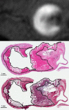
Comparison of carotid MPRAGE and IPH detected on histology. Top: Representative MPRAGE positive plaque in a patient undergoing subsequent endarterectomy. Middle: H&E stain demonstrating recent IPH (solid line) outlined by a pathologist. Bottom: PTAH positive staining (solid line) also indicates fibrin deposition and recent IPH.
Figure 4
Figure 4
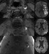
Left: MPRAGE hyperintense signal demonstrating IPH in ICA plaque in the three separate patients. From top to bottom: large, moderate and small IPH is present in the left ICA. Right: Corresponding DWI images showing infarction in each patient’s IPH positive left ICA distribution.
Figure 5
Figure 5
Pathway analysis demonstrating predictors of carotid IPH and carotid-source stroke.

References

    1. Lloyd-Jones D, Adams RJ, Brown TM, et al. Heart disease and stroke statistics—2010 update: a report from the American Heart Association. Circulation. 2010;121(7):e46–e215. - PubMed
    1. McPhee JT, Schanzer A, Messina LM, Eslami MH. Carotid artery stenting has increased rates of postprocedure stroke, death, and resource utilization than does carotid endarterectomy in the United States, 2005. J Vasc Surg. 2008;48(6):1442–1450. 1450 e1441. - PubMed
    1. Carotid surgery versus medical therapy in asymptomatic carotid stenosis. The CASANOVA Study Group. Stroke. 1991;22(10):1229–1235. - PubMed
    1. Endarterectomy for asymptomatic carotid artery stenosis. Executive Committee for the Asymptomatic Carotid Atherosclerosis Study. JAMA. 1995;273(18):1421–1428. - PubMed
    1. Risk of stroke in the distribution of an asymptomatic carotid artery. The European Carotid Surgery Trialists Collaborative Group. Lancet. 1995;345(8944):209–212. - PubMed

LinkOut - more resources