Review
. 2014 Apr 2:2:10.
doi: 10.1186/2052-8426-2-10. eCollection 2014.

The macrophage: a therapeutic target in HIV-1 infection

Affiliations
Review

The macrophage: a therapeutic target in HIV-1 infection

Amit Kumar et al. Mol Cell Ther. .

Abstract

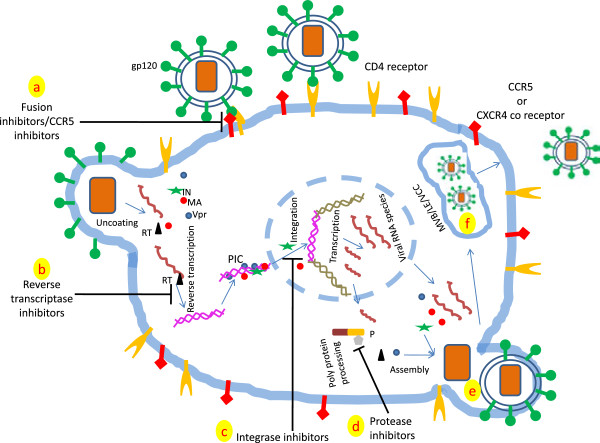
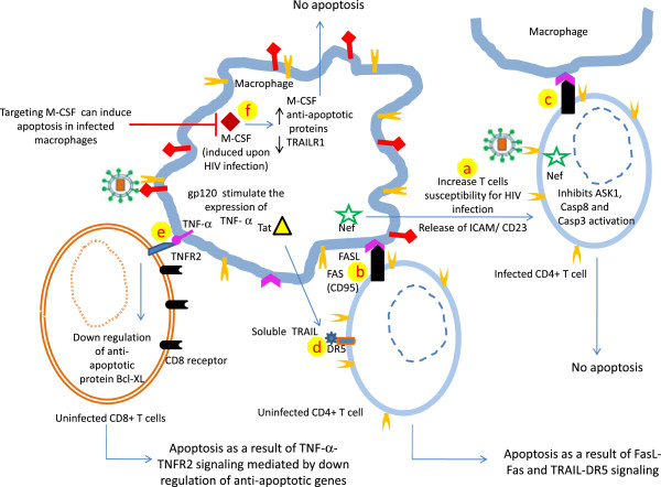
Human immunodeficiency virus (HIV) is still a serious global health concern responsible for more than 25 million deaths in last three decades. More than 34 million people are living with HIV infection. Macrophages and CD4+ T cells are the principal targets of HIV-1. The pathogenesis of HIV-1 takes different routes in macrophages and CD4+ T cells. Macrophages are resistant to the cytopathic effect of HIV-1 and produce virus for longer periods of time. In addition, macrophages being present in every organ system thus can disseminate virus to the different anatomical sites leading to the formation of viral sanctuaries. Complete cure of HIV-1 needs better understanding of viral pathogenesis in these reservoirs and implementation of knowledge into robust therapeutic products. In this review we will focus on the unique relationship between HIV-1 and macrophages. Furthermore, we will describe how successful antiretroviral therapy (ART) is in suppressing HIV and novel molecular and cellular strategies against HIV-1 in macrophages.

Keywords: Antiretroviral therapy; HIV-1; Latency; Macrophages; Nef; Tat; Vpr.


Figures

Figure 1
Figure 1
Depicting key events of HIV-1 life cycle targeted by anti-retroviral drugs. The anti-retroviral drugs target four critical steps of the viral life cycle which are fusion (or entry) of virion in the susceptible cell, reverse transcription, integration of proviral DNA into host chromatin and polyprotein processing by viral encoded protease. Depending upon the steps they target, the anti-retroviral drugs are termed as fusion (entry) inhibitors (a), reverse transcriptase inhibitors (b), integrase inhibitors (c) and protease inhibitors (d). Targeting single step at a time usually results in the emergence of resistant mutants. ART is formulation of these inhibitors which suppresses HIV-1 growth to a significant extent. Please note that virus assembly in macrophages takes place at both plasma membrane (e) as well as in virus containing compartments (f)[47]. Only key proteins involved in HIV-1 life cycle in macrophage have been shown. Abbreviations: RT- reverse transcriptase, MA- matrix protein, IN- Integrase, Vpr-Viral protein R, P- virus encoded protease, PIC- Pre-integration complex, MVB- multi vesicular bodies, LE- late endosomes and VCC- virus containing compartment.
Figure 2
Figure 2
Relationship between macrophages and T lymphocytes in HIV-1 infection. Macrophages harboring HIV-1 play an important role in HIV pathogenesis. Nef stimulates the release of soluble factors ICAM and CD23 which makes uninfected CD4+ T cells more susceptible to HIV infection, thereby favoring the expansion of the viral reservoir (a). In addition, Nef induces the expression of Fas ligand (FasL, CD95L) on HIV-infected cells. Interaction of CD95L and its receptor (Fas) present on uninfected CD4+ T cells results in apoptosis (b). On the other hand in infected CD4+ T cells, Nef inhibits the expression of proteins involved in apoptosis including ASK1, caspase 8 and caspase 3 (c), protects infected CD4+ T cells from cell death and further expands the viral reservoir. HIV regulatory protein Tat stimulates the production and release of TRAIL from the infected macrophages. TRAIL binds with its receptor (DR5) present on uninfected CD4+ T cells and induces apoptosis (d). Furthermore, gp120 interaction with CXCR4 receptor increases the expression of TNF-α on macrophages which interacts with TNFR2 present on CD8+ T cells. This interaction results in the down regulation of the anti-apoptotic protein Bcl-XL and ultimately leads to apoptosis (e). Moreover, HIV infection in macrophages is known to induce macrophage colony stimulating factor (M-CSF) which inhibits the expression of TRAILR1 on macrophages and upregulates the expression of anti-apoptotic proteins (f), favoring the resistance to apoptosis of infected macrophages. Therefore, targeting M-CSF has been suggested to increase apoptosis in infected macrophages.

References

    1. Iordanskiy S, Santos S, Bukrinsky M. Nature, nurture and HIV: the effect of producer cell on viral physiology. Virology. 2013;443:208–213. - PMC - PubMed
    1. Abbas W, Herbein G. T-cell signaling in HIV-1 infection. Open Virol J. 2013;7:57–71. - PMC - PubMed
    1. Ackerman AL, Cresswell P. Cellular mechanisms governing cross-presentation of exogenous antigens. Nat Immunol. 2004;5:678–684. - PubMed
    1. Koppensteiner H, Brack-Werner R, Schindler M. Macrophages and their relevance in Human Immunodeficiency Virus Type I infection. Retrovirology. 2012;9:82. - PMC - PubMed
    1. Crowe SM, Mills J, Kirihara J, Boothman J, Marshall JA, McGrath MS. Full-length recombinant CD4 and recombinant gp120 inhibit fusion between HIV infected macrophages and uninfected CD4-expressing T-lymphoblastoid cells. AIDS Res Hum Retroviruses. 1990;6:1031–1037. - PubMed