Review
. 2015 Apr:31:124-39.
doi: 10.1016/j.sbi.2015.04.002. Epub 2015 Jun 5.

The chromatin fiber: multiscale problems and approaches

Affiliations
Review

The chromatin fiber: multiscale problems and approaches

Gungor Ozer et al. Curr Opin Struct Biol. 2015 Apr.

Abstract

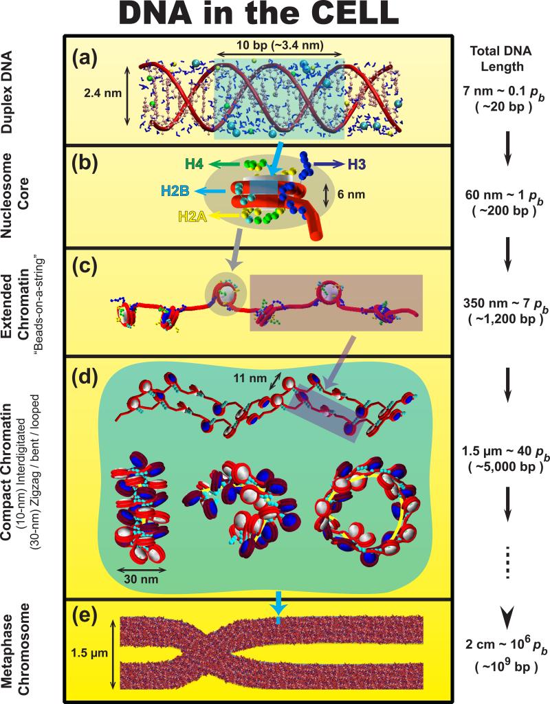
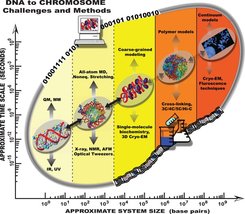
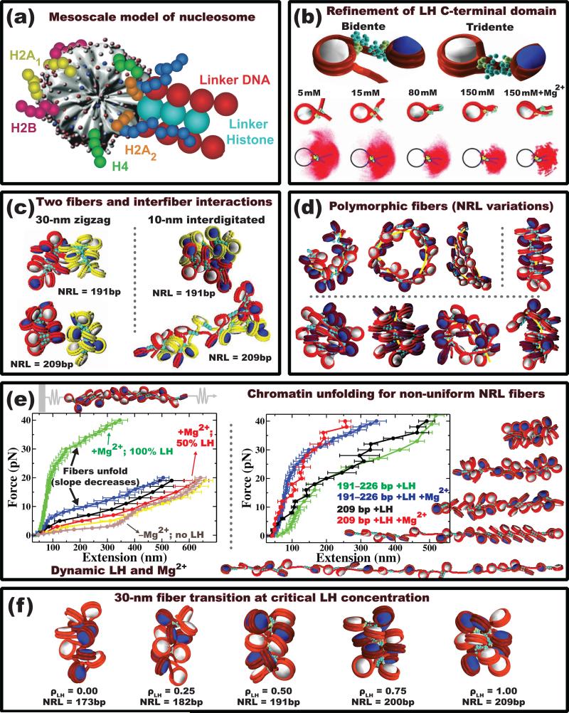
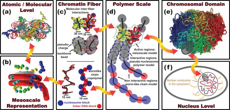
The structure of chromatin, affected by many factors from DNA linker lengths to posttranslational modifications, is crucial to the regulation of eukaryotic cells. Combined experimental and computational methods have led to new insights into its structural and dynamical features, from interactions due to the flexible core histone tails or linker histones to the physical mechanism driving the formation of chromosomal domains. Here we present a perspective of recent advances in chromatin modeling techniques at the atomic, mesoscopic, and chromosomal scales with a view toward developing multiscale computational strategies to integrate such findings. Innovative modeling methods that connect molecular to chromosomal scales are crucial for interpreting experiments and eventually deciphering the complex dynamic organization and function of chromatin in the cell.


Figures

Figure 1
Figure 1
DNA's many levels of folding [99]. Various colored backgrounds/masks are used to illustrate the extent of DNA compaction from one stage to the next. (a) DNA is relatively straight for length scales smaller than its persistence length (~ 0.15pb ≈ 15 bp). (b) In eukaryotic cells, DNA wraps around a core of eight histone proteins to form the nucleosome, the fundamental unit in chromatin. (c) Linker DNAs connect consecutive nucleosomes to form chromatin fibers, whose structures are unknown. The stretched 10-nm fiber, “beads-on-a-string”, is observed at low salt concentrations or when applying stretching forces to unfold the polymer. (d) At physiological conditions with multivalent ions and binding proteins such as linker histones, chromatin fibers condense. This can lead to side-by-side inter-digitated 10-nm fibers [11] (top), canonical 30-nm zigzag fibers with solenoid bent linker DNA motifs (bottom left), bent and looped fibers (bottom right) [12••]. The precise spontaneous secondary structure of chromatin depends on the cell type and other internal and external factors, and is still under debate [6,49••]. (e) Chromosomes are made up of dense chromatin fibers, shown here in the metaphase stage. Nuclear arrangements of chromatin in interphase stages are thought to be less ordered and more diverse (See polymer model in Fig. 4). The blue strip of size ~80×120 nm on the metaphase chromosome of size ~1.5×10 μm helps illustrate the enormous compaction of DNA in the cell.
Figure 2
Figure 2
Chromatin challenges and techniques on multiple spatial (base pair) and temporal (seconds) scales. Along the diagonal, representative systems are illustrated. Major computational (top) and experimental (bottom) techniques are listed, and the strength of connection between them is indicated by the boldness of the vertical arrows. Similarly, the boldness of the arrows along the diagonal reflect the strength of the connection between each pair of scales. Although inevitably losing some details during parameterization, the information transferred from high-resolution models generally enhances the overall validity of the coarser models. For instance, some of the current mesoscale models of the nucleosome are parameterized using all-atom simulations, and shown to produce many experimentally observed features of chromatin (Fig. 3). At the polymer level, existing models have not yet been connected to higher-resolution models and instead used to interpret specific experimental data. Thus, more work is required on higher scales for example to develop a computationally efficient polymer model that can be parameterized with various cell-specific information such as linker DNA length, linker histone presence, or monovalent/divalent salt concentration. See proposed multiscale connections in Fig. 4. Metaphase chromosome image credit: Wessex Reg. Genetics Centre. Wellcome Images.
Figure 3
Figure 3
Recent findings on chromatin fibers using variations of the Schlick group's mesoscale model. (a) The mesoscale model of nucleosome is composed of an irregularly shaped core with uniformly distributed 300 Debye-Hückel charges, 10 flexible core histone tails representing the C-termini of H2A and the N-termini of H2A/H2B/H3/H4, and linker DNA beads described as a worm-like chain; it can also include a linker histone attached to the dyad axis of the nucleosome [68,5]. (b) A refined model captures the spontaneous condensation of the linker histone C-terminal domain upon nucleosome binding [69••]. The side view trajectories of the nucleosome exiting/entering linker DNA show increasing formation of the nucleosome stem as a function of the monovalent salt concentration and the presence of divalent salts in the environment. At low salt, the nucleosome adopts a bidente configuration (top left), where the linker histone interacts with the nucleosome core and the entry linker DNA, leading to the formation of extended fibers. At high salt, the nucleosome adopts a tridente configuration (top right), where the linker histone interacts with the nucleosome core and both linker DNAs, promoting the stem formation and overall fiber compaction. (c) Two chromatin fibers (red and yellow) can interact in various ways. Low chromatin densities favor the formation of segregated canonical zigzag 30-nm fibers with weak inter-fiber interactions. Denser chromatin environments lead to heteromorphic and metastable 10-nm interdigitated fibers stabilized by stronger inter-fiber interactions [11]. (d) Nonuniform (alternating) DNA linker lengths (measured in units of nucleosome repeat lengths, NRL) increase the structural diversity of chromatin fibers [12••]. Fibers with one short linker DNA length (26 bp) can form bent-ladder like structures (top left); fibers with medium to long linker DNAs (35 to 79 bp) and moderate NRL variations, NRL (see full details in Ref. [12••]), mostly adopt canonical zigzag conformations (top right); long linker length (>44 bp) fibers with large NRL variations are highly polymorphic as to adopt various stable equilibrium conformations ranging from canonical to slightly bent to intra-looped nature. (e) Various chromatin unfolding simulations help interpret force/extension curves measured by single-molecule manipulation experiments. These have helped interpret the effect of linker histones, divalent Mg2+ ions, and non-uniform nucleosome repeat lengths (NRL) on chromatin unfolding. Here, a strong negative correlation between forced extension and linker histone concentration is captured (left) [73]. Fibers with nonuniform NRLs exhibit a smoother structural transition than uniform NRL fibers (right) [75]. (f) The empirical relationship between linker histone concentration and NRL was recently associated with formation of compact 30-nm fibers. The critical linker histone concentration, ρLH (ratio of linker histones per nucleosome), for this transition increases linearly with the NRL [74].
Figure 4
Figure 4
Proposed dynamic multiscale model connecting atomic to nuclear chromatin levels. (a) All-atom models of nucleosomes provide atomistic details of nucleosome complexes. (b) Such knowledge can be transferred into mesoscale models to build coarse-grained representations of the nucleosome core particle (NCP) interacting with many internal and external elements as found in cellular environment. (c) Because coarse-grained models indicate that chromatin fibers are highly polymorphic— adopting canonical zigzag (30-nm), interdigitated “beads-on-a-string” (10-nm), hairpin-like, bent, and looped conformations [12••]—further coarse graining could describe these various fibers to investigate diverse phase space and structural dynamics of chromosomal and nuclear domains observed in vivo and in vitro experiments. (d) These mesoscale fibers can then be represented as active regions within lower resolution polymer models that capture crucial physical properties of the nuclear environment. (e) By incorporating cell specific information to the all-atom details, different polymer models can be constructed to describe human interphase chromatin [85] (polymer model in Fig. 2) or mitotic chromatin [87••] (Fig. 4e), for example. (f) At the nuclear level, the chromatin polymer organization could be treated as a viscoelastic fluid incorporating passive fluctuations and ATP-active regions [92]. The red arrows emphasize the dynamic information transfer required, zooming in and out of each level depending on the chromatin properties examined. Such a combined multiscale model could ultimately define an interactive framework to identify the synergy between molecular events and chromatin reorganization, like heterochromatin formation due to specific epigenetic marks.

References

    1. Strahl BD, Allis CD. The language of covalent histone modifications. Nature. 2000;403:41–45. - PubMed
    1. Jenuwein T, Allis CD. Translating the histone code. Science. 2001;293:1074–1080. - PubMed
    1. Rando OJ. Combinatorial complexity in chromatin structure and function: revisiting the histone code. Curr Opin Genet Dev. 2012;222:148–155. - PMC - PubMed
    1. Schlick T, Hayes J, Grigoryev S. Toward convergence of experimental studies and theoretical modeling of the chromatin fiber. J Biol Chem. 2012;287:5183–5191. - PMC - PubMed
    1. Grigoryev S, Arya G, Correll S, Woodcock C, Schlick T. Evidence for heteromorphic chromatin fibers from analysis of nucleosome interactions. Proc Natl Acad Sci USA. 2009;106:13317–13322. [Pioneering cross-linking experiment combined with mesoscale modeling shows that the two different models—zigzag and solenoid fibers—could be merged into one heteromorphic compact fiber with enhanced i±1 internucleosome interactions due to small percentage (Ȉ20%) of linker DNA bending. Such a fiber can be dominant in moderate divalent ion concentrations.] - PMC - PubMed

Publication types

LinkOut - more resources