. 2015 Jun 9;11(6):e1004960.
doi: 10.1371/journal.ppat.1004960. eCollection 2015 Jun.

TRIM32 Senses and Restricts Influenza A Virus by Ubiquitination of PB1 Polymerase

Affiliations

TRIM32 Senses and Restricts Influenza A Virus by Ubiquitination of PB1 Polymerase

Bishi Fu et al. PLoS Pathog. .

Abstract

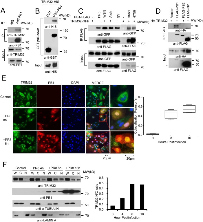
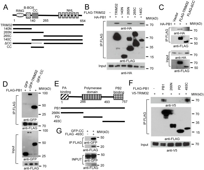
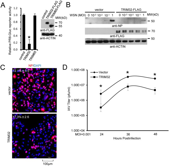
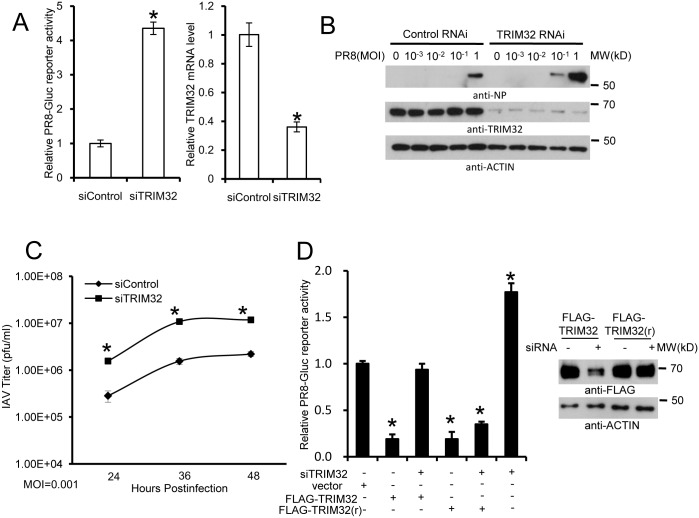
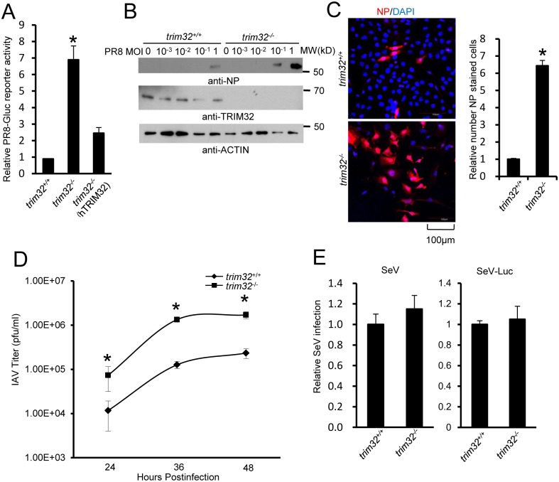
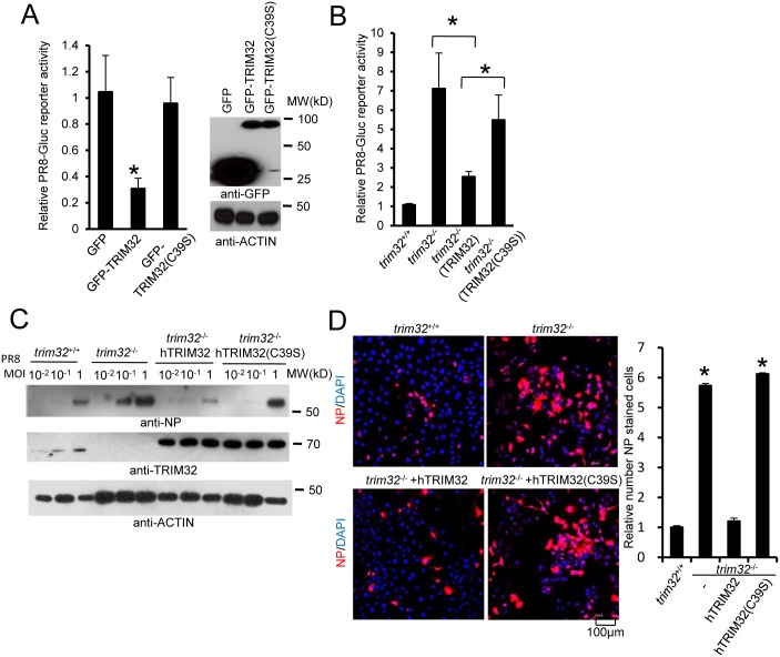
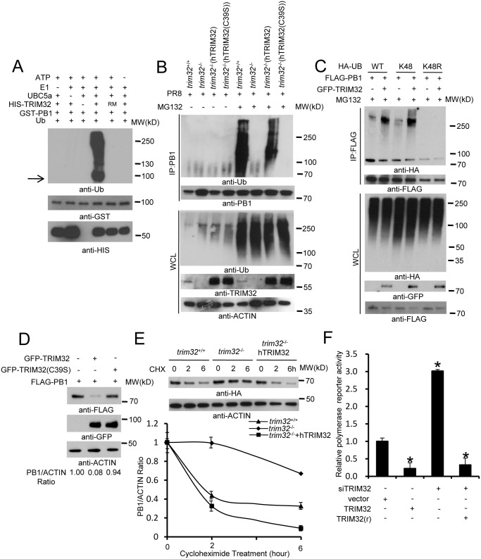
Polymerase basic protein 1 (PB1) is the catalytic core of the influenza A virus (IAV) RNA polymerase complex essential for viral transcription and replication. Understanding the intrinsic mechanisms which block PB1 function could stimulate development of new anti-influenza therapeutics. Affinity purification coupled with mass spectrometry (AP-MS) was used to identify host factors interacting with PB1. Among PB1 interactors, the E3 ubiquitin ligase TRIM32 interacts with PB1 proteins derived from multiple IAV strains. TRIM32 senses IAV infection by interacting with PB1 and translocates with PB1 to the nucleus following influenza infection. Ectopic TRIM32 expression attenuates IAV infection. Conversely, RNAi depletion and knockout of TRIM32 increase susceptibility of tracheal and lung epithelial cells to IAV infection. Reconstitution of trim32-/- mouse embryonic fibroblasts with TRIM32, but not a catalytically inactive mutant, restores viral restriction. Furthermore, TRIM32 directly ubiquitinates PB1, leading to PB1 protein degradation and subsequent reduction of polymerase activity. Thus, TRIM32 is an intrinsic IAV restriction factor which senses and targets the PB1 polymerase for ubiquitination and protein degradation. TRIM32 represents a model of intrinsic immunity, in which a host protein directly senses and counters viral infection in a species specific fashion by directly limiting viral replication.


Conflict of interest statement

The authors have declared that no competing interests exist.

Figures

Fig 1
Fig 1. TRIM32 interacts and translocates with influenza A virus PB1 protein.
(A) Primary human tracheal epithelial cells were infected with 0.1 MOI PR8 IAV for 16 hr. Whole cell lysates (WCL) were subjected to immunoprecipitation (IP) and immunoblotting with indicated antibodies to detect endogenous interactions. Molecular weights (MW) are indicated. (B) GST pull down of bacterially expressed GST-PB1 and HIS-TRIM32. (C) FLAG-tagged PB1 from 6 different influenza A strains [PR8, A/Puerto Rico/8/1934 (H1N1); WSN, A/WSN/1933 (H1N1); Aichi, A/Aichi/2/1968 H3N2; NY, A/New York/1682/2009 (H1N1); A/Vietnam/1194/2004 (H5N1); A/Anhui/1/2013 (H7N9)] were co-expressed with TRIM32-GFP in HEK293 cells. After 48 hr, cell lysates were immunoprecipitated with anti-FLAG and probed as indicated. As input controls, WCL were immunoblotted. (D) TRIM32 fused with HA epitope was co-transfected into HEK293 cells with PR8 derived FLAG-tagged PB1, PB2 or NP. After 48 hr, WCL were immunoprecipitated with anti-FLAG antibody and blotted with indicated reagents. TRIM32 can appear as a doublet on Western blots. (E) Primary human tracheal epithelial cells were infected with 0.01 MOI PR8 strain IAV for 8 or 16 hr and stained with anti-TRIM32 (green), anti-PB1 (red) and DAPI nuclear stain (blue). Right panel shows quantitated TRIM32-PB1 colocalization data. (F) A549 cells were infected with 0.01 MOI IAV PR8 strain for the indicated times, whole cell lysates (W) or cytosolic (C) and nuclear (N) fractions were extracted and blotted as indicated. Right panel depicts the densitometric ratio of nuclear to cytoplasmic PB1.
Fig 2
Fig 2. Domain requirements for TRIM32-PB1 interaction.
(A) Schematic representation of TRIM32 protein domains and the individual TRIM32 deletion mutants investigated in this study. (B) Full length and various TRIM32 deletion mutants were fused with FLAG epitope and co-transfected with HA-PB1 into HEK293 cells. WCL were immunoprecipitated with anti-FLAG antibody and blotted with indicated reagents. (C) Full length and TRIM32 with a deleted CC segment were fused with FLAG epitope and co-transfected with HA-PB1 into HEK293 cells. WCL were immunoprecipitated with anti-FLAG antibody and blotted with indicated reagents. (D) Full length and TRIM32 CC-containing segment (residues 140–265) were tagged with GFP and co-transfected with FLAG-PB1 into HEK293 cells. WCL were immunoprecipitated with anti-FLAG antibody and blotted with the indicated reagents. (E) Schematic representation of IAV PB1 protein domains and PB1 deletion mutants investigated in this study. (F) Full length and various PB1 mutants (PR8) were fused with FLAG epitope and co-transfected with V5-TRIM32 into HEK293 cells. WCL were immunoprecipitated with anti-FLAG antibody and blotted with indicated reagents. (G) TRIM32 CC fragment (residues 140–265) was cotransfected with C-terminal PB1 fragment (residues 493–757) into HEK293 cells. Immunoprecipitation and immunoblotting were performed with indicated reagents.
Fig 3
Fig 3. TRIM32 attenuates influenza A virus infection.
(A) A549 cells were transfected with control vector, TRIM32-FLAG or TRIM65-FLAG. After 36 hr cells were infected with 0.01 MOI PR8-Gluc for 16 hr and then Gaussia luciferase activity was examined. An asterisk indicates P<0.01. (B) A549 stable cell lines carrying vector or TRIM32-FLAG were infected with indicated MOI of WSN strain IAV for 16 hr. WCL were blotted with indicated reagents. (C) A549 cells stably transfected with control vector or TRIM32-FLAG were infected with 0.01 MOI PR8 for 8 hr and stained with anti-NP (red) and DAPI (blue). The percentage of NP stained cells is indicated. (D) A549 stable cell lines transfected with vector or TRIM32-FLAG were infected with 0.001 MOI of WSN IAV for the indicated times. Supernatant was titered on MDCK cells and plaques were enumerated. Asterisk indicates P<0.05.
Fig 4
Fig 4. RNAi depletion of TRIM32 promotes influenza virus infection.
(A) Primary human tracheal epithelial cells were transfected with scrambled control or TRIM32 siRNA duplex #1. After 24 hr cells were infected with 0.01 MOI PR8-Gluc for 16 hr. The relative luciferase activity was examined. An asterisk indicates P<0.01. Right panel displays knockdown efficiency by qPCR. (B) A549 cells were transfected with control siRNA or TRIM32 siRNA#1. After 24 hr cells were infected with indicated MOI of PR8 IAV. WCL were blotted with the indicated antibodies. (C) Tracheal epithelial cells transfected with control or TRIM32 siRNA were infected with 0.001 MOI of WSN IAV for the indicated times. Culture supernatants containing IAV were titered on MDCK cells and plaques were enumerated. An asterisk indicates P<0.05. (D) HEK293 cells were transfected with TRIM32 siRNA and wild type TRIM32 or a TRIM32 rescue construct. After 24 hr cells were infected with 0.01 MOI PR8-Gluc for 16 hr. The relative luciferase signal is shown. An asterisk indicates P<0.05. Western blot shows knockdown efficiency.
Fig 5
Fig 5. TRIM32 deficiency increases susceptibility to influenza A virus infection.
(A) Trim32 +/+, trim32 -/- and trim32 -/- MEF transfected (24 hr) with human TRIM32 were infected with 0.1 MOI PR8-Gluc. The relative luciferase signal is shown. An asterisk indicates significant difference (P<0.05) for trim32 -/- vs. either trim32 +/+ or TRIM32 reconstituted trim32 -/- MEF. (B) Trim32 +/+ and trim32 -/- MEF were infected with indicated MOI of PR8 IAV. After 16 hr, WCL were blotted with indicated reagents. (C) Trim32 +/+ and trim32 -/- MEF cells were infected with PR8 IAV for 8 hr, then cells were stained with anti-NP (red) and DAPI (blue). The right panel shows the relative ratio of NP stained cells. An asterisk indicates P<0.01. (D) Trim32 +/+ and trim32 -/- MEF were infected with 0.01 MOI of WSN IAV for the indicated times. Supernatants were titered on MDCK cells and pfu were enumerated. An asterisk indicates P<0.05. (E) Trim32 +/+ and trim32 -/- MEF were infected with wild type Sendai virus (SeV) or luciferase reporter Sendai virus (SeV-Luc) for 16 hr. Sendai infection was detected by staining with anti-Sendai antibody or luciferase assay. Relative numbers of Sendai virus infected cells or luciferase activities are presented.
Fig 6
Fig 6. E3 ligase activity is indispensable for TRIM32-dependent against influenza A virus.
(A) HEK293 cells were transfected with GFP, GFP-TRIM32 or GFP-TRIM32(C39S). After 24 hr, cells were infected with 0.01 MOI PR8-Gluc for reporter assay. The relative luciferase signal is shown. Asterisk indicates P<0.05 (GFP-TRIM32 vs. either GFP or GFP-TRIM32(C39S) transfected cells). (B) Trim32 +/+, trim32 -/- or trim32 -/- MEF reconstituted with TRIM32 and TRIM32(C39S) were infected with 0.1 MOI PR8-Gluc. The relative luciferase signal is shown. An asterisk indicates P<0.05. (C) Trim32 +/+, trim32 -/- or trim32 -/- MEF reconstituted with TRIM32 and TRIM32 (C39S) were infected with designated MOI of IAV PR8 for 16 hr. WCL were blotted with indicated reagents. (D) Trim32 +/+, trim32 -/- or trim32 -/- reconstituted with TRIM32 and TRIM32(C39S) MEF were infected with 0.1 MOI PR8 for 16 hr, then cells were stained with anti-NP (red) and DAPI. The right panel shows the relative ratio of NP stained cells. An asterisk indicates P<0.01.
Fig 7
Fig 7. TRIM32 limits viral infection by targeting PB1 for ubiquitination.
(A) In vitro ubiquitination of PB1 by TRIM32 plus E1, E2 (UBCH5A), ATP and ubiquitin (Ub). GST-tagged PB1, HIS-tagged TRIM32 and the catalytically dead TRIM32(C39S) RING mutant (RM) were purified from bacteria. The arrow indicates approximate PB1 position. (B) Trim32 +/+, trim32 -/- or TRIM32 and TRIM32(C39S) reconstituted trim32 -/- MEF were infected with 0.1 MOI PR8 IAV for 16 hr, then treated with 10 μg/ml MG132. After 6 hr cell lysates were immunoprecipitated with anti-PB1 and probed as indicated. Whole cell lysates are presented as input controls. (C) HEK293 stably transfected cell lines carrying FLAG-PB1 were transiently transfected with either HA-tagged wild type, K48 only or K48R ubiquitin along with GFP-TRIM32. Cells were treated for 6 hr with 10 μg/ml MG132, then immunoprecipitated with anti-FLAG and probed with the indicated antibodies. (D) GFP-TRIM32 or TRIM32(C39S) were transfected into HEK293 cells stably transfected with FLAG-PB1. Immunoblotting was performed with indicated reagents. Quantitative Western blotting was used to determine PB1 levels, which were used to calculate the ratio of PB1 to actin. (E) Trim32 +/+, trim32 -/- and trim32 -/- MEF reconstituted with TRIM32 were transfected with PB1-HA for 48 hr, then treated with 20 μg/ml cycloheximide for 0, 2 or 6 hr. WCL were blotted with indicated reagents. Quantitative Western blotting was used to derive data presented in lower panel. (F) HEK293 cells were transiently transfected with a plasmid cocktail containing PR8 PB1, PB2, PA, NP expression plasmids plus a polymerase I plasmid expressing an influenza virus-like RNA coding for the reporter protein firefly luciferase, Renilla luciferase control and TRIM32 siRNA along with TRIM32 wild type or rescue mutant for 48 hr. The relative luciferase signal is shown. An asterisk indicates P<0.05.

References

    1. Pflug A, Guilligay D, Reich S, Cusack S (2014) Structure of influenza A polymerase bound to the viral RNA promoter. Nature 516: 355–360. 10.1038/nature14008 - DOI - PubMed
    1. Reich S, Guilligay D, Pflug A, Malet H, Berger I, et al. (2014) Structural insight into cap-snatching and RNA synthesis by influenza polymerase. Nature 516: 361–366. 10.1038/nature14009 - DOI - PubMed
    1. Ghanem A, Mayer D, Chase G, Tegge W, Frank R, et al. (2007) Peptide-mediated interference with influenza A virus polymerase. J Virol 81: 7801–7804. - PMC - PubMed
    1. He X, Zhou J, Bartlam M, Zhang R, Ma J, et al. (2008) Crystal structure of the polymerase PA(C)-PB1(N) complex from an avian influenza H5N1 virus. Nature 454: 1123–1126. 10.1038/nature07120 - DOI - PubMed
    1. Obayashi E, Yoshida H, Kawai F, Shibayama N, Kawaguchi A, et al. (2008) The structural basis for an essential subunit interaction in influenza virus RNA polymerase. Nature 454: 1127–1131. 10.1038/nature07225 - DOI - PubMed

Publication types

MeSH terms