. 2015 Jun;71(Pt 6):1238-56.
doi: 10.1107/S1399004715005210. Epub 2015 May 14.

In meso in situ serial X-ray crystallography of soluble and membrane proteins

Affiliations

In meso in situ serial X-ray crystallography of soluble and membrane proteins

Chia Ying Huang et al. Acta Crystallogr D Biol Crystallogr. 2015 Jun.

Abstract

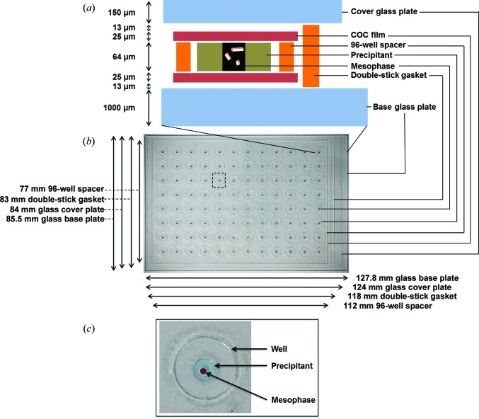
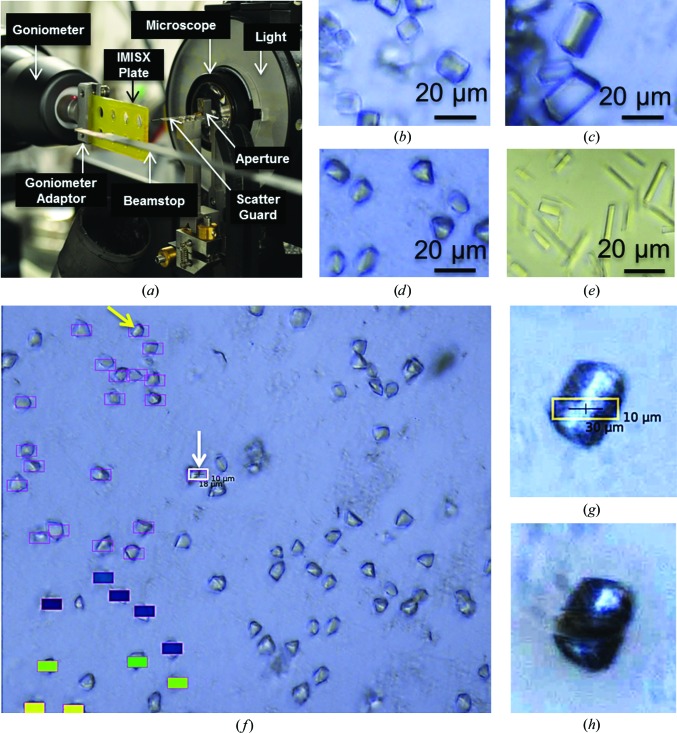
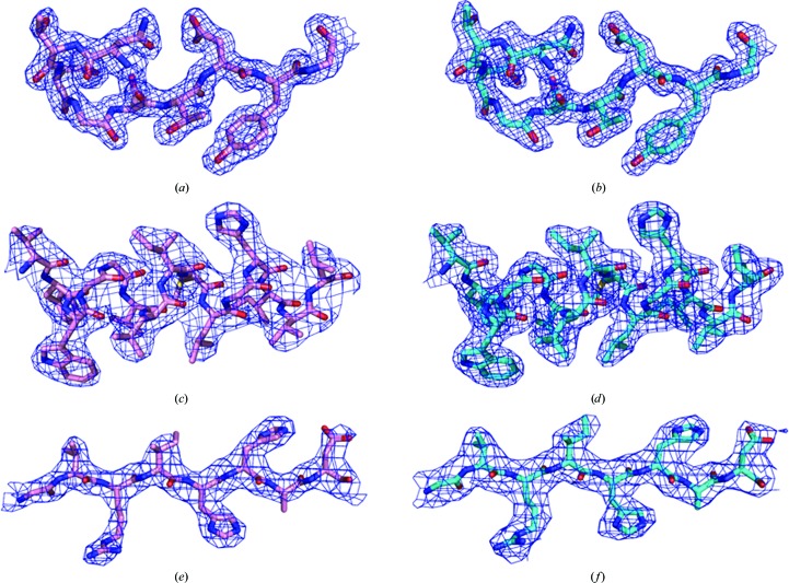
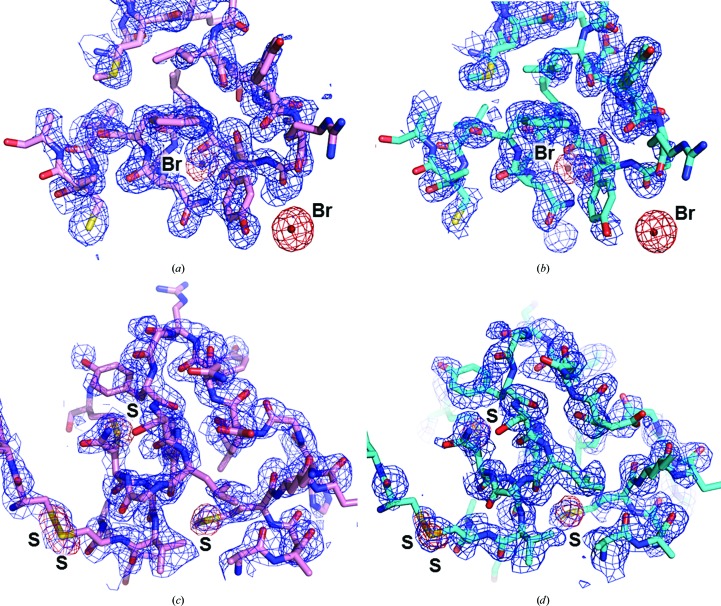
The lipid cubic phase (LCP) continues to grow in popularity as a medium in which to generate crystals of membrane (and soluble) proteins for high-resolution X-ray crystallographic structure determination. To date, the PDB includes 227 records attributed to the LCP or in meso method. Among the listings are some of the highest profile membrane proteins, including the β2-adrenoreceptor-Gs protein complex that figured in the award of the 2012 Nobel Prize in Chemistry to Lefkowitz and Kobilka. The most successful in meso protocol to date uses glass sandwich crystallization plates. Despite their many advantages, glass plates are challenging to harvest crystals from. However, performing in situ X-ray diffraction measurements with these plates is not practical. Here, an alternative approach is described that provides many of the advantages of glass plates and is compatible with high-throughput in situ measurements. The novel in meso in situ serial crystallography (IMISX) method introduced here has been demonstrated with AlgE and PepT (alginate and peptide transporters, respectively) as model integral membrane proteins and with lysozyme as a test soluble protein. Structures were solved by molecular replacement and by experimental phasing using bromine SAD and native sulfur SAD methods to resolutions ranging from 1.8 to 2.8 Å using single-digit microgram quantities of protein. That sulfur SAD phasing worked is testament to the exceptional quality of the IMISX diffraction data. The IMISX method is compatible with readily available, inexpensive materials and equipment, is simple to implement and is compatible with high-throughput in situ serial data collection at macromolecular crystallography synchrotron beamlines worldwide. Because of its simplicity and effectiveness, the IMISX approach is likely to supplant existing in meso crystallization protocols. It should prove particularly attractive in the area of ligand screening for drug discovery and development.

Keywords: AlgE; PepTSt; bromine SAD; experimental phasing; in meso; in situ; lipid cubic phase; membrane protein; mesophase; serial crystallography; sulfur SAD.


Figures

Figure 1
Figure 1
Schematic (a) and photographic image (b) of a double-sandwich 96-well IMISX plate. The schematic is not drawn to scale. An expanded view of one of the wells is shown in (c). To make the mesophase and precipitant more obvious, they were prepared with Sudan Red and blue food dye (Goodalls Blue Colouring containing Brilliant Blue FCF E133 and Carmoisine E122), respectively. The mesophase and precipitant volumes are 100 and 600 nl, respectively. The well diameter is 6 mm.
Figure 2
Figure 2
Experimental setup for IMISX data collection and images of crystals grown in IMISX plates. (a) A view of a section of an IMISX plate in the goniometer positioned for SX data collection on beamline PXII (X10SA) at the SLS. (be) Crystals of lysozyme (b), lysozyme–bromide (c), PepTSt (d) and AlgE (e) in COC wells removed from IMISX plates as viewed through the high-resolution on-axis microscope. (f) Screenshot of PepTSt crystals in a well from an IMISX plate as viewed through the on-axis microscope during SX data collection. Crystals measuring ∼10 × 10 µm (yellow arrow) are clearly visible in these in situ samples using the high-resolution microscope, which greatly facilitates crystal picking. ‘Hand-picking’ of crystals is performed at the click of a mouse with the SLS software DA+ and involves simply positioning a rectangular box (white; white arrow) on the crystal of interest. In this instance, the beam dimensions are 18 × 10 µm. Open boxes correspond to crystals due for data collection. Filled boxes identify crystals that have already been exposed and are colour-coded by the number of reflections detected at that particular site of exposure. (g, h) Images of a lysozyme crystal before (g) and after (h) SX data collection. The position of the beam on the crystal and the size of the beam are shown in (g). Beam damage to the crystal caused by a 0.5 s exposure at 2.2 × 10¹² (12 keV) photons s−1 at RT is clearly visible in (h). The crystal used in this demonstration of radiation damage is large by comparison with those used for IMISX.
Figure 3
Figure 3
A comparison of electron-density maps for measurements made by the IMISX method at room temperature (a, c, e) and by the conventional method using harvested crystals at 100 K (b, d, f) for lysozyme (a, b), PepTSt (c, d) and AlgE (e, f). Residues Asn46–Gly54, Val88–Leu102 and Gly222–Asp229 are shown for lysozyme, PepTSt and AlgE, respectively. The 2F oF c maps are shown as blue meshes contoured at 1σ. The resolution of the corresponding data are 1.8, 2.8 and 2.8 Å for lysozyme, PepTSt and AlgE at room temperature, respectively. At 100 K, the corresponding resolution values are 1.8, 2.3 and 2.9 Å, respectively. Stick models show N atoms (blue), O atoms (red) and C atoms (pink at room temperature, light blue at 100 K).
Figure 4
Figure 4
A comparison of the initial electron-density maps obtained by bromine SAD (a, b) and sulfur SAD (c, d) phasing for measurements made with lysozyme by the IMISX method at room temperature (a, c) and by the conventional method using harvested crystals in loops at 100 K (b, d). Residues Ala9–Ala32 are shown for the bromine SAD data (a, b) and residues Ala9–Ala32, Met105, Cys115 and Lys116 for the sulfur SAD data. The initial 2F oF c map obtained after density modification with SHELXE was contoured at 1σ and is shown as a blue mesh. The anomalous difference maps contoured at 5σ are shown as a red mesh. Br and S atoms are labelled. The final model is shown in stick representation. The resolutions of the corresponding data are 2.0 and 1.8 Å for sulfur SAD and bromine SAD at room temperature, respectively. At 100 K, the corresponding values are 2.0 and 1.8 Å, respectively.
Figure 5
Figure 5
Observed and predicted distributions of reflection multiplicity in IMISX data sets recorded from lysozyme, PepTSt and AlgE crystals at room temperature. Blue, multiplicity observed for IMISX data set. Red, binomial distribution of multiplicity predicted for a defined effective crystal rotation range. Green, multiplicity observed for a native lysozyme data set collected by conventional crystallography with a single crystal. (a) Lysozyme native SX data sets recorded with 113 crystals and an effective rotation range of 0.86°. (b) Lysozyme bromine SAD SX data sets recorded with 239 crystals and an effective rotation range of 1.66°. (c) Lysozyme sulfur SAD SX data sets recorded with 992 crystals and an effective rotation range of 1.55°. (d) PepTSt native SX data sets recorded with 572 crystals and an effective rotation range of 0.42°. (e) AlgE native SX data sets recorded with 175 crystals and an effective rotation range of 0.76°.

Comment in

References

    1. Adams, P. D., Grosse-Kunstleve, R. W., Hung, L.-W., Ioerger, T. R., McCoy, A. J., Moriarty, N. W., Read, R. J., Sacchettini, J. C., Sauter, N. K. & Terwilliger, T. C. (2002). Acta Cryst. D58, 1948–1954. - PubMed
    1. Aherne, M., Lyons, J. A. & Caffrey, M. (2012). J. Appl. Cryst. 45, 1330–1333. - PMC - PubMed
    1. Axford, D. et al. (2012). Acta Cryst. D68, 592–600. - PMC - PubMed
    1. Barends, T. R. M., Foucar, L., Botha, S., Doak, R. B., Shoeman, R. L., Nass, K., Koglin, J. E., Williams, G. J., Boutet, S., Messerschmidt, M. & Schlichting, I. (2014). Nature (London), 505, 244–247. - PubMed
    1. Bingel-Erlenmeyer, R., Olieric, V., Grimshaw, J. P. A., Gabadinho, J., Wang, X., Ebner, S., Isenegger, A., Schneider, R., Schneider, J., Glettig, W., Pradervand, C., Panepucci, E. H., Tomizaki, T., Wang, M. & Schulze-Briese, C. (2011). Cryst. Growth Des. 11, 916–923.

Publication types

MeSH terms

Substances