. 2015 Jun 9;10(6):e0129603.
doi: 10.1371/journal.pone.0129603. eCollection 2015.

HIF-1α Promotes Epithelial-Mesenchymal Transition and Metastasis through Direct Regulation of ZEB1 in Colorectal Cancer

Affiliations

HIF-1α Promotes Epithelial-Mesenchymal Transition and Metastasis through Direct Regulation of ZEB1 in Colorectal Cancer

Wenjing Zhang et al. PLoS One. .

Abstract

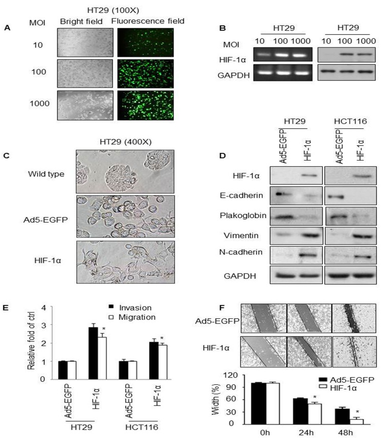
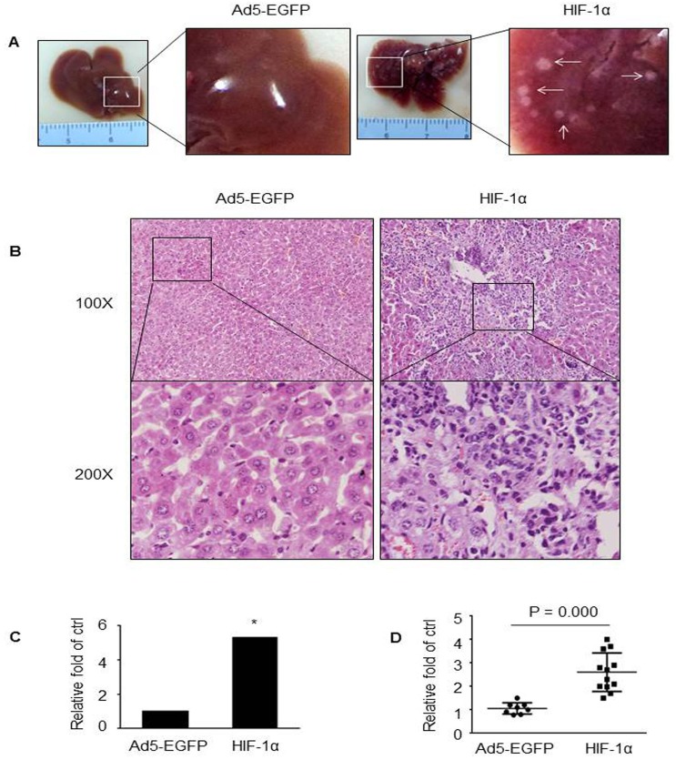
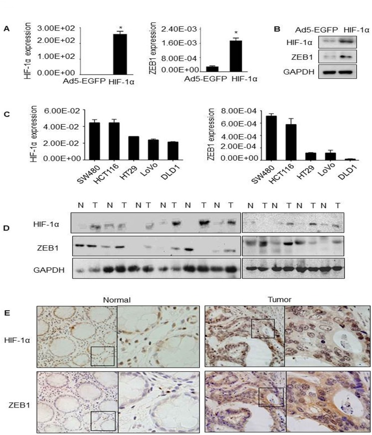
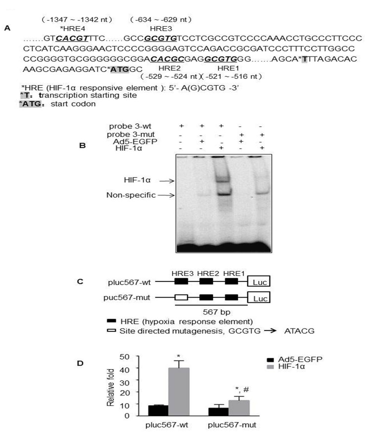
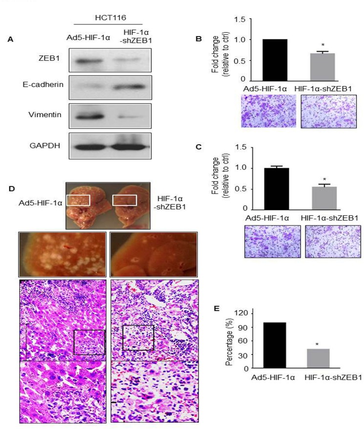
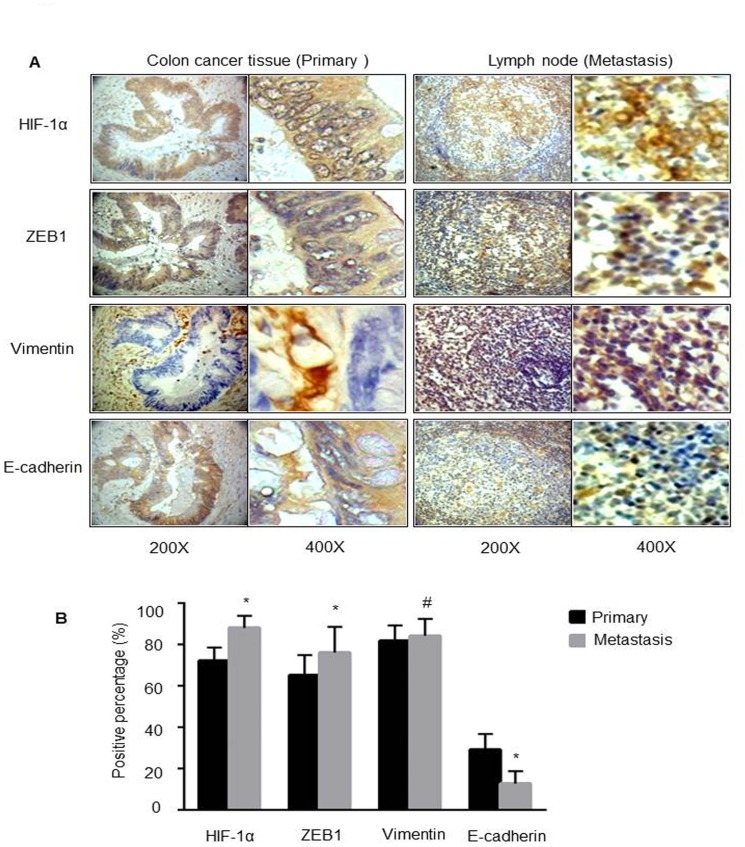
It is well recognized that hypoxia-inducible factor 1 alpha (HIF-1α) is involved in cancer metastasis, chemotherapy and poor prognosis. We previously found that deferoxamine, a hypoxia-mimetic agent, induces epithelial-mesenchymal transition (EMT) in colorectal cancer. Therefore, here we explored a new molecular mechanism for HIF-1α contributing to EMT and cancer metastasis through binding to ZEB1. In this study, we showed that overexpression of HIF-1α with adenovirus infection promoted EMT, cell invasion and migration in vitro and in vivo. On a molecular level, HIF-1α directly binding to the proximal promoter of ZEB1 via hypoxia response element (HRE) sites thus increasing the transactivity and expression of ZEB1. In addition, inhibition of ZEB1 was able to abrogate the HIF-1α-induced EMT and cell invasion. HIF-1α expression was highly correlated with the expression of ZEB1 in normal colorectal epithelium, primary and metastatic CRC tissues. Interestingly, both HIF-1α and ZEB1 were positively associated with Vimentin, an important mesenchymal marker of EMT, whereas negatively associated with E-cadherin expression. These findings suggest that HIF-1α enhances EMT and cancer metastasis by binding to ZEB1 promoter in CRC. HIF-1α and ZEB1 are both widely considered as tumor-initiating factors, but our results demonstrate that ZEB1 is a direct downstream of HIF-1α, suggesting a novel molecular mechanism for HIF-1α-inducing EMT and cancer metastasis.


Conflict of interest statement

Competing Interests: The authors have declared that no competing interests exist.

Figures

Fig 1
Fig 1. Overexpression of HIF-1α induced EMT and metastasis in CRC cell lines.
(A) HT29 cells were transduced with Ad5-HIF-1α at the indicated MOI values for 48 hours. Fluorescence intensity and bright field at the same area were observed at the original magnification as 100X. (B) mRNA (left) and protein (right) expressions of HIF-1α were detected using RT-PCR and western blot, respectively. GAPDH was used as the loading control. (C) Cell morphology of wildtype HT29 or HT29 cells transduced with the indicated adenoviruses (Original magnification = 400X). (D) Western blot analysis of HIF-1α, E-cadherin, Plakoglobin, Vimentin and N-cadherin in adenovirus-infected HT29 and HCT116 cells. GAPDH was used as the internal control. (E) Fold change of invasion and migration in the indicated cells. Quantification of the results was shown in the bar graph with means ± SD. *P < 0.05. (F) Wound healing assay. Cell monolayers were scratched with a pipette tip and images were taken 0, 24 and 48 hours after wound formation. *P < 0.05.
Fig 2
Fig 2. HIF-1α overexpression promoted liver metastasis in vivo.
Representative photographic pictures (A) and H&E staining (B) of liver of BALB/c mice five weeks after subsplenic injection of HT29 cells transduced with Ad5-HIF-1α or control viruses. White arrows indicated metastatic nodules. (C) The comparison of liver metastases between HT29-Ad5-HIF-1α mice (N = 12) and control mice (N = 8). *P < 0.05. (D) Confirmation of the overexpression of HIF-1α from metastatic nodules in mice injected with Ad5-HIF-1α by qPCR, compared with those injected with control viruses.
Fig 3
Fig 3. Regulation of HIF-1α on ZEB1 and their expressions in CRC.
(A) Endogenous expression levels of HIF-1α and ZEB1 in the indicated five cell lines were detected by qPCR with GAPDH used as the internal control. (B&C) HT29 cells were transduced with Ad5-HIF-1α or control viruses for 48 hours, following by qPCR and western blot to detect the expression of HIF-1α and ZEB1. GAPDH was used as the internal control. (D) HIF-1α and ZEB1 protein levels in matched nonneoplastic/cancerous colorectal tissues. N: normal; T: tumor. The level of each protein was normalized against GAPDH. (E) Representative pictures of IHC staining on human CRC formalin-fixed paraffin-embedded samples for HIF-1α and ZEB1. Left panel showed the adjacent normal colorectal tissues. Right panel showed CRC specimens. Original magnification, 200X.
Fig 4
Fig 4. Regulation of ZEB1 by HIF-1α through HRE-3.
(A) Schematic representation of the proximal promoter (~ 3500 nt upstream) of ZEB1 gene. HRE: hypoxia response element. (B) Oligonucleotides for EMSA were wildtype (lane 1–3) and mutant (lane 4–5) probe 3 from the ZEB1 promoter, which contained a consensus HRE-3. Nuclear extracts prepared from HT29-Ad5-HIF-1α (lane 3 and 5) or control cells (lane 2 and 4) were incubated with [γ-32P] ATP-labelled probe before electrophoresis. Negative control was performed with wildtype probe without nuclear extract (lane 1). (C) Schematic representation of the promoter region of ZEB1 and the report constructs used in adenovirus-infected experiments. The constructs contained wildtype (pluc567-wt) or mutant (pluc567-mut) HRE-3 located -634 ~ -630 nt upstream of the transcription start site of ZEB1. (D) Activation of plu567 or pluc567-mut in Ad5-HIF-1α- or Ad5-EGFP-infected HT29 cells (N = 3 replicate experiments). *, # P < 0.05.
Fig 5
Fig 5. ZEB1 inhibition abolished HIF-1α-induced EMT and metastasis.
(A) Western blot analysis of ZEB1, E-cadherin and Vimentin in HCT116-Ad5-HIF-1α or HCT116-HIF-1α-shZEB1 cells. GAPDH was used as the loading control. Invasion (B) and migration (C) assays were performed in the same two cell lines. The representative images of invaded cells in the inserts of transwell chambers were shown at original magnification = 200X. *P < 0.05. (D) Representative photographic pictures and H&E staining of liver of BALB/c mice 5 weeks after subsplenic injection of HCT116-Ad5-HIF-1α or HCT116-HIF-1α-shZEB1 cells. (E) Quantification of the average numbers of metastatic foci in the livers of mice (N = 5). *P < 0.05.
Fig 6
Fig 6. Expression statuses of HIF-1α, ZEB1, Vimentin and E-cadherin in tissue specimens.
Representative images of IHC (A) and quantification of positive tumor cells (B) in both primary CRC specimens and the according metastatic lymph node. Original magnification, 200X. *P < 0.05 and # P > 0.05.

References

    1. Kalluri R, Weinberg RA. The basics of epithelial-mesenchymal transition. J Clin Invest 2009, 119:1420–1428. 10.1172/JCI39104 - DOI - PMC - PubMed
    1. Zeisberg M, Neilson EG. Biomarkers for epithelial-mesenchymal transitions. J Clin Invest 2009, 119:1429–1437. 10.1172/JCI36183 - DOI - PMC - PubMed
    1. Le Bras GF, Taubenslag KJ, Andl CD. The regulation of cell-cell adhesion during epithelial-mesenchymal transition, motility and tumor progression. Cell Adh Migr 2012, 6:365–373. 10.4161/cam.21326 - DOI - PMC - PubMed
    1. Higgins DF, Kimura K, Bernhardt WM, Shrimanker N, Akai Y, Hohenstein B, et al. Hypoxia promotes fibrogenesis in vivo via HIF-1 stimulation of epithelial-to-mesenchymal transition. J Clin Invest 2007, 117:3810–3820. - PMC - PubMed
    1. Choi JY, Jang YS, Min SY, Song JY. Overexpression of MMP-9 and HIF-1alpha in Breast Cancer Cells under Hypoxic Conditions. J Breast Cancer 2011, 14:88–95. 10.4048/jbc.2011.14.2.88 - DOI - PMC - PubMed

Publication types

MeSH terms