

doi: 10.1107/S2053230X15007025.
Epub 2015 May 22.
X-ray structure of cyanide-bound bovine heart cytochrome c oxidase in the fully oxidized state at 2.0 Å resolution
Affiliations
- PMID: 26057802
- PMCID: PMC4461337
- DOI: 10.1107/S2053230X15007025
Item in Clipboard
X-ray structure of cyanide-bound bovine heart cytochrome c oxidase in the fully oxidized state at 2.0 Å resolution
Acta Crystallogr F Struct Biol Commun.
2015 Jun.
Abstract
The X-ray structure of cyanide-bound bovine heart cytochrome c oxidase in the fully oxidized state was determined at 2.0 Å resolution. The structure reveals that the peroxide that bridges the two metals in the fully oxidized state is replaced by a cyanide ion bound in a nearly symmetric end-on fashion without significantly changing the protein conformation outside the two metal sites.
Keywords: cytochrome c oxidase; membrane protein.
Figures
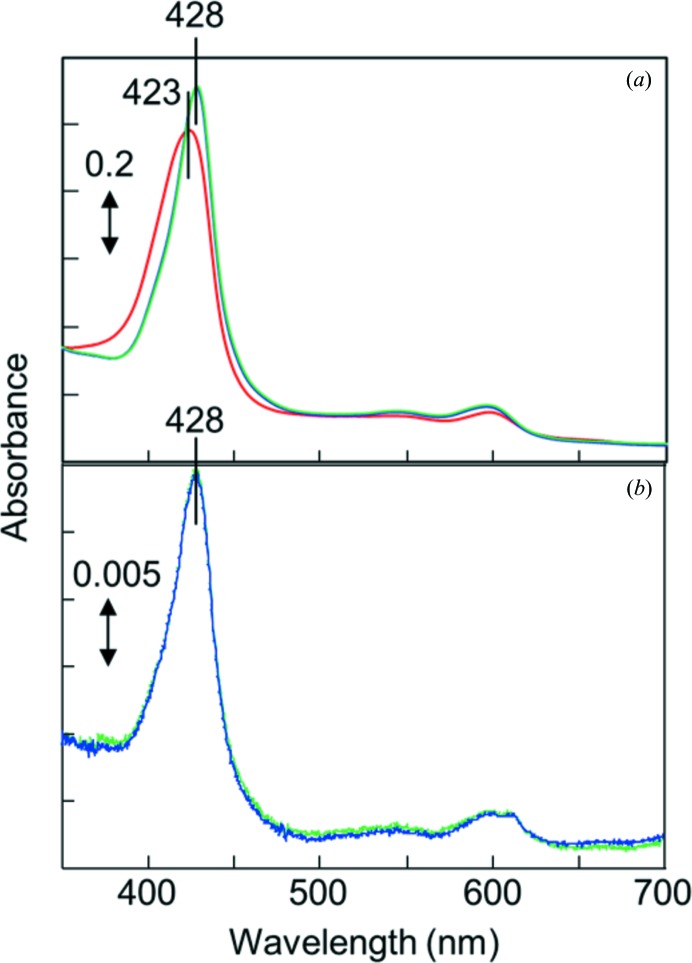
Absorption spectra of CcO. (a) Spectra of a solution containing 10 µM CcO in the fully oxidized resting state in 100 mM sodium phosphate buffer pH 7.4 containing 0.2%(w/v) n-decyl β-d -maltoside (red line) and 2 h (blue line) or 1 d (green line) after the addition of 5 mM potassium cyanide. (b) Spectra of a solution containing dissolved CcO crystals after CN− treatment in 100 mM sodium phosphate buffer pH 7.4 and 0.2%(w/v) n-decyl β-d -maltoside (blue line) and 2 h after the addition of 5 mM potassium cyanide (green line).
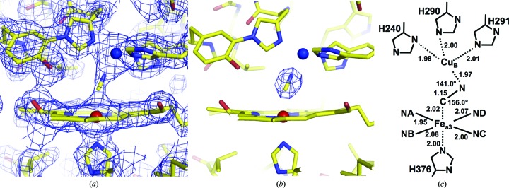
Structure of the oxygen-reduction centre containing CN−. (a) MR/DM electron-density map drawn at the 1.5σ level. (b) (F
o − F
c) electron-density map drawn at the 9.0σ level. (c) Schematic description of the CN−-bound metal sites, giving distances (in Å) and angles.

Structure of the cavity containing the water molecule that forms a hydrogen bond to the OH of Tyr244. The cavity is surrounded by His240, Tyr244, Val287, His290, Thr309 and Ile312. The side-chain structure of Ile312 exhibits multiple conformations, the major and minor components of which are coloured yellow and green, respectively. The distance between the O atom of the water and Cδ1 of the major component of Ile312 is 2.02 Å, whereas that between the O atom and Cδ1 of the minor component of Ile312 is 3.93 Å.
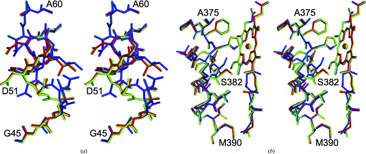
Comparison of the structure of CN−-bound oxidized CcO with those of the reduced (PDB entry 2eij ) and oxidized (PDB entry 2dyr ) enzymes. The structures of CN−-bound oxidized CcO and reduced CcO were superposed on that of oxidized CcO by a least-squares method using Coot (Emsley et al., 2010 ▶). The CN−-bound oxidized CcO, the reduced CcO and the oxidized CcO are coloured green, blue and red, respectively. The three structures for (a) residues 45–60 and (b) residues 375–390 of subunit I and haem a are shown as stereoscopic pairs.
References
Publication types
MeSH terms
Substances
Associated data
- Actions
LinkOut - more resources
Full Text Sources
