Review
. 2015 Nov;88(Pt B):101-107.
doi: 10.1016/j.freeradbiomed.2015.05.034. Epub 2015 Jun 7.

Structural basis of Keap1 interactions with Nrf2

Affiliations
Review

Structural basis of Keap1 interactions with Nrf2

Peter Canning et al. Free Radic Biol Med. 2015 Nov.

Abstract

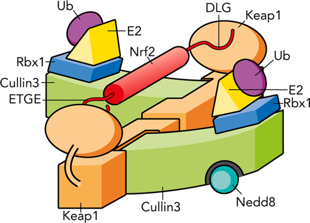
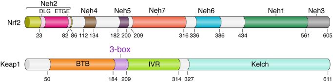
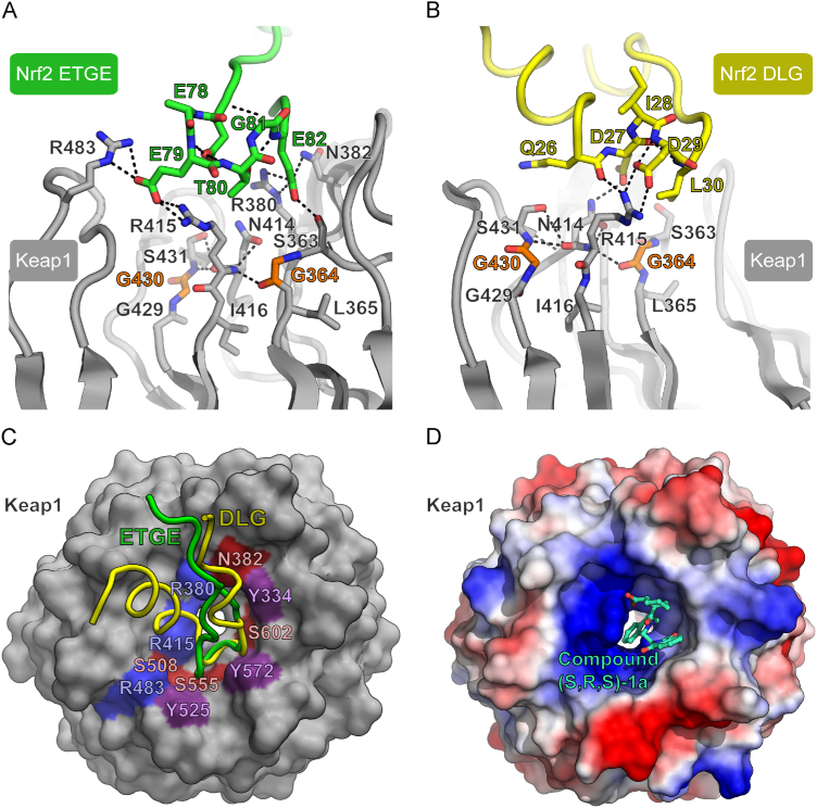
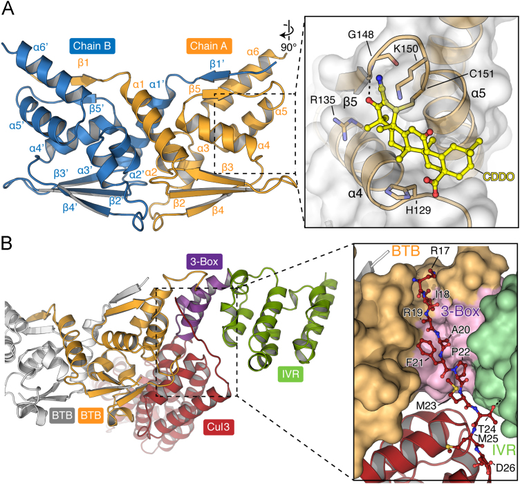
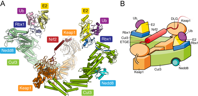
Keap1 is a highly redox-sensitive member of the BTB-Kelch family that assembles with the Cul3 protein to form a Cullin-RING E3 ligase complex for the degradation of Nrf2. Oxidative stress disables Keap1, allowing Nrf2 protein levels to accumulate for the transactivation of critical stress response genes. Consequently, the Keap1-Nrf2 system is extensively pursued for the development of protein-protein interaction inhibitors that will stabilize Nrf2 for therapeutic effect in conditions of neurodegeneration, inflammation, and cancer. Here we review current progress toward the structure determination of Keap1 and its protein complexes with Cul3, Nrf2 substrate, and small-molecule antagonists. Together the available structures establish a rational three-dimensional model to explain the two-site binding of Nrf2 as well as its efficient ubiquitination.

Keywords: BTB; Cullin; Free radicals; Keap1; Kelch; Nrf2; Ubiquitin.


Figures

None
Graphical abstract
Fig. 1
Fig. 1
Domain architecture of the Keap1 and Nrf2 proteins. Domain boundaries and residue numbers are shown for the human proteins.
Fig. 2
Fig. 2
Binding of Nrf2 to Keap1. (A) Selected side-chain interactions are shown in the complex of human Keap1 and the Nrf2 ETGE motif (PDB 2FLU). Kelch domain positions with known somatic cancer mutations (G364C and G430C) are shown in orange; other Keap1 and Nrf2 interface residues are shown in gray and green, respectively. (B) Selected side-chain interactions in the DLG motif complex with mouse Keap1 (PDB 3WN7). DLG peptide residues are colored yellow; Keap1 residues are colored as in (A). (C) Comparison of the binding of the ETGE (green) and DLG (yellow) peptides. Colored areas on the Keap1 surface indicate the main interacting residues (blue, basic; red, polar; purple, hydrophobic). (D) Structural basis for Keap1 inhibition by small molecules targeting the Kelch domain. The electrostatic potential of the protein surface reveals a basic patch around the Nrf2 binding site. A bound small-molecule inhibitor is shown from PDB 4L7B (chain B) .
Fig. 3
Fig. 3
BTB dimerization and Cul3 binding. (A) Ribbon representation of the Keap1 BTB dimer (PDB 4CXI). The inset shows the binding mode of the antagonist bardoxolone/CDDO (PDB 4CXT), which forms a covalent bond to the side chain of Cys151. (B) Model of the Cul3 interface, based on the crystal structure of the KLHL11–Cul3 complex (PDB 4AP2). Cul3 is shown in red, whereas the BTB, 3-box, and IVR domains are colored as shown in Fig. 1. The second BTB subunit is colored gray. The inset shows the interaction between the residues of the Cul3 N-terminal extension and the 3-box of KLHL11.
Fig. 4
Fig. 4
Model of the fully assembled CRL3 complex. (A) Structural model assembled as previously described . A predicted helix in the Neh2 region of Nrf2 is modeled between the bound DLG and ETGE sites. (B) Schematic illustration of the complex shown in (A).

References

    1. Moi P., Chan K., Asunis I., Cao A., Kan Y.W. Isolation of NF-E2-related factor 2 (Nrf2), a NF-E2-like basic leucine zipper transcriptional activator that binds to the tandem NF-E2/AP1 repeat of the beta-globin locus control region. Proc. Natl. Acad. Sci. USA. 1994;91:9926–9930. - PMC - PubMed
    1. Sykiotis G.P., Bohmann D. Stress-activated cap’n’collar transcription factors in aging and human disease. Sci. Signaling. 2010;3 - PMC - PubMed
    1. Hayes J.D., Dinkova-Kostova A.T. The Nrf2 regulatory network provides an interface between redox and intermediary metabolism. Trends Biochem. Sci. 2014;39:199–218. - PubMed
    1. Suzuki T., Motohashi H., Yamamoto M. Toward clinical application of the Keap1–Nrf2 pathway. Trends Pharmacol. Sci. 2013;34:340–346. - PubMed
    1. Giudice A., Montella M. Activation of the Nrf2–ARE signaling pathway: a promising strategy in cancer prevention. Bioessays. 2006;28:169–181. - PubMed

Publication types

MeSH terms

Substances