. 2015 Jun 8;27(6):864-76.
doi: 10.1016/j.ccell.2015.05.004.

Inhibition of the Mitochondrial Protease ClpP as a Therapeutic Strategy for Human Acute Myeloid Leukemia

Affiliations

Inhibition of the Mitochondrial Protease ClpP as a Therapeutic Strategy for Human Acute Myeloid Leukemia

Alicia Cole et al. Cancer Cell. .

Abstract

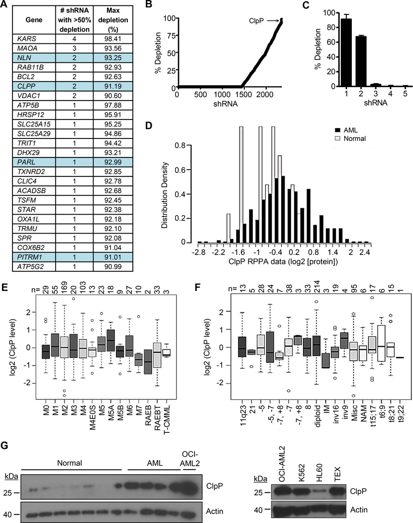
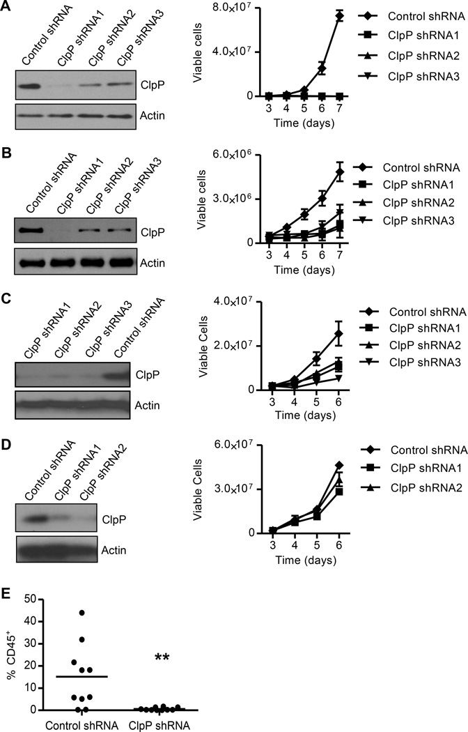
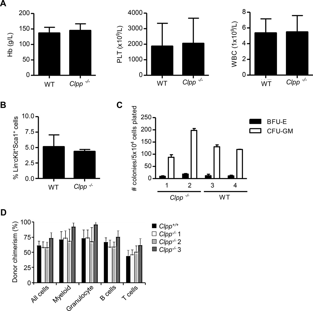
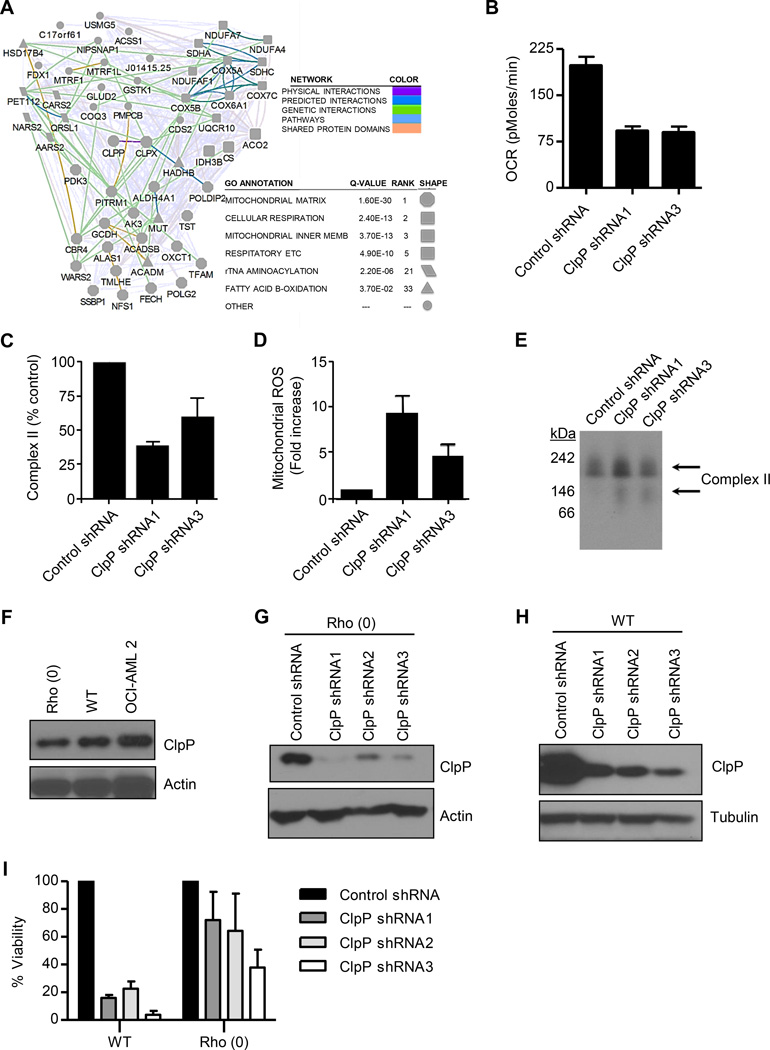
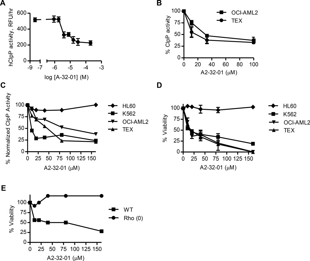
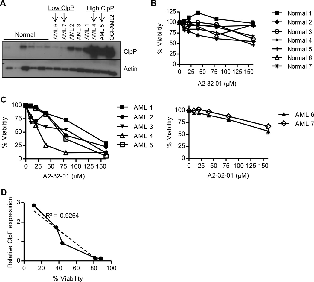
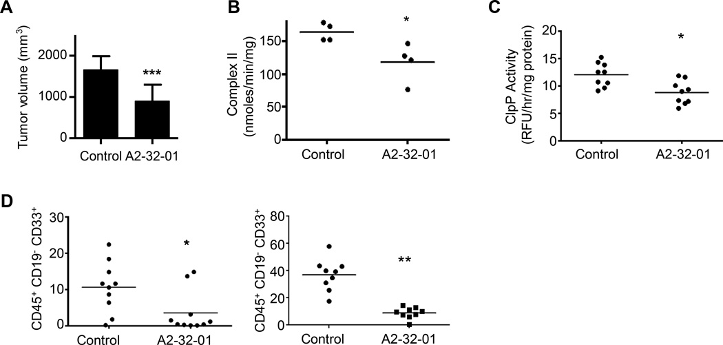
From an shRNA screen, we identified ClpP as a member of the mitochondrial proteome whose knockdown reduced the viability of K562 leukemic cells. Expression of this mitochondrial protease that has structural similarity to the cytoplasmic proteosome is increased in leukemic cells from approximately half of all patients with AML. Genetic or chemical inhibition of ClpP killed cells from both human AML cell lines and primary samples in which the cells showed elevated ClpP expression but did not affect their normal counterparts. Importantly, Clpp knockout mice were viable with normal hematopoiesis. Mechanistically, we found that ClpP interacts with mitochondrial respiratory chain proteins and metabolic enzymes, and knockdown of ClpP in leukemic cells inhibited oxidative phosphorylation and mitochondrial metabolism.


Figures

Figure 1
Figure 1. Genetic screen identifies ClpP as essential for the viability of leukemia cells
(A) The list of top 25 shRNAs targeting the mitochondrial proteome. (B) The percent depletion in rank order of the 2422 shRNA targeting 496 members of the mitochondrial proteome is shown. (C) The percent depletion of individual clones in the library targeting ClpP. (D) Expression of ClpP measured in total cell lysates from 511 primary AML cases and 21 samples of normal CD34+ hematopoietic cells using a reverse phase protein array. (E, F) Expression of ClpP in primary AML cases classified based on their morphologic FAB subtype (E) or cytogenetic abnormalities (F). (G) Expression of ClpP protein in total cell lysates prepared from normal hematopoietic cells, primary AML samples, and indicated cell lines by immunoblotting. Error bars represent standard deviation. The box represents the first and third quartiles, whiskers represent the range, the center line represents the median, and the circles outliers. See also Figure S1 and Table S1.
Figure 2
Figure 2. Knockdown of ClpP reduces the growth and viability of AML cells
(A – D) The expression of ClpP and Actin in TEX (A), OCI-AML2 (B), K562 (C), and HL60 (D) cells four days after transduced with lentiviral vectors expressing control shRNA or one of the three shRNAs targeting ClpP were detected by immunoblotting. Viable cell counts of cells seed four days after transduction are shown as mean ± SD. One of three representative experiments is shown. (E) The percentage of human CD45+ cells in the non-injected femur of sublethally irradiated NOD/SCID-GF mice determined by flow cytometry (n = 10 per group). ** p = 0.005, by t-test. Bar represents mean engraftment.
Figure 3
Figure 3. ClpP knockout mice are viable with normal hematopoiesis
(A) Mean ± SD hemoglobin (Hb), platelet (PLT), and white blood cell (WBC) counts from peripheral blood samples of WT and Clpp−/− mice (n = 4 per group). (B) The frequency of Lin-cKit+Sca1+ hematopoietic cells in the bone marrow of WT and Clpp−/− mice. Mean ± SD percentages were determined by flow cytometry. (C) Erythroid and myeloid colony formation from BFU-E and CFU-GM of hematopoietic cells from WT and Clpp−/− mice (n = 3 per group) plated in vitro under standard conditions. The mean ± SD colony counts are shown. (D) The mean ± SD proportions of CD45.2 cells from WT or Clpp−/− mice transplanted in sublethally irradiated congenic W41/W41 CD45.1 mice measured by flow cytometry. See also Figure S2.
Figure 4
Figure 4. ClpP regulates mitochondrial metabolism
(A) Network of proteins that interacted with WT or catalytically inactive ClpP. (B) Effects of ClpP knockdown on basal oxygen consumption (OCR) in OCI-AML2 cells 4 days after transduction with ClpP shRNA or control sequences. Mean ± SD cell counts from one of three representative experiments are shown. (C) Effects of ClpP knockdown on complex II activity in OCI-AML2 cells 4 days after transduction with ClpP shRNA or control sequences. Percent mean ± SD enzymatic activity from one of 3 representative experiments is shown. (D) Effects of ClpP knockdown on mitochondrial ROS production in OCI-AML2 cells 4 days after transduction with ClpP shRNA or control sequences. Mean ± SD mitochondrial ROS positive cells from one of three representative experiments are shown. (E) Effect of ClpP knockdown in OCI-AML2 cells four days after infection with virus expressing shRNA targeting ClpP or control sequences on SDHA expression and migration on a native gel determined by immunoblotting. (F) Expression of ClpP in total cell lysates prepared from WT and Rho (0) 143B cells and OCI-AML2 cells. (G, H) The expression of ClpP and β-actin were detected by immunoblotting in WT (G) and Rho (0) 143B (H) cells transduced with individual shRNA targeting ClpP or control sequences. (I) The number of viable WT and Rho (0) 143B cells was measured by trypan blue staining 12 days after transduction as described in panel F and G. One of 3 representative experiments showing the percent mean ± SD viable cells relative to cells infected with control shRNA is presented. See also Figure S3 and Table S2.
Figure 5
Figure 5. A2-32-01 inhibits ClpP and is cytotoxic to AML cells
(A) Effect of A2-32-01 on the ability of recombinant human ClpP and ClpX to cleave the fluorogenic substrate casein-FITC. Mean ± SD. (B) Effect of A2-32-01 on ClpP activity measured in mitochondrial lysates from OCI-AML2 and TEX cells after 2 hr based on cleavage of the fluorogenic substrate Suc-LY-AMC. Percent mean ± SD. (C) Effect of 48 hr A2-32-01 treatment on the activity of ClpP in HL60, K562, OCI-AML2, and TEX cells based on the cleavage of the fluorogenic substrate Suc-LY-AMC. Normalized data are shown. (D) Effect of 48 hr A2-32-01 treatment on the viability of HL60, K562, OCI-AML2, and TEX cells. Percent mean ± SD of viable cells was measured by trypan blue staining. (E) Effect of 48 hr A2-32-01 treatment on the viability of WT and Rho (0) 143B cells. Percent of viable cells was measured by trypan blue staining. See also Figure S4.
Figure 6
Figure 6. ClpP expression correlates with sensitivity to A2-32-01 in primary AML cells
(A) Expression of ClpP and Actin in primary normal hematopoietic cells, primary AML patient samples, and OCI-AML2 cells was determined by immunoblotting. (B, C) Effect of A2-32-01 on the viability of primary normal hematopoietic cells (B) and primary AML cells (C) assessed after a 48 hour period of exposure. Mean ± SD percent of viable cells was measured by Annexin V/PI staining and flow cytometry. (D) Correlation analysis of ClpP expression and cell viability of primary AML cells. Cell viability was measured by Annexin-V/PI staining 48 hours after treatment with 80 µM of A2-32-01.
Figure 7
Figure 7. A2-32-01 shows anti-AML activity in xenograft models of human leukemia
(A) Effect of i.p. injection of 300 mg/kg of A2-32-01 or corn oil vehicle control twice daily for 5 of 7 days on tumor volume when OCI-AML2 cells were xenografted into SCID mice (n = 10 mice per group). Mean ± SD. *** p < 0.001 by t-test. (B) Respiratory chain complex II activity in tumors from SCID mice xenografted with OCI-AML2 cells and treated with i.p. injection of 300 mg/kg A2-32-01 or corn oil vehicle control daily for 5 days (n = 4 mice per group). * p = 0.03, by t-test. (C) ClpP activity measured by the cleavage of the fluorogenic substrate casein-FITC in mitochondrial lysates in tumors from SCID mice xenografted with OCI-AML2 cells and treated with i.p. injection of 300 mg/kg A2-32-01 or corn oil vehicle control daily for 4 days (n = 9 mice per group). * p = 0.005, by t-test. (D) Human leukemia cell engraftment in sublethally irradiated female NOD/SCID mice and treated with A2-32-01 (300 mg/kg by i.p. injection daily) or vehicle control (n = 10 per group) 3 of 7 days for 4 weeks was measured by flow cytometric analysis. The panels show results from two independent experiments. * p = 0.02, ** p < 0.0001, as determined by t-test. Horizontal bars indicate the mean in panels B–D. See also Figure S5.

Comment in

References

    1. Aldridge JE, Horibe T, Hoogenraad NJ. Discovery of genes activated by the mitochondrial unfolded protein response (mtUPR) and cognate promoter elements. PLoS One. 2007;2:e874. - PMC - PubMed
    1. Choi H, Larsen B, Lin ZY, Breitkreutz A, Mellacheruvu D, Fermin D, Qin ZS, Tyers M, Gingras AC, Nesvizhskii AI. SAINT: probabilistic scoring of affinity purification-mass spectrometry data. Nat Methods. 2011;8:70–73. - PMC - PubMed
    1. Conlon BP, Nakayasu ES, Fleck LE, LaFleur MD, Isabella VM, Coleman K, Leonard SN, Smith RD, Adkins JN, Lewis K. Activated ClpP kills persisters and eradicates a chronic biofilm infection. Nature. 2013;503:365–370. - PMC - PubMed
    1. Corydon TJ, Bross P, Holst HU, Neve S, Kristiansen K, Gregersen N, Bolund L. A human homologue of Escherichia coli ClpP caseinolytic protease: recombinant expression, intracellular processing and subcellular localization. Biochem J. 1998;331(Pt 1):309–316. - PMC - PubMed
    1. Dan S, Naito M, Tsuruo T. Selective induction of apoptosis in Philadelphia chromosome-positive chronic myelogenous leukemia cells by an inhibitor of BCR - ABL tyrosine kinase, CGP 57148. Cell Death Differ. 1998;5:710–715. - PubMed

Publication types