. 2015 Jun 10:15:107.
doi: 10.1186/s12862-015-0378-1.

Insights into the evolution of enzyme substrate promiscuity after the discovery of (βα)₈ isomerase evolutionary intermediates from a diverse metagenome

Affiliations

Insights into the evolution of enzyme substrate promiscuity after the discovery of (βα)₈ isomerase evolutionary intermediates from a diverse metagenome

Lianet Noda-García et al. BMC Evol Biol. .

Abstract

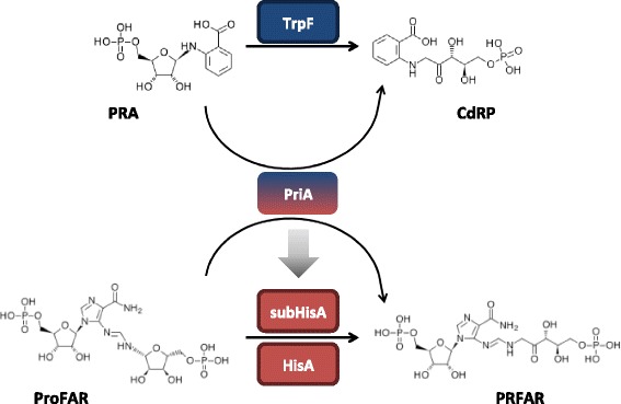
Background: Current sequence-based approaches to identify enzyme functional shifts, such as enzyme promiscuity, have proven to be highly dependent on a priori functional knowledge, hampering our ability to reconstruct evolutionary history behind these mechanisms. Hidden Markov Model (HMM) profiles, broadly used to classify enzyme families, can be useful to distinguish between closely related enzyme families with different specificities. The (βα)8-isomerase HisA/PriA enzyme family, involved in L-histidine (HisA, mono-substrate) biosynthesis in most bacteria and plants, but also in L-tryptophan (HisA/TrpF or PriA, dual-substrate) biosynthesis in most Actinobacteria, has been used as model system to explore evolutionary hypotheses and therefore has a considerable amount of evolutionary, functional and structural knowledge available. We searched for functional evolutionary intermediates between the HisA and PriA enzyme families in order to understand the functional divergence between these families.

Results: We constructed a HMM profile that correctly classifies sequences of unknown function into the HisA and PriA enzyme sub-families. Using this HMM profile, we mined a large metagenome to identify plausible evolutionary intermediate sequences between HisA and PriA. These sequences were used to perform phylogenetic reconstructions and to identify functionally conserved amino acids. Biochemical characterization of one selected enzyme (CAM1) with a mutation within the functionally essential N-terminus phosphate-binding site, namely, an alanine instead of a glycine in HisA or a serine in PriA, showed that this evolutionary intermediate has dual-substrate specificity. Moreover, site-directed mutagenesis of this alanine residue, either backwards into a glycine or forward into a serine, revealed the robustness of this enzyme. None of these mutations, presumably upon functionally essential amino acids, significantly abolished its enzyme activities. A truncated version of this enzyme (CAM2) predicted to adopt a (βα)6-fold, and thus entirely lacking a C-terminus phosphate-binding site, was identified and shown to have HisA activity.

Conclusion: As expected, reconstruction of the evolution of PriA from HisA with HMM profiles suggest that functional shifts involve mutations in evolutionarily intermediate enzymes of otherwise functionally essential residues or motifs. These results are in agreement with a link between promiscuous enzymes and intragenic epistasis. HMM provides a convenient approach for gaining insights into these evolutionary processes.


Figures

Fig. 1
Fig. 1
Enzymatic activities of the HisA / PriA enzyme superfamily. HisA is a ubiquitous enzyme that catalyzes the conversion of N’-[(5-phosphoribosyl) formimino]-5-aminoimidazole-4-carboxamide ribonucleotide (ProFAR) into N’-[(5′-phosphoribulosyl) formimino]-5-aminoimidazole-4-carboxamide ribonucleotide (PRFAR). PriA, exclusively found in the Actinobacteridae class of the high G + C Gram positive bacteria, evolved from HisA to catalyze, in addition to the HisA activity, the conversion of phosphoribosyl anthranilate (PRA) into 1-[(2-carboxyphenyl)amino]-1-deoxyribulose 5-phosphate (CdRP). This latter activity is the same as that catalyzed by the ubiquitous TrpF enzyme. subHisA has recently evolved from PriA (indicated with a gray arrow), and it is found exclusively in certain, but not all, Corynebacterium species. As with HisA, subHisA is mono-functional enzyme
Fig. 2
Fig. 2
Performance of HMM profiles against UniProt and CAMERA databases. HisA, PriA, subHisA and ‘Transition Zone’ sequences are shown as red, blue, orange and green dots, respectively. UniProt codes corresponding to sequences removed to construct the HMM refined profile are indicated. The functionally characterized evolutionary intermediates from CAMERA database are marked as CAM1 and CAM2
Fig. 3
Fig. 3
Phylogenetic analysis of HisA / PriA evolutionary intermediates. A Bayesian phylogenic tree of HisA homologs is shown. Minimum branch support is 0.7. HisA, PriA and intermediate sequences, as CAM1, are grouped in different clades, labeled in red, blue and green, respectively. The key enzymes HisA_Afer (C7LZ82) and PriA_Blon (Q8G4S5) are also shown. The evolutionary intermediate, reflected at the codon level in the N-PBS motif, is shown. Functionally analyzed proteins, and their first N-PBS amino acid codon usage, are marked with an asterisk
Fig. 4
Fig. 4
Comparison of steady-state enzyme kinetics. A graphical comparison of the catalytic efficiencies (k cat/K M) of wild type and mutant enzymes that were characterized is shown. Values for ProFAR (x axis) and PRA (y axis) isomerase activities, expressed as log10, are compared. Data from HisA_Afer (red square), PriA_Blon (blue circle), CAM1 (green circle), CAM1_A81S (green circle with a black border and inner black circle) and CAM1_A81G (green circle with a black border and inner black cross, this study), as well as from PriA_Scoe (blue circle) and the Ser81Thr mutant of PriA from S. coelicolor, labeled as PriA_Scoe (blue circle with a black border and inner black circle) (data obtained from [38]), is included
Fig. 5
Fig. 5
Structural analysis of CAM1 and CAM2. a. Structural homology model of CAM1, corresponding to a full (βα)8-barrel, and CAM2. The (βα)2 subunit missing in CAM2 is shown in gray. N and C-terminal tails are shown in magenta, loops, alpha helix and beta sheet are marked in green, red and yellow, respectively. b. Structural superposition of CAM2 (gray backbone), CAM2_204, CAM2_R1 and CAM2_R2, where N and C-terminal tails are shown in blue, red, green and orange, respectively. c. Sequence alignment of the 12-amino acid C-terminal variable region of CAM2, CAM2_204 (equivalent to CAM1), CAM2_R1 and CAM2_R2. The pairwise RMSD between CAM1 and CAM2, CAM2 and CAM2_R1, CAM2 and CAM2_R2, and CAM2 and CAM2_204 was 1.73 Å, 1.91 Å, 1.41 Å, 1.73 Å, respectively

References

    1. Khersonsky O, Roodveldt C, Tawfik DS. Enzyme promiscuity: evolutionary and mechanistic aspects. Curr Opin Chem Biol. 2006;10(5):498–508. doi: 10.1016/j.cbpa.2006.08.011. - DOI - PubMed
    1. Khersonsky O, Tawfik DS. Enzyme promiscuity: a mechanistic and evolutionary perspective. Annu Rev Biochem. 2010;79:471–505. doi: 10.1146/annurev-biochem-030409-143718. - DOI - PubMed
    1. Notebaart RA, Szappanos B, Kintses B, Pal F, Gyorkei A, Bogos B, et al. Network-level architecture and the evolutionary potential of underground metabolism. Proc Natl Acad Sci U S A. 2014;111(32):11762–7. doi: 10.1073/pnas.1406102111. - DOI - PMC - PubMed
    1. Jensen RA. Enzyme recruitment in evolution of new function. Annu Rev Microbiol. 1976;30:409–25. doi: 10.1146/annurev.mi.30.100176.002205. - DOI - PubMed
    1. Soskine M, Tawfik DS. Mutational effects and the evolution of new protein functions. Nat Rev Genet. 2010;11(8):572–82. doi: 10.1038/nrg2808. - DOI - PubMed

Publication types

LinkOut - more resources