. 2015 Jun 10:10:14.
doi: 10.1186/s13064-015-0041-y.

Netrin-1 directs dendritic growth and connectivity of vertebrate central neurons in vivo

Affiliations

Netrin-1 directs dendritic growth and connectivity of vertebrate central neurons in vivo

Anastasia N Nagel et al. Neural Dev. .

Abstract

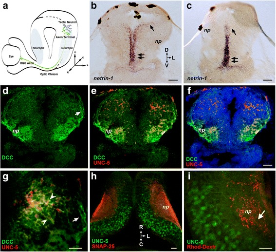
Background: Netrins are a family of extracellular proteins that function as chemotropic guidance cues for migrating cells and axons during neural development. In the visual system, netrin-1 has been shown to play a key role in retinal ganglion cell (RGC) axon growth and branching at the target, where presynaptic RGC axons form partnerships with the dendrites of tectal neurons. However, the signals that guide the connections between RGC axons and their postsynaptic partners are yet unknown. Here, we explored dynamic cellular mechanisms by which netrin-1 influences visual circuit formation, particularly those that impact postsynaptic neuronal morphology and connectivity during retinotectal wiring.

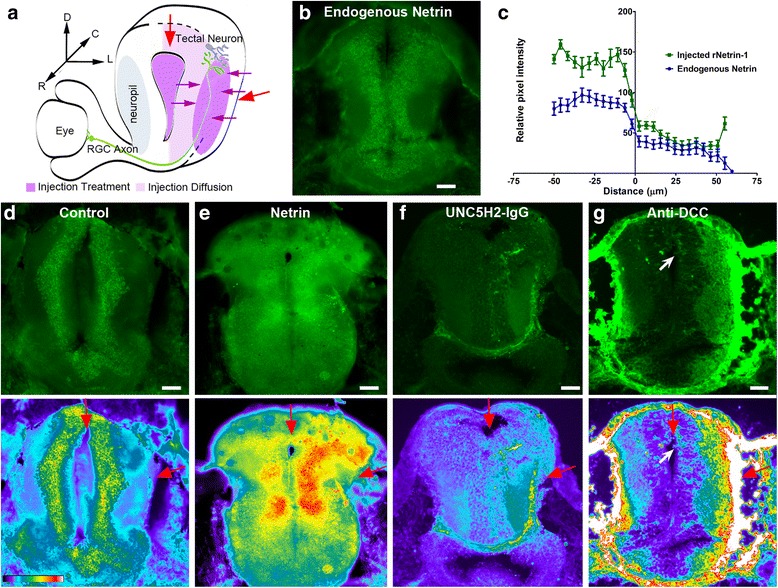
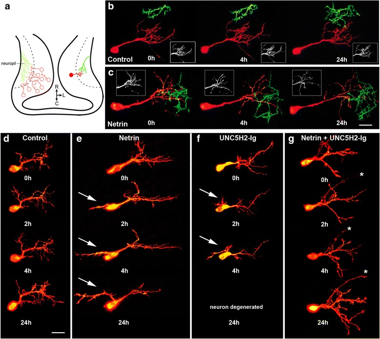
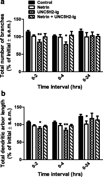
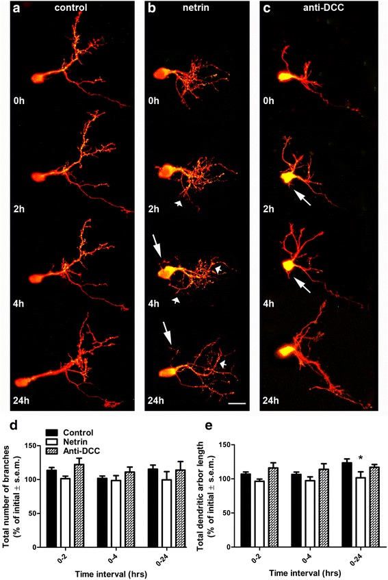
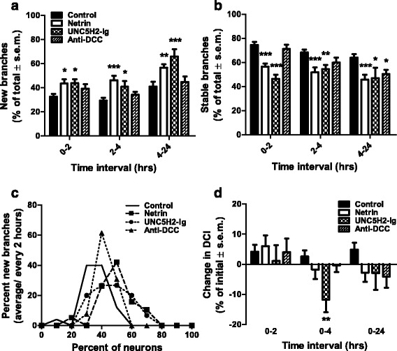
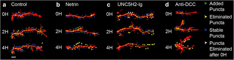
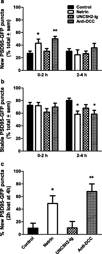
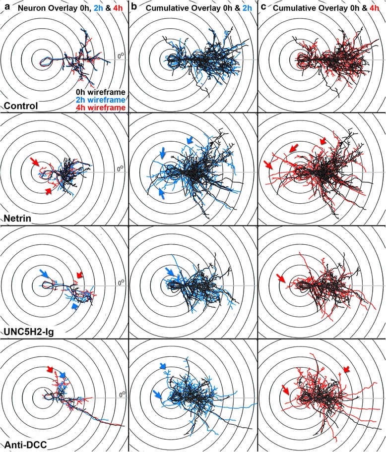
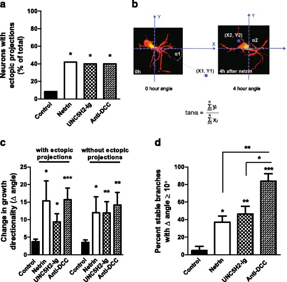
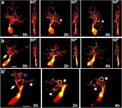
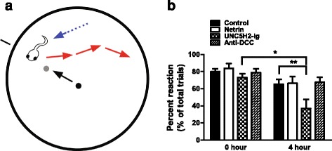
Results: Time-lapse in vivo imaging of individual Xenopus laevis optic tectal neurons co-expressing tdTomato and PSD95-GFP revealed rapid remodeling and reorganization of dendritic arbors following acute manipulations in netrin-1 levels. Effects of altered netrin signaling on developing dendritic arbors of tectal neurons were distinct from its effects on presynaptic RGC axons. Within 4 h of treatment, tectal injection of recombinant netrin-1 or sequestration of endogenous netrin with an UNC-5 receptor ectodomain induced significant changes in the directionality and orientation of dendrite growth and in the maintenance of already established dendrites, demonstrating that relative levels of netrin are important for these functions. In contrast, altering DCC-mediated netrin signaling with function-blocking antibodies induced postsynaptic specialization remodeling and changed growth directionality of already established dendrites. Reducing netrin signaling also decreased avoidance behavior in a visually guided task, suggesting that netrin is essential for emergent visual system function.

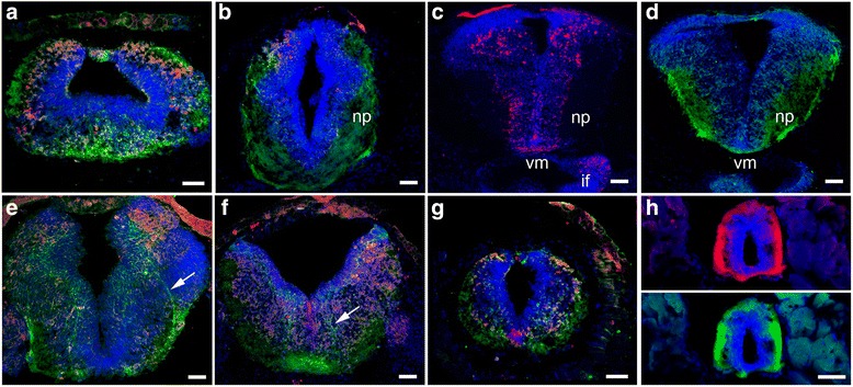
Conclusions: These in vivo findings together with the patterns of expression of netrin and its receptors reveal an important role for netrin in the early growth and guidance of vertebrate central neuron dendritic arbors. Collectively, our studies indicate that netrin shapes both pre- and postsynaptic arbor morphology directly and in multiple ways at stages critical for functional visual system development.


Figures

Fig. 1
Fig. 1
Expression of netrin-1 and of its receptors DCC and UNC-5 in stage 45 Xenopus optic tectum. a Schematic of coronal section of Xenopus retinotectal circuit. RGC axons (green) travel from the contralateral eye to connect with tectal neurons in the neuropil (blue). b, c In situ hybridization with Xenopus-specific antisense netrin-1 probes. Coronal sections of the midbrain at the level of the optic tectum show ventral-high (double arrows) to dorsal-low (arrow) netrin-1 mRNA expression along the ventricle wall. d–g Coronal and h, i horizontal sections show DCC and UNC-5 expression. d–g Co-immunostaining illustrates the differential distribution of UNC-5 (red) and DCC (green) immunoreactivity. d DCC immunoreactivity (green) is localized to the cell bodies in the dorsal tectum and proximal dendrites and to incoming axons near the dorsal neuropil (arrow). The tectal neuropil (np) is also positive for DCC. The low- (e, f) and high- (g) magnification coronal images show UNC-5 (red) and DCC (green) co-localization, with UNC-5 being localized to a subset of cells that also expresses DCC (g, arrowheads). f Counterstaining with DAPI (blue) serves to distinguish nuclear staining from cytoplasmic UNC-5 (red) and DCC (green) expression in tectal cells. h UNC-5 immunoreactivity (green) is localized to a subset of cell bodies in the dorsal area of the tectum and area adjacent to the tectal neuropil identified by immunostaining with antibodies to the presynaptic protein SNAP-25 (red). i Anterograde labeling with rhodamine dextran shows that RGC axons (red) terminate in the areas of the tectal neuropil (arrow) where UNC-5 immunopositive neurons localize (green). D dorsal, V ventral, C caudal, R rostral, L lateral, np neuropil. Scale bars: 50 μm in b–f, 20 μm in g, 20 μm in h–i
Fig. 2
Fig. 2
Specific patterns of DCC and UNC-5 expression in the X. laevis central nervous system. Immunostaining with antibodies to UNC-5 (red) and DCC (green) revealed specific patterns of expression of the netrin-1 receptors in stage 45 tadpoles. a–g UNC-5 (red) and DCC (green) immunoreactivity in the forebrain (a), pre-tectum (b), caudal tectum (e), hindbrain (f), and rostral spinal cord (g) demonstrate a specific pattern of expression for each of these receptors within subpopulations of neurons in the central nervous system. c UNC-5 immunostaining (red) localizes to subpopulations of neurons in the dorsal tectum, lateral-ventral midbrain, ventral midline (vm), and infundibulum (if). d DCC immunoreactivity (green) is localized in dorsal tectal neuron cell bodies and processes in the tectum and ventral midline, as well as in the tectal neuropil (np). e, f Note the specificity of immunostaining and co-localization of UNC-5 and DCC expression in subpopulations of cells in the caudal tectum (e) and hindbrain (f) and the localization of DCC receptors to discrete fiber tracts (arrows). g, h UNC-5 (red) and DCC (green) immunoreactivity in the rostral (g) and caudal (h) spinal cord is localized to fiber tracts and ventral midline in agreement with published observations in Xenopus and other species (for review, see [, , –45]). DCC immunoreactivity in the spinal cord is similar when staining with antibodies directed against the extracellular (g) or intracellular (h, bottom) domains of DCC. Counterstaining with DAPI (blue) serves to distinguish nuclear staining from UNC-5 (red) and DCC (green) expression in cell bodies and fiber tracts. Scale bars: 50 μm
Fig. 3
Fig. 3
Protein diffusion after treatment. a Schematic of coronal view of stage 45 Xenopus retinotectal circuit depicting injection sites (red arrows) and spread of injected proteins (violet color). b Coronal section at the level of the optic tectum immunostained with antibodies to netrin-1. Note endogenous netrin immunoreactivity in cell body layer and neuropil. c–g Sections at the level of the optic tectum of tadpoles injected with vehicle, recombinant netrin-1, UNC5H2-Ig, or anti-DCC were immunostained to examine the spread of the injected proteins after treatment. c Quantitative analysis of fluorescence intensity in sections of uninjected tadpoles (Endogenous Netrin) or tadpoles injected with recombinant netrin (Injected rNetrin-1). The relative levels of netrin within the cell body layer and the neuropil are illustrated by the average pixel intensity values along the medial-to-lateral axis of the tectum. The zero value in the X-axis corresponds to the cell body layer-neuropil boundary; negative X-coordinates represent distance from the boundary to the ventricle while positive X-coordinates represent distance from the boundary to the lateral-most neuropil. n = 10 brain sections per group, from four tadpole brains per group, with three 20-pixel-wide line scans quantified per section. Error bars represent the standard error of the mean. d–g Sample coronal sections of tadpoles injected with vehicle (d), recombinant netrin-1 (e), UNC5H2-Ig (f), or anti-DCC (g) immunostained with chick antibodies to netrin-1 and Alexa 488 secondary antibodies to chick IgG (top; d, e) or stained with Alexa 488 secondary antibodies to human IgG (top; f) or mouse IgG (top; g). The pseudo-color images in d–g (bottom) show the relative intensity of the Alexa fluor 488 fluorescence. Pixel intensity values ranged from 0 (black) to 255 (white) as illustrated by the color-scale bar (d, bottom). Note the increased immunofluorescence in the cell body layer and neuropil of netrin-1-treated tadpoles (e) when compared to vehicle-injected controls (d) and with endogenous netrin-1 expression (b). In f and g, the relatively higher fluorescence intensity in the hemisphere that received the injection (red arrows) and the diffusion patterns of the proteins are more evident in the pseudo-color images. In g, white arrows point to fluorescently labeled cells in the injected tectal hemisphere. Scale bars in b, d–g: 50 μm
Fig. 4
Fig. 4
Rapid remodeling of dendritic arbors upon acute manipulations in netrin signaling. a Schematic diagram of a stage 45 Xenopus tectal midbrain (horizontal view). Tectal neurons (red) make dendritic connections with contralateral RGC axons (green) within the tectal neuropil. b, c Sample RGC axons and tectal neurons, visualized by expression of GFP and tdTomato, respectively, in control (b) and netrin-treated (c) tadpoles. Note change in tectal neuron dendritic architecture evident at 4 and 24 h after netrin-1 treatment (inserts). d–g Confocal projections of representative tectal neurons co-expressing tdTomato (red) and PSD95-GFP (green) in tadpoles injected with control vehicle solution (d), Netrin (e), UNC5H2-Ig (f), or Netrin + UNC5H2-Ig (g). Note the emergence of an alternative primary dendrite (arrow) growing towards the midline in neurons exposed to netrin-1 or UNC5H2-Ig. Tadpoles treated with netrin + UNC5H2-Ig appeared identical to controls. Axons of tectal neurons are labeled by the asterisks. Scale bars: 20 μm
Fig. 5
Fig. 5
Altering endogenous netrin levels decreases dendrite branch number and total dendritic arbor length. Effects of tectal microinjection of netrin, UNC5H2-Ig, or netrin + UNC5H2-Ig on total dendrite branch number (a) and length (b). Netrin-1 and UNC5H2-Ig altered tectal neuron morphology with a different time scale. Note that exogenous netrin-1 treatment decreased dendrite arbor length at 24 h, while the UNC5H2-Ig treatment that sequesters endogenous netrin induced a transient but significant decrease in branch number at the 0- to 2- and 0- to 4-h imaging intervals when compared to all other treatments. Co-treatment with netrin + UNC5H2-Ig did not influence branch number or length. Values are expressed as percent change from the initial 0-h imaging session. Two-way ANOVA with Bonferroni multiple comparison test; *p < 0.05, **p < 0.01. Error bars indicate SEM
Fig. 6
Fig. 6
Blocking DCC signaling induces changes in dendritic arbor shape without altering total branch number or length. a–c Confocal projections of representative tectal neurons co-expressing tdTomato (red) and PSD95-GFP (green) in tadpoles injected with control vehicle solution (a), netrin-1 (b), or function-blocking antibodies to DCC (c). While control neurons branch, elaborate, and add PSD95-GFP puncta (a), neurons in tadpoles treated with netrin-1 undergo dynamic remodeling of existing branches (b). Short arrows point to dendrites with altered directions of growth. Neurons in tadpoles treated with anti-DCC (c) also appear to change dendritic arbor direction and form small basal projections at 2 and 4 h post-injection (long arrows). Scale bars: 20 μm. d, e Comparison of effects of netrin and anti-DCC on total branch number (d) and dendritic arbor length (e). Note that only netrin-1 treatment decreased arbor length at 24 h (e), but neither netrin nor anti-DCC affects the total number of branches (d). Two-way ANOVA with Bonferroni multiple comparison test; *p < 0.05. Error bars indicate SEM
Fig. 7
Fig. 7
Acute manipulations in endogenous netrin levels induce rapid changes in dendrite remodeling. a, b Effects of netrin-1, UNC5H2-Ig, or anti-DCC treatments on new branch addition (a) and branch stabilization (b). Note that while netrin-1 and UNC5H2-Ig increased branch addition and decreased branch stabilization throughout the 24-h imaging period, the anti-DCC treatment influenced the stability of branches at the 4- to 24-h interval only. c Relative proportion of neurons with different branch addition rates. A significant shift in the distribution of neurons that responded with increased branch addition rates was observed after netrin-1 and UNC5H2-Ig treatments. Values are expressed as percent change from total branches. d Relative change in DCI values is shown for each group at all imaging intervals. Note that neurons in UNC5H2-Ig-treated tadpoles significantly decreased their complexity by 4 h compared to controls. Two-way ANOVA with Bonferroni multiple comparison test; *p < 0.05, **p < 0.01, ***p < 0.001. Error bars indicate SEM
Fig. 8
Fig. 8
Altered netrin-1 levels and DCC signaling impact postsynaptic cluster remodeling. a–d Confocal projections of single branches from representative tectal neurons co-expressing tdTomato (red) and PSD95-GFP (green) from control (a), netrin (b), UNC5H2-Ig (c), or Anti-DCC (d) groups before and after treatment. Dynamic remodeling of postsynaptic specializations is illustrated by the addition (green arrowheads) and elimination (yellow arrowheads) of PSD95-GFP clusters. Blue arrowheads denote puncta that remained stable from one observation interval to the next; white arrowheads denote puncta that were present at the initial observation time point but were eliminated (yellow) at 2 h. Scale bar: 20 μm
Fig. 9
Fig. 9
Postsynaptic cluster addition and stabilization are modulated by alterations in netrin signaling. a, b Effects of netrin-1, UNC5H2-Ig, or anti-DCC treatments on postsynaptic cluster remodeling were quantified as the proportion of PSD95-GFP puncta that were added (a) and remained stable (b) within the 0–2 and 2–4 observation intervals. Note that significantly more PSD95-GFP puncta were between 0 and 2 h (a), while fewer were stable between 2 and 4 h (b) following netrin-1 or anti-DCC treatment when compared to controls. c To determine the relative stability of newly added postsynaptic clusters, we quantified relative proportion of PSD95-GFP puncta added over the 0- to 2-h interval that were lost in the subsequent 2- to 4-h interval for a subset of randomly selected neurons for each group (n = 4). PSD95-GFP puncta added from 0 to 2 h were significantly less stable in the netrin-1- or anti-DCC-treated neurons. Statistical significance was by one-way ANOVA and with unpaired t-tests. Significance when compared to control is *p < 0.05, **p < 0.01. Error bars indicate SEM
Fig. 10
Fig. 10
Overlays of sample neurons at 0, 2, and 4 h illustrate changes in dendritic arbor morphology in response to treatment and between imaging intervals. a Confocal stacks of individual neurons from control, netrin-1-, UNC5H2-Ig-, and anti-DCC-treated tadpoles were reconstructed with MetaMorph creating three-dimensional wireframes of each stack. Wireframes were color-coded based on imaging time point (black, 0 h; blue, 2 h; red, 4 h), overlapped, and aligned over Scholl concentric circles with the primary dendrite placed at a 0° angle (X-axis; gray line). Dynamic changes in dendritic morphology every 2 h over a 4-h imaging period are illustrated by the emergence of blue (2 h) or red branches (4 h) from under the black wireframe (0 h). b, c Cumulative wireframes from a subset of seven neurons per condition better illustrate the dynamic changes in growth between the 0- and 2-h imaging interval (b), and the 0- and 4-h imaging interval (c), for each treatment group. Large arrows point to sample ectopic branches newly extended at the time point indicated by the color of the arrow (blue, 2 h; red, 4 h). Short arrows point to already established branches that changed their directionality of growth at the time point indicated by the color of the arrow (blue, 2 h; red, 4 h)
Fig. 11
Fig. 11
Perturbations in tectal netrin levels or signaling alter dendritic arbor directionality. a Proportion of neurons that developed ectopic basal projections within the 24-h period in each group. b Angle analysis performed on tectal neuron arbors sums all branch points to produce a net vector. The angle change was calculated from the tangents of arbors from 0 to 4 h. c The change in dendritic arbor directionality is shown as the difference in angle for neurons from 0 to 4 h and was measured both including (with) and excluding (without) ectopic projections. d Proportion of stable branches with net angle change. The percentage of stable branches that individually changed their angle by at least 10° was calculated for a subset of randomly selected neurons (n = 4). The individual branch tip vectors for each branch were compared from 0 to 4 h to calculate the angle change. Note that a larger proportion of stable branches altered their angle in neurons following anti-DCC treatment when compared to all other groups. Statistical significance was by Kruskal-Wallis Friedman with Dunn’s multiple comparison test. Significance when compared to control is *p < 0.05, **p < 0.01, ***p < 0.001. Error bars indicate SEM
Fig. 12
Fig. 12
Individual branches change their orientation of growth in response to altered netrin levels. a, b The maximum projections of each confocal z-stack of two sample neurons at the 0-, 2-, and 4-h imaging time points, and the corresponding 90° view of each three-dimensional z-stack, illustrate the dynamic changes in growth and directionality of individual dendrites in response to acute netrin-1 treatment. The neuron in a corresponds to that shown in Fig. 5b. b’ For the sample neuron in b, a single primary dendrite and its individual secondary branches of the same branch can be discerned in the higher magnification images by selecting and projecting only the z-planes from each confocal stack that include that branch. By isolating the individual dendrite from the rest of the dendritic arbor, one can better differentiate the change in the direction of growth of the primary dendrite (short white arrows) that took place while some of its secondary branches were pruned (double blue arrows) or changed their direction of growth (green arrow) and others were maintained. Scale bars: 20 μm
Fig. 13
Fig. 13
Sequestration of endogenous netrin-1 with UNC-5 ectodomain affects swimming behavior in a visually guided task. a Schematic of the visual avoidance task viewed from above. Stage 45 tadpoles swim in the 60-mm open field (blue arrow and dotted line) while the Matlab program projects an image on the monitor where the petri dish rests. The black line outside the field represents the vector the 0.3-mm dot (small black circle) will travel. Every 30 s, the 0.3-mm dot appears in the center and is directed towards the black line to intercept the tadpole (black arrow). The tadpole’s response to the advancing stimuli (gray circle) is video recorded and typically results in the tadpole changing its swimming velocity and/or direction (red arrows). b Reaction to the presentation of a moving visual stimulus for tadpoles before treatment (0 h) and 4 h after treatment with vehicle solution (control), netrin-1, anti-DCC, or UNC5H2-Ig is shown as the percent of trials in which tadpoles showed an avoidance response. Tadpoles injected with UNC5H2-Ig had decreased avoidance responses to the presentation of the stimulus 4 h post-injection. Two-way, repeated measures ANOVA with Bonferroni multiple comparison test; *p < 0.05, **p < 0.01. Error bars indicate SEM

References

    1. Lai Wing Sun K, Correia JP, Kennedy TE. Netrins: versatile extracellular cues with diverse functions. Development. 2011;138(11):2153–69. doi: 10.1242/dev.044529. - DOI - PubMed
    1. Finci LI, Kruger N, Sun X, Zhang J, Chegkazi M, Wu Y, et al. The crystal structure of netrin-1 in complex with DCC reveals the bifunctionality of netrin-1 as a guidance cue. Neuron. 2014;83(4):839–49. doi: 10.1016/j.neuron.2014.07.010. - DOI - PMC - PubMed
    1. Xu K, Wu Z, Renier N, Antipenko A, Tzvetkova-Robev D, Xu Y, et al. Neural migration. Structures of netrin-1 bound to two receptors provide insight into its axon guidance mechanism. Science. 2014;344(6189):1275–9. doi: 10.1126/science.1255149. - DOI - PMC - PubMed
    1. Chan SS, Zheng H, Su MW, Wilk R, Killeen MT, Hedgecock EM, et al. UNC-40, a C. elegans homolog of DCC (deleted in colorectal cancer), is required in motile cells responding to UNC-6 netrin cues. Cell. 1996;87(2):187–95. doi: 10.1016/S0092-8674(00)81337-9. - DOI - PubMed
    1. Keino-Masu K, Masu M, Hinck L, Leonardo ED, Chan SS, Culotti JG, et al. Deleted in colorectal cancer (DCC) encodes a netrin receptor. Cell. 1996;87(2):175–85. doi: 10.1016/S0092-8674(00)81336-7. - DOI - PubMed

Publication types