. 2015 Jun 9;6(3):e00775.
doi: 10.1128/mBio.00775-15.

Genome-Wide Identification of Klebsiella pneumoniae Fitness Genes during Lung Infection

Affiliations

Genome-Wide Identification of Klebsiella pneumoniae Fitness Genes during Lung Infection

Michael A Bachman et al. mBio. .

Abstract

Klebsiella pneumoniae is an urgent public health threat because of resistance to carbapenems, antibiotics of last resort against Gram-negative bacterial infections. Despite the fact that K. pneumoniae is a leading cause of pneumonia in hospitalized patients, the bacterial factors required to cause disease are poorly understood. Insertion site sequencing combines transposon mutagenesis with high-throughput sequencing to simultaneously screen thousands of insertion mutants for fitness defects during infection. Using the recently sequenced K. pneumoniae strain KPPR1 in a well-established mouse model of pneumonia, insertion site sequencing was performed on a pool of >25,000 transposon mutants. The relative fitness requirement of each gene was ranked based on the ratio of lung to inoculum read counts and concordance between insertions in the same gene. This analysis revealed over 300 mutants with at least a 2-fold fitness defect and 69 with defects ranging from 10- to >2,000-fold. Construction of 6 isogenic mutants for use in competitive infections with the wild type confirmed their requirement for lung fitness. Critical fitness genes included those for the synthesis of branched-chain and aromatic amino acids that are essential in mice and humans, the transcriptional elongation factor RfaH, and the copper efflux pump CopA. The majority of fitness genes were conserved among reference strains representative of diverse pathotypes. These results indicate that regulation of outer membrane components and synthesis of amino acids that are essential to its host are critical for K. pneumoniae fitness in the lung.

Importance: Klebsiella pneumoniae is a bacterium that commonly causes pneumonia in patients after they are admitted to the hospital. K. pneumoniae is becoming resistant to all available antibiotics, and when these infections spread to the bloodstream, over half of patients die. Since currently available antibiotics are failing, we must discover new ways to treat these infections. In this study, we asked what genes the bacterium needs to cause an infection, since the proteins encoded by these genes could be targets for new antibiotics. We identified over 300 genes that K. pneumoniae requires to grow in a mouse model of pneumonia. Many of the genes that we identified are found in K. pneumoniae isolates from throughout the world, including antibiotic-resistant forms. If new antibiotics could be made against the proteins that these genes encode, they may be broadly effective against K. pneumoniae.


Figures

FIG 1
FIG 1
InSeq analysis indicates that fitness genes are important during pneumonia based on differential transposon read counts between the inoculum and mouse lung. Mice (n = 5) inoculated with 1.4 × 10⁶ CFU of a pool of ~25,000 transposon mutants for 24 h were sacrificed and cultured on LB agar for CFU (A) or for DNA extraction, Illumina sequencing of transposon junctions, and read alignment with the KPPR1 genome to map and count transposon insertions. The locations and frequencies of transposon insertions in ilvD (B) and VK055_468 (C) are shown.
FIG 2
FIG 2
Competitive infection against the wild-type strain confirms the fitness defect of mutants selected by InSeq. Insertion-deletion mutants of six genes selected and one gene not selected by CEDER analysis (VK055_468) of InSeq fitness were constructed using Lambda Red recombinase and used to coinfect mouse lungs with the wild type (WT), or WT carrying pACYC184 where noted, at a 1:1 ratio in a total inoculum of 1 × 10⁶ CFU. Mean competitive index (log10) for each mutant compared to the wild type at 24 h is shown. *, P < 0.05; **, P < 0.01; ***, P < 0.001, by one-sample t test on log10-transformed data (≥4 mice per group); ###, P < 0.001 by two-sample t test.
FIG 3
FIG 3
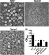
The rfaH mutant is deficient in extracellular capsule. (A to C) India ink staining of K. pneumoniae KPPR1 (WT) (A) and isogenic rfaH (B) and aroE (C) mutants after overnight culture in LB broth is shown by phase-contrast microscopy, in which the presence of capsule is indicated by an area of negative staining around the bacteria. Magnification, ×1,000. (D) Mucoviscosity, as measured by optical density (OD600) after centrifugation for 5 min at 1,000 × g with a starting turbidity of 1.0, is shown as the mean and standard deviation from 6 biological replicates. *, P < 0.0001 by ANOVA with Fisher’s LSD test compared to the wild type; #, P < 0.0001 compared to both WT carrying pACYC and the rfaH mutant carrying pRfaH.
FIG 4
FIG 4
The K. pneumoniae rfaH and aroE mutants are susceptible to serum killing. Viability of K. pneumoniae KPPR1 (WT) and isogenic mutants, without (A) or containing pACYC184 with or without rfaH (B), after a 3-h incubation in human serum is shown as mean log10 CFU/ml ± standard deviation from at least two replicates per group. *, P < 0.0001 by ANOVA with Fisher’s LSD test compared to the wild type.
FIG 5
FIG 5
The K. pneumoniae rfaH mutant has a mild growth defect in heat-inactivated serum. K. pneumoniae KPPR1 (WT) and isogenic mutant growth (log10 CFU/ml) after overnight incubation of a 1 × 10³-CFU/ml inoculum in RPMI medium supplemented with 10% heat-inactivated serum is shown as the mean ± standard deviation from 4 independent experiments. *, P < 0.05; **, P < 0.01; ****, P < 0.0001, by ANOVA with Fisher’s LSD test.
FIG 6
FIG 6
K. pneumoniae ilvC and ilvD mutants are unable to replicate in minimal medium without branched-chain amino acids. Optical density (OD600) of K. pneumoniae KPPR1 (WT) and ilvC and ilvD mutants in M9 minimal medium with or without 10 mM isoleucine (I) and valine (V) is shown as the mean from triplicate samples representative of 3 independent experiments.
FIG 7
FIG 7
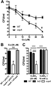
The K. pneumoniae copA mutant has increased sensitivity to copper. (A) Viability of a 1 × 10⁸-CFU inoculum of K. pneumoniae WT and copA mutant after an 18-h incubation in M9 broth with concentrations of cupric sulfate indicated is shown as mean ± standard deviation of results from at least 3 independent experiments. Based on a linear regression model (modeling log-transformed CFU as a function of the Cu concentration, group [WT or copA], and interaction between concentration and group), the copA mutant has a significantly faster-decreasing rate over concentrations than the wild type (#). (B) Viability of K. pneumoniae harboring pACYC184 (pACYC) or pACYC184 with copA (pCopA) after incubation in 25 µM CuSO4 is shown as the mean ± standard deviation from three independent experiments. #, P < 0.0001 compared to both strains by ANOVA with Fisher’s LSD test; ns, not significant. (C) Viability of K. pneumoniae mutants as indicated in 25 µM CuSO4 with or without 10 mM isoleucine and valine is shown as the mean ± standard deviation from at least three independent experiments. ****, P < 0.0001 by ANOVA with Fisher’s LSD.
FIG 8
FIG 8
K. pneumoniae conserved genes are fitness factors during pneumonia. (A) The number of genes shared between KPPR1 (orange), pneumonia isolate MGH78578 (green), pyogenic liver abscess isolate NTUH-K2044 (pink), carbapenem-resistant isolate NJST258_1 (blue), and plant endophyte Kp342 (yellow) is shown with the number of genes in each quadrant (labeled by Roman numerals) indicated as calculated by CloVR comparative software. (B) Based on InSeq analysis, 60 of 69 genes with a fitness factor of >10 were conserved in all five isolates (green), 2 were present in human isolates but not Kp342 (yellow), and 2 were shared with hypervirulent NTUH-K2044 (red).

References

    1. Podschun R, Ullmann U. 1998. Klebsiella spp. as nosocomial pathogens: epidemiology, taxonomy, typing methods, and pathogenicity factors. Clin Microbiol Rev 11:589–603. - PMC - PubMed
    1. Maroncle N, Balestrino D, Rich C, Forestier C. 2002. Identification of Klebsiella pneumoniae genes involved in intestinal colonization and adhesion using signature-tagged mutagenesis. Infect Immun 70:4729–4734. doi:10.1128/IAI.70.8.4729-4734.2002. - DOI - PMC - PubMed
    1. Hsieh PF, Lin TL, Lee CZ, Tsai SF, Wang JT. 2008. Serum-induced iron-acquisition systems and TonB contribute to virulence in Klebsiella pneumoniae causing primary pyogenic liver abscess. J Infect Dis 197:1717–1727. doi:10.1086/588383. - DOI - PubMed
    1. Centers for Disease Control and Prevention 2013. Antibiotic resistance threats in the United States. Centers for Disease Control and Prevention, Atlanta, GA.
    1. Bachman MA, Oyler JE, Burns SH, Caza M, Lépine F, Dozois CM, Weiser JN. 2011. Klebsiella pneumoniae yersiniabactin promotes respiratory tract infection through evasion of lipocalin 2. Infect Immun 79:3309–3316. doi:10.1128/IAI.05114-11. - DOI - PMC - PubMed

Publication types

Substances

LinkOut - more resources