. 2015 Mar 7:10:3.
doi: 10.1186/s13029-015-0033-7. eCollection 2015.

Membrainy: a 'smart', unified membrane analysis tool

Affiliations

Membrainy: a 'smart', unified membrane analysis tool

Matthew Carr et al. Source Code Biol Med. .

Abstract

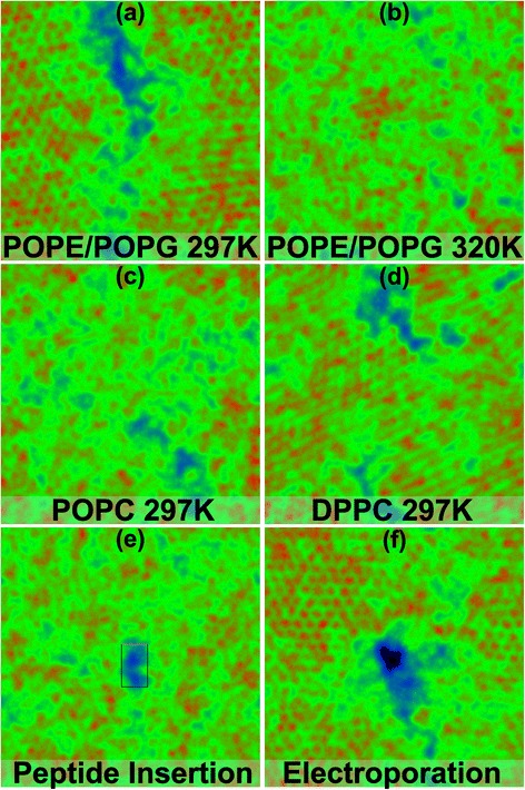
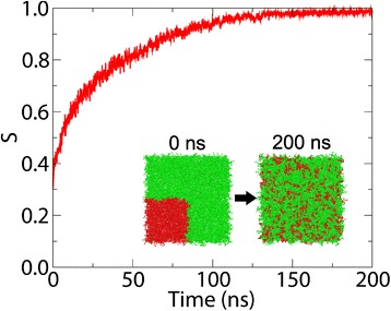
Background: The study of biological membranes using Molecular Dynamics has become an increasingly popular means by which to investigate the interactions of proteins, peptides and potentials with lipid bilayers. These interactions often result in changes to the properties of the lipids which can modify the behaviour of the membrane. Membrainy is a unified membrane analysis tool that contains a broad spectrum of analytical techniques to enable: measurement of acyl chain order parameters; presentation of 2D surface and thickness maps; determination of lateral and axial headgroup orientations; measurement of bilayer and leaflet thickness; analysis of the annular shell surrounding membrane-embedded objects; quantification of gel percentage; time evolution of the transmembrane voltage; area per lipid calculations; and quantification of lipid mixing/demixing entropy.

Results: Each analytical component within Membrainy has been tested on a variety of lipid bilayer systems and was found to be either comparable to or an improvement upon existing software. For the analytical techniques that have no direct comparable software, our results were confirmed with experimental data.

Conclusions: Membrainy is a user-friendly, intelligent membrane analysis tool that automatically interprets a variety of input formats and force fields, is compatible with both single and double bilayers, and capable of handling asymmetric bilayers and lipid flip-flopping. Membrainy has been designed for ease of use, requiring no installation or configuration and minimal user-input to operate.

Keywords: Area per lipid; Asymmetric bilayer; Bilayer/Leaflet thickness; Double bilayer; Headgroup orientations; Lipid flip-flopping; Membrane analysis; Mixing/Demixing entropy; Molecular dynamics; Order parameters.


Figures

Figure 1
Figure 1
Annular shell order parameters. The order parameters of saturated lipid tails from an annular shell analysis of MinD-MTS, an amphipathic helical peptide inserted into a POPE/POPG (3:1) double bilayer at 300K. The shell order parameters are shown in black, along with two control groups: the red plot uses the option built into Membrainy to ignore all gel lipids, which produces a more accurate control group for this peptide as it resides in a fluid region of the bilayer; and the blue plot contains both gel and fluid lipids. The differences between the black and red plots indicate the presence of splayed lipid tails in the annular shell, whereas the blue plot is sampling the wrong phase of lipids and provides an inaccurate comparison to the lipids within the annular shell.
Figure 2
Figure 2
Evolution of the TMV and membrane thickness. A POPE/POPG (3:1) double bilayer was subject to an ion imbalance of +20, achieving an initial TMV of -2.65 V. Within 5 ns, the TMV lowers to -2.35 V as the bilayers expand laterally and experience a thickness reduction due to electrostriction. At 15 ns, a transient water pore formed through electroporation, allowing ions to travel through the pore in opposite directions. This resulted in a rapid loss of the initial ion imbalance which incurs a sharp drop in TMV. By 17 ns, the TMV is insufficient to maintain electrostriction, allowing the bilayer thickness to increase.
Figure 3
Figure 3
Lipid flip-flopping. TMV and leaflet symmetry measurements of a POPE/POPG (3:1) double bilayer undergoing electroporation over 30 ns. A value of -2 in leaflet symmetry indicates a single flip-flop from the anodic to the cathodic leaflet. A pore was formed within 5 ns, which saw both POPE and POPG lipids from the anodic leaflet form the toroidal structure of the pore. After 15 ns, the POPE lipids within the pore return to the anodic leaflet while additional POPG lipids translocate to the cathodic leaflet. By 30 ns, one POPE lipid had flip-flopped from both leaflets (producing a symmetry of 0) and five POPG lipids had flip-flopped to the cathodic leaflet. This suggests that POPG lipids are more susceptible to flip-flopping towards the cathodic leaflet through transient water pores when under the influence of a TMV.
Figure 4
Figure 4
2D surface maps. These maps depict leaflets taken from a variety of lipid bilayer simulations. Red hexagonally packed dots represents gel clusters and black areas indicate the presence of a pore or hole in the leaflet. (a) and (b) depict POPE/POPG (3:1) bilayers at two temperatures, where (a) is near the transition temperature and contains ∼53% gel, and (b) is in the fluid phase and contains ∼14% gel. (c) and (d) depict POPC and DPPC bilayers at 297 K, containing ∼16% and ∼85% gel respectively. These percentages correspond with the correct phase of each bilayer as 297 K is above the transition temperature for POPC and below that of DPPC. The DPPC map also reveals a smudged appearance to the gel clusters which is indicative of lipids in the tilted Lβ phase. (e) depicts an inserted MinD-MTS peptide in a POPE/POPG (3:1) bilayer at 300 K. (f) depicts a leaflet containing a transient water pore established through electroporation in a POPE/POPG (3:1) bilayer at 297 K.
Figure 5
Figure 5
Mixing entropy. The mixing entropy of a POPE/POPC (3:1) bilayer over 200 ns, scaled such that S max=1. The bilayer is initialised such that POPC lipids (shown in red) encompass the lower left quadrant of the bilayer and the remaining bilayer contains POPE lipids (shown in green), creating a perfectly demixed system. An initial entropy of 0.3 is observed, which increases as the lipid types mix together. By 150 ns, the resulting entropy settles just below the theoretical maximum entropy, indicating a perfectly mixed system.

References

    1. van Meer G, Voelker DR, Feigenson GW. Membrane lipids: where they are and how they behave. Nat Rev Mol Cell Biol. 2008;9:112–24. doi: 10.1038/nrm2330. - DOI - PMC - PubMed
    1. Bloom M, Evans E, Mouritsen OG. Physical properties of the fluid lipid-bilayer component of cell membranes: a perspective. Q Rev Biophys. 1991;24:293–397. doi: 10.1017/S0033583500003735. - DOI - PubMed
    1. Penkett S, Flook A, Chapman D. Physical studies of phospholipids. ix. nuclear resonance studies of lipid-water systems. Chem Phys Lipids. 1968;2:273–90. doi: 10.1016/0009-3084(68)90004-2. - DOI
    1. Schindler H, Seelig J. Epr spectra of spin labels in lipid bilayers. J Chem Phys. 1973;59:1841–50. doi: 10.1063/1.1680269. - DOI
    1. De Kruijff B, Verkley A, Van Echteld C, Gerritsen W, Mombers C, Noordam P, et al. The occurrence of lipidic particles in lipid bilayers as seen by 31P NMR and freeze-fracture electron-microscopy. Biochimica et Biophysica Acta (BBA)-Biomembranes. 1979;555:200–9. doi: 10.1016/0005-2736(79)90160-3. - DOI - PubMed

LinkOut - more resources