Case Reports
. 2015 Apr 21;12(2):e7349.
doi: 10.5812/iranjradiol.7349. eCollection 2015 Apr.

Evaluation of Enteroneovesical Fistula by 64-Detector CT Enterography: A Case Report

Affiliations
Case Reports

Evaluation of Enteroneovesical Fistula by 64-Detector CT Enterography: A Case Report

Oktay Algin et al. Iran J Radiol. .

Abstract

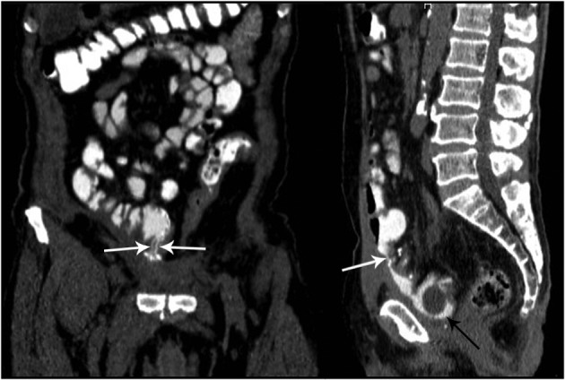
Enterovesical fistula is an abnormal communication between the bladder and the intestine. The accurate localization of leakage is important for accurate treatment planning. Some imaging techniques can not demonstrate the fistula; therefore, choosing the appropriate imaging technique is necessary. CT enterography (CTE) is a new technique for evaluation of the small bowel and the entire abdomen. CTE examination with multi-detector CT (MDCT) enables us to get excellent quality reformatted images with high spatial resolution. We report a patient with neobladder and enteroneovesical fistula. We showed the exact location of the fistula and its' association with the bowels and neobladder by CTE. The aim of this report is to show that CTE can be a new and effective modality in the detection of enteroneovesical fistulas and to discuss the efficacy of CTE in the detection and evaluation of enterovesical fistula referring to the literature. In conclusion, CTE may be a useful, sensitive, effective, and non-invasive technique for the evaluation of enteroneovesical fistula, leakage from the anastomose sides, and other extraintestinal complications such as urinary tract obstruction or abscess formation.

Keywords: Bladder Cancer; Fistula; Ileum; Urography.


Figures

Figure 1.
Figure 1.. A 64-year-old man who has feces exiting from the transurethral catheter after radical cystectomy and neobladder operation. The cystography images do not show evidence of enteroneovesical leakage or fistula.
Figure 2.
Figure 2.. Coronal (left) and sagittal (right) CTE images. Ileoneovesical fistula tracts are clearly visible on the images (white arrows). The neobladder and foley catheter balloon are also seen (black arrow).

References

    1. Kavanagh D, Neary P, Dodd JD, Sheahan KM, O'Donoghue D, Hyland JM. Diagnosis and treatment of enterovesical fistulae. Colorectal Dis. 2005;7(3):286–91. doi: 10.1111/j.1463-1318.2005.00786.x. - DOI - PubMed
    1. Hong SS, Kim AY, Kwon SB, Kim PN, Lee MG, Ha HK. Three-dimensional CT enterography using oral gastrografin in patients with small bowel obstruction: comparison with axial CT images or fluoroscopic findings. Abdom Imaging. 2010;35(5):556–62. doi: 10.1007/s00261-009-9567-y. - DOI - PubMed
    1. Huprich JE, Fletcher JG, Alexander JA, Fidler JL, Burton SS, McCullough CH. Obscure gastrointestinal bleeding: evaluation with 64-section multiphase CT enterography--initial experience. Radiology. 2008;246(2):562–71. doi: 10.1148/radiol.2462061920. - DOI - PubMed
    1. Paulsen SR, Huprich JE, Fletcher JG, Booya F, Young BM, Fidler JL, et al. CT enterography as a diagnostic tool in evaluating small bowel disorders: review of clinical experience with over 700 cases. Radiographics. 2006;26(3):641–57. doi: 10.1148/rg.263055162. - DOI - PubMed
    1. Macari M, Megibow AJ, Balthazar EJ. A pattern approach to the abnormal small bowel: observations at MDCT and CT enterography. AJR Am J Roentgenol. 2007;188(5):1344–55. doi: 10.2214/AJR.06.0712. - DOI - PubMed

Publication types

LinkOut - more resources