Comparative Study
. 2015 Jun 10;10(6):e0127992.
doi: 10.1371/journal.pone.0127992. eCollection 2015.

Comparative Characterization of Cells from the Various Compartments of the Human Umbilical Cord Shows that the Wharton's Jelly Compartment Provides the Best Source of Clinically Utilizable Mesenchymal Stem Cells

Affiliations
Comparative Study

Comparative Characterization of Cells from the Various Compartments of the Human Umbilical Cord Shows that the Wharton's Jelly Compartment Provides the Best Source of Clinically Utilizable Mesenchymal Stem Cells

Arjunan Subramanian et al. PLoS One. .

Abstract

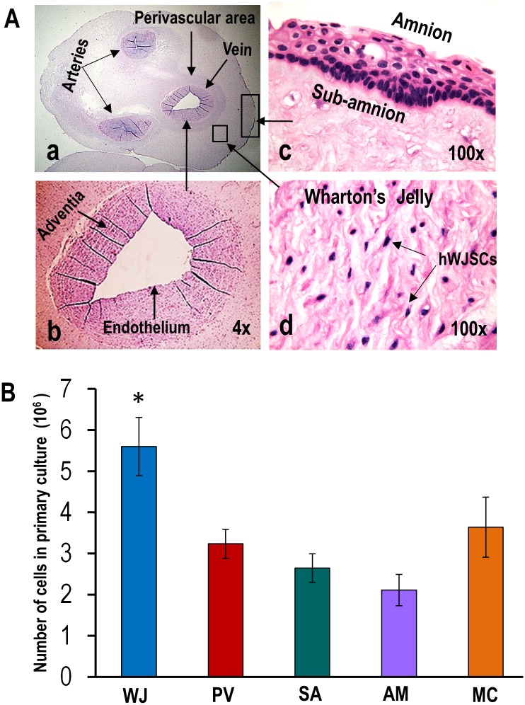
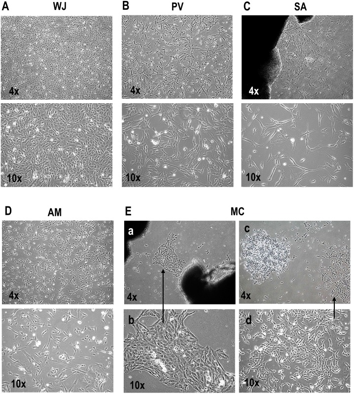
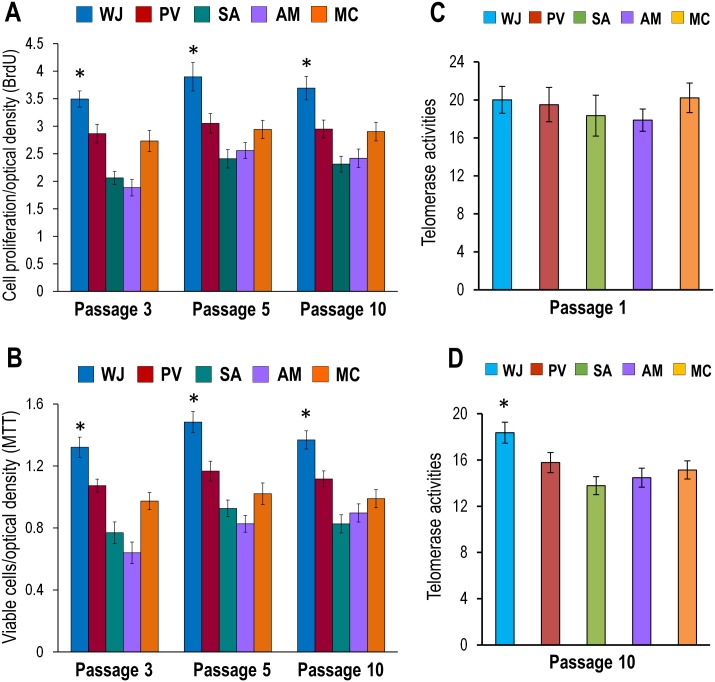
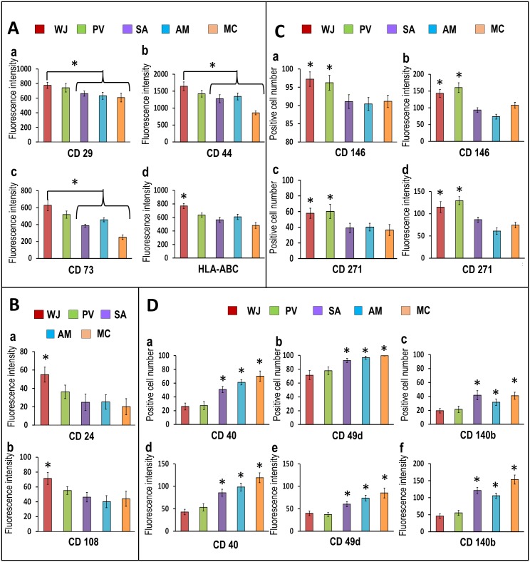
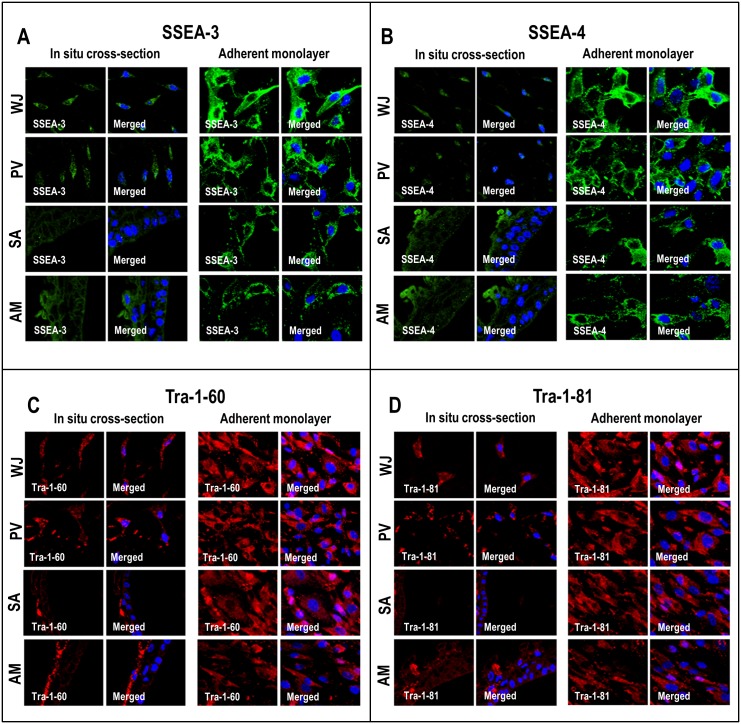
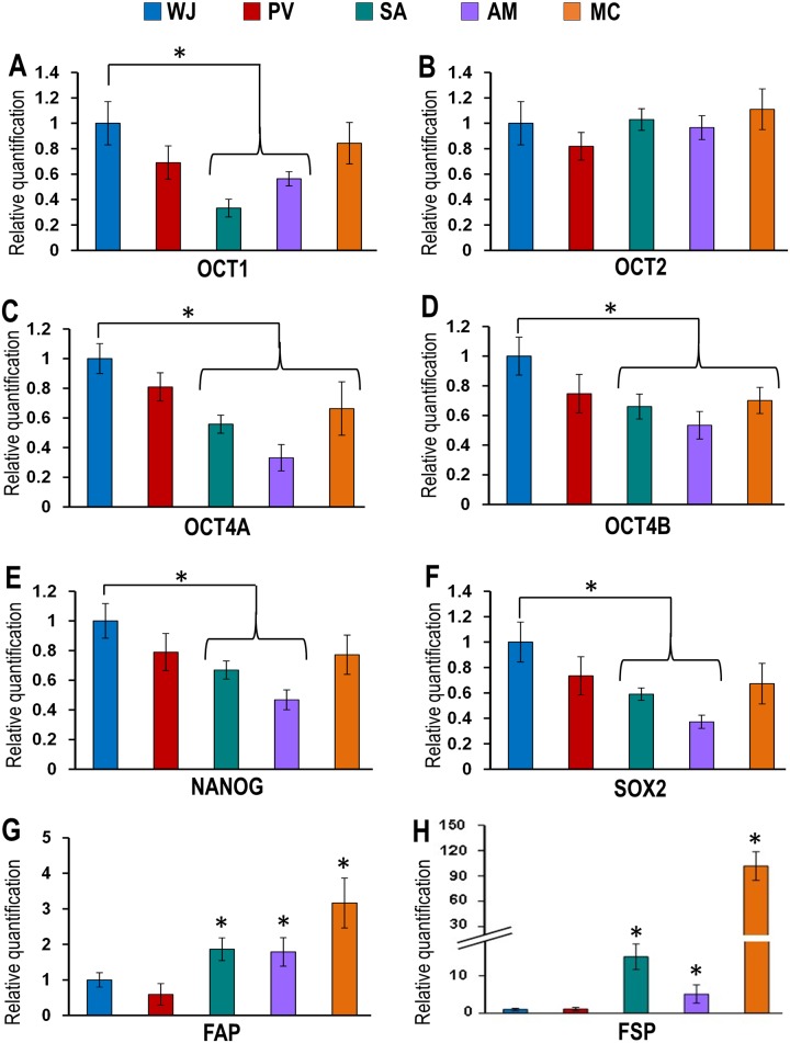
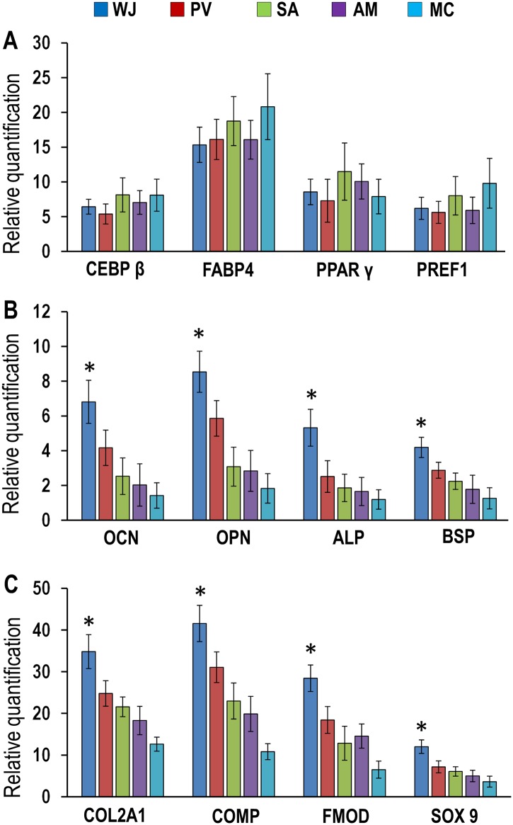
The human umbilical cord (UC) is an attractive source of mesenchymal stem cells (MSCs) with unique advantages over other MSC sources. They have been isolated from different compartments of the UC but there has been no rigorous comparison to identify the compartment with the best clinical utility. We compared the histology, fresh and cultured cell numbers, morphology, proliferation, viability, stemness characteristics and differentiation potential of cells from the amnion (AM), subamnion (SA), perivascular (PV), Wharton's jelly (WJ) and mixed cord (MC) of five UCs. The WJ occupied the largest area in the UC from which 4.61 ± 0.57 x 10⁶ /cm fresh cells could be isolated without culture compared to AM, SA, PV and MC that required culture. The WJ and PV had significantly lesser CD40+ non-stem cell contaminants (26-27%) compared to SA, AM and MC (51-70%). Cells from all compartments were proliferative, expressed the typical MSC-CD, HLA, and ESC markers, telomerase, had normal karyotypes and differentiated into adipocyte, chondrocyte and osteocyte lineages. The cells from WJ showed significantly greater CD24+ and CD108+ numbers and fluorescence intensities that discriminate between MSCs and non-stem cell mesenchymal cells, were negative for the fibroblast-specific and activating-proteins (FSP, FAP) and showed greater osteogenic and chondrogenic differentiation potential compared to AM, SA, PV and MC. Cells from the WJ offer the best clinical utility as (i) they have less non-stem cell contaminants (ii) can be generated in large numbers with minimal culture avoiding changes in phenotype, (iii) their derivation is quick and easy to standardize, (iv) they are rich in stemness characteristics and (v) have high differentiation potential. Our results show that when isolating MSCs from the UC, the WJ should be the preferred compartment, and a standardized method of derivation must be used so as to make meaningful comparisons of data between research groups.


Conflict of interest statement

Competing Interests: The authors have declared that no competing interests exist.

Figures

Fig 1
Fig 1
(A) (a-d) In situ H & E histological cross-sections of the human umbilical cord showing the various regions [Wharton’s jelly (WJ), perivascular area (PV), subamnion (SA), amnion (AM)] from which MSCs were derived. Note individual MSCs (hWJSCs) lying in gelatinous matrix of WJ. (B) Histogram showing significantly greater numbers of MSCs (hWJSCs) in the WJ in primary culture compared to PV, SA, AM and mixed cord cultures (MC) (*p<0.05).
Fig 2
Fig 2. (A-E) Morphology of MSCs derived from WJ, PV, SA, AM and MC in primary culture.
(A-D) The MSCs from WJ showed one type of stellate epithelioid-like morphology while those from the PV, SA and AM showed one type of short fibroblast-like morphology. (E) The MSCs from MC cultures showed cell islands with two types of morphology (a-b) epithelioid-like and (c-d) short fibroblast-like.
Fig 3
Fig 3. (A-D) Cell proliferation, viability and telomerase levels of MSCs derived from WJ, PV, SA, AM and MC.
(A) BrdU assays showed significantly greater proliferation rates at passages 3, 5 and 10 in cells from WJ compared to PV, SA, AM and MC (*p<0.05). (B) MTT assays showed significantly greater cell viability at passages 3, 5 and 10 in cells from WJ compared PV, SA, AM and MC (*p<0.05). (C) qRT-PCR analysis for telomerase showed similar levels of telomerase activity with no significant differences (p>0.05) in early passages (P1) for cells derived from WJ, PV, SA, AM and MC but (D) at late passages (P10) the telomerase levels for WJ were significantly greater than PV, SA, AM and MC (*p<0.05).
Fig 4
Fig 4. (A-D) CD marker analysis of cells derived from WJ, PV, SA, AM and MC.
(A) (a-d) and (B) (a-b) The mean fluorescence intensities were significantly greater in cells from WJ compared to PV, SA, AM and MC for CD29, CD44, CD73 and HLA-ABC and for CD24 and CD108 (*p<0.05). (C) The percentages of positive cells (a,c) and mean fluorescence intensities (b,d) for CD146 and CD271 were significantly greater in cells from WJ and PV compared to SA, AM and MC (*p<0.05). (D) SA, AM and MC showed significantly greater percentages of positive cells (a,b,c) and mean fluorescence intensities (d,e,f) for the fibroblast markers CD40, CD49d and CD140b compared WJ and PV (*p<0.05).
Fig 5
Fig 5. (A-D) Confocal microscopic analysis of embryonic stem cell (ESC) markers in cells of in situ cross-sections and adherent monolayers.
WJ, PV, SA and AM were positive for SSEA-3, SSEA-4, Tra-1-60 and Tra-1-81.
Fig 6
Fig 6. (A-F) qRT-PCR for embryonic stem cell (ESC) pluripotent genomic markers in cells derived from the WJ, PV, SA, AM and MC.
Cells derived from WJ expressed significantly greater levels of the pluripotency genes OCT1, OCT4A, OCT4B, NANOG and SOX2 compared to cells from SA, AM and MC. (*p<0.05). (G-H) Cells from SA, AM and MC showed significantly greater levels of expression of the fibroblast-related genes FAP and FSP compared to WJ and PV (*p<0.05).
Fig 7
Fig 7. (A-C) Differentiation of cells from WJ, PV, SA, AM and MC into adipogenic, osteogenic and chondrogenic lineages.
Cells from WJ, PV, SA, AM and MC stained (A) Oil Red O positive for adipocytes, (B) Von Kossa positive for osteocytes and (C) Alcian blue positive for chondrocytes when exposed to their respective differentiation media for each of the lineages. Adipocyte colonies stained with Oil Red O were similar in number with no differences in staining intensity between WJ, PV, SA, AM and MC. However, Von Kossa and Alcian blue stained cells for osteocytes and chondrocytes were greater in numbers and staining intensities in WJ compared to PV, SA, AM and MC.
Fig 8
Fig 8. (A-C) Expression levels of adipogenic, osteogenic and chodrogenic genomic markers in differentiated WJ, PV, SA, AM and MC cells.
(A) The expression levels of the adipogenic-related genes CEBPβ, FABP4, PPARγ and PREF1 were increased for WJ, PV, SA, AM and MC compared to their respective controls with no significant differences between them. (B) The expression levels of the osteogenic-related genes osteocalcin (OCN), osteopontin (OPN), alkaline phosphatase (ALP) and bone sialoprotein (BSP) were significantly greater for WJ compared to PV, SA, AM and MC (*p<0.05). (C) The expression levels of the chondrogenic-related genes collagen type II (COL2A1), cartilage oligomeric matrix protein (COMP), fibromodulin (FMOD) and sex determining region Y-box 9 (SOX 9) were significantly greater for WJ compared to PV, SA, AM and MC (*p<0.01).

References

    1. Rao MS, Mattson MP. Stem cells and aging: expanding the possibilities. Mechanisms of ageing and development. 2001;122(7):713–34. . - PubMed
    1. Baksh D, Yao R, Tuan RS. Comparison of proliferative and multilineage differentiation potential of human mesenchymal stem cells derived from umbilical cord and bone marrow. Stem cells. 2007;25(6):1384–92. 10.1634/stemcells.2006-0709 . - DOI - PubMed
    1. Sarugaser R, Lickorish D, Baksh D, Hosseini MM, Davies JE. Human umbilical cord perivascular (HUCPV) cells: a source of mesenchymal progenitors. Stem cells. 2005;23(2):220–9. 10.1634/stemcells.2004-0166 . - DOI - PubMed
    1. Ilancheran S, Michalska A, Peh G, Wallace EM, Pera M, Manuelpillai U. Stem cells derived from human fetal membranes display multilineage differentiation potential. Biology of reproduction. 2007;77(3):577–88. 10.1095/biolreprod.106.055244 . - DOI - PubMed
    1. Troyer DL, Weiss ML. Concise review: Wharton's jelly-derived cells are a primitive stromal cell population. Stem cells. 2008;26(3):591–9. Epub 2007/12/11. 10.1634/stemcells.2007-0439 ; PubMed Central PMCID: PMCPmc3311226. - DOI - PMC - PubMed

Publication types