. 2015 Jun 10;10(6):e0128655.
doi: 10.1371/journal.pone.0128655. eCollection 2015.

1,25 Dihydroxyvitamin D3 Inhibits TGFβ1-Mediated Primary Human Cardiac Myofibroblast Activation

Affiliations

1,25 Dihydroxyvitamin D3 Inhibits TGFβ1-Mediated Primary Human Cardiac Myofibroblast Activation

Anna Meredith et al. PLoS One. .

Abstract

Aims: Epidemiological and interventional studies have suggested a protective role for vitamin D in cardiovascular disease, and basic research has implicated vitamin D as a potential inhibitor of fibrosis in a number of organ systems; yet little is known regarding direct effects of vitamin D on human cardiac cells. Given the critical role of fibrotic responses in end stage cardiac disease, we examined the effect of active vitamin D treatment on fibrotic responses in primary human adult ventricular cardiac fibroblasts (HCF-av), and investigated the relationship between circulating vitamin D (25(OH)D3) and cardiac fibrosis in human myocardial samples.

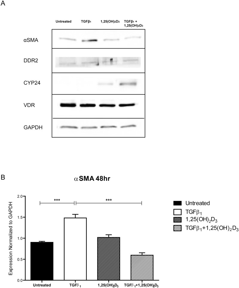
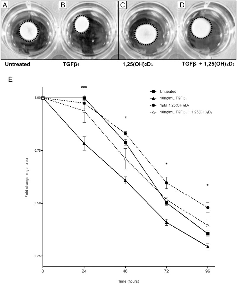
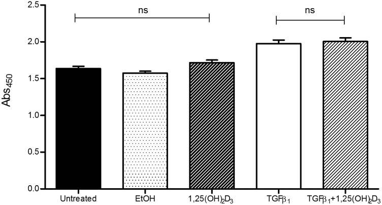
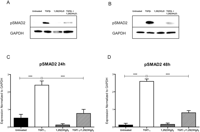
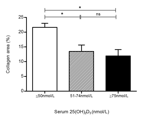
Methods and results: Interstitial cardiac fibrosis in end stage HF was evaluated by image analysis of picrosirius red stained myocardial sections. Serum 25(OH)D3 levels were assayed using mass spectrometry. Commercially available HCF-av were treated with transforming growth factor (TGF)β1 to induce activation, in the presence or absence of active vitamin D (1,25(OH)2D3). Functional responses of fibroblasts were analyzed by in vitro collagen gel contraction assay. 1,25(OH)2D3 treatment significantly inhibited TGFβ1-mediated cell contraction, and confocal imaging demonstrated reduced stress fiber formation in the presence of 1,25(OH)2D3. Treatment with 1,25(OH)2D3 reduced alpha-smooth muscle actin expression to control levels and inhibited SMAD2 phosphorylation.

Conclusions: Our results demonstrate that active vitamin D can prevent TGFβ1-mediated biochemical and functional pro-fibrotic changes in human primary cardiac fibroblasts. An inverse relationship between vitamin D status and cardiac fibrosis in end stage heart failure was observed. Collectively, our data support an inhibitory role for vitamin D in cardiac fibrosis.


Conflict of interest statement

Competing Interests: The authors have declared that no competing interests exist.

Figures

Fig 1
Fig 1. Vitamin D treatment inhibits expression of TGFβ1-mediated α-smooth muscle actin.
A) Representative Western blot of cells 48 hours after treatment. Expression of the discoidin domain receptor 2 (DDR2) is present in human primary adult ventricular cardiac fibroblasts (HCF-av). CYP24 expression was upregulated 48 hours after treatment with 1,25(OH)2D3±TGFβ1. Expression of α-smooth muscle actin (αSMA) was upregulated 48 hours after treatment with TGFβ1, and significantly reduced with 1,25(OH)2D3 co-treatment. B) Densitometry data generated from Western blots of HCF-av cells 48 hours after treatment with TGFβ1±1,25(OH)2D3, and normalized to GAPDH. All data represent mean±SEM. p-values were calculated using one way analysis of variance with a Bonferroni multiple comparison test. *p<0.05, ***<p<0.001.
Fig 2
Fig 2. Vitamin D inhibits TGFβ1-mediated myofibroblast contraction.
Representative images of 48 hour timepoint from collagen gel contraction assay are shown in A-D. A) Untreated HCF-av; B) HCF-av treated with TGFβ1; C) HCF-av treated with 1,25(OH)2D3; D) HCF-av treated with TGFβ1 + 1,25(OH)2D3. A time course of gel contraction over 96 hours is shown in (E). Active vitamin D treatment significantly inhibited TGFβ1-induced gel contraction at all time points post-treatment. All data points represent mean ± SEM. p-values calculated using two way repeated measures analysis of variance with Bonferroni multiple comparison test. *p<0.05, ***p<0.001. F and G) Confocal images of HCF-av at 48 hours following treatment. Stress fibers were stained with phalloidin. HCF-av treated with TGFβ1(F) demonstrate increased presence of clearly defined stress fibers (white) as compared with cells treated with TGFβ1 + 1,25(OH)2D3 (G). Scale bar: 70μm.
Fig 3
Fig 3. Vitamin D does not inhibit TGFβ1-mediated cellular proliferation.
Evaluation of proliferation rates in our treatment groups revealed no significant change in cellular proliferation between cells treated with active vitamin D and TGFβ1 or TGFβ1 alone. Proliferation was increased in the presence of TGFβ1. All data represent mean ± SEM. p-values were calculated using one way analysis of variance with a Bonferroni multiple comparison test.
Fig 4
Fig 4. Vitamin D reduces TGFβ1-mediated phosphorylation of SMAD2.
Representative Western blot images of HCF-av treated for 24 hours (A) and 48 hours (B) with TGFβ1 in the presence and absence of 1,25(OH)2D3, which demonstrate a reduction in pSMAD2 with active vitamin D treatment. Densitometry of Western blot data shows significantly increased phosphorylation of SMAD2 at both 24 hours (C) and 48 hours (D) following treatment with TGFβ1, which is significantly reduced with co-treatment with 1,25(OH)2D3. All data represent mean ± SEM. p-values were calculated using one way analysis of variance with a Bonferroni multiple comparison test. ***p<0.001, ****p<0.0001.
Fig 5
Fig 5. Circulating levels of vitamin D are positively associated with myocardial fibrosis in heart failure patients.
Collagen area as a fraction of total myocardial tissue area was significantly higher in patients with vitamin D deficiency as compared with vitamin D insufficient or sufficient patients. There was no difference in collagen area in patients without vitamin D deficiency. All data represent mean ± SEM. p-values were calculated using one way analysis of variance with a Bonferroni multiple comparison test. *p<0.05.

References

    1. Go AS, Mozaffarian D, Roger VL, Benjamin EJ, Berry JD, Borden WB, et al. Heart disease and stroke statistics—2013 update: a report from the American Heart Association. Circulation 2013;127:e6–e245. 10.1161/CIR.0b013e31828124ad - DOI - PMC - PubMed
    1. Neubauer S. The failing heart—an engine out of fuel. N Engl J Med 2007;356:1140–1151. - PubMed
    1. Kane CJ, Hebda PA, Mansbridge JN, Hanawalt PC. Direct evidence for spatial and temporal regulation of transforming growth factor beta 1 expression during cutaneous wound healing. J Cell Physiol 1991;148:157–173. - PubMed
    1. Lijnen PJ, Petrov VV, Fagard RH. Induction of cardiac fibrosis by transforming growth factor-beta(1). Mol Genet Metab 2000;71:418–435. - PubMed
    1. Ikeuchi M, Tsutsui H, Shiomi T, Matsusaka H, Matsushima S, Wen J, et al. Inhibition of TGF-beta signaling exacerbates early cardiac dysfunction but prevents late remodeling after infarction. Cardiovasc Res 2004;64:526–535. - PubMed

Publication types