. 2015 Jul 20;6(20):18094-104.
doi: 10.18632/oncotarget.4093.

rRNA synthesis inhibitor, CX-5461, activates ATM/ATR pathway in acute lymphoblastic leukemia, arrests cells in G2 phase and induces apoptosis

Affiliations

rRNA synthesis inhibitor, CX-5461, activates ATM/ATR pathway in acute lymphoblastic leukemia, arrests cells in G2 phase and induces apoptosis

Sandeep S Negi et al. Oncotarget. .

Abstract

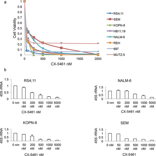
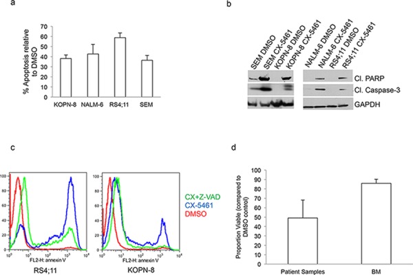
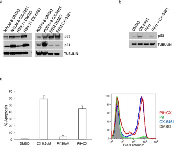
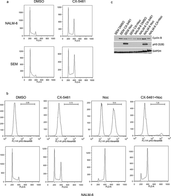
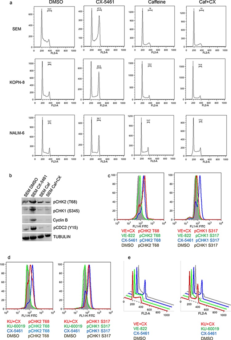
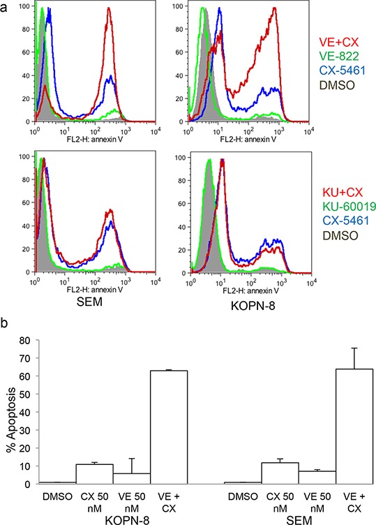
Ribosome biogenesis is a fundamental cellular process and is elevated in cancer cells. As one of the most energy consuming cellular processes, it is highly regulated by signaling pathways in response to changing cellular conditions. Many of the regulators of this process are aberrantly activated in various cancers. Recently two novel rRNA synthesis inhibitors, CX-5461 and BMH-21, have been shown to selectively kill cancer cells while sparing normal cells. Here, we tested the effectiveness of pre-rRNA synthesis inhibitor CX-5461 on acute lymphoblastic leukemia cells with different cytogenetic abnormalities. Acute lymphoblastic leukemia cells are more sensitive to rRNA synthesis inhibition compared to normal bone marrow cells. CX-5461 treated cells undergo caspase-dependent apoptosis independent of their p53 status. More-over, CX5461, activates checkpoint kinases and arrests cells in G2 phase of cell cycle. Finally, overcoming this G2 arrest by inhibiting ATR kinase leads to robust cell killing. These results show that CX-5461 can be even more potent in combination with ATR inhibitors.

Keywords: ATM/ATR pathway; CX-5461; G2/M arrest; acute lymphoblastic leukemia; rRNA synthesis.


Conflict of interest statement

CONFLICTS OF INTEREST

Authors declare no conflict of interest.

Figures

Figure 1
Figure 1. CX-5461 inhibits growth in acute lymphoblastic leukemia (ALL) cells
a. All eight ALL cell lines showed marked decrease in proliferation after a 3 day treatment with CX-5461. b. 3 h treatment with CX-5461 reduced 45S pre-rRNA transcript in a dose dependent manner. Transcript levels were measured using quantitative PCR and normalized to the expression of GAPDH and Actin. (a, b) Experiments were repeated three times and error bars represent +/− S.D.
Figure 2
Figure 2. CX-5461 induces caspase dependent apoptosis in ALL cells
a. Annexin V was used to measure apoptosis in ALL cell lines.% apoptosis relative to DMSO treated control is plotted. Histograms show the values (mean ± S.D.) of three independent experiments. b. Cells were treated with 0.25 μM CX-5461 for 1 day. Cleaved caspase-3, cleaved PARP and GAPDH antibodies were used for western blot. c. RS4;11 and KOPN-8 cells were pre-treated with pan-caspase inhibitor Z-VAD-FMK followed by 2 days of treatment with 0.5 μM CX-5461. Annexin V was used to measure apoptosis. One of two representative experiments is shown. d. Patient samples (n = 6) or normal bone marrow (n = 3) were treated with 1 μM CX-5461 or DMSO for 2 days and apoptosis was measured with Annexin V staining. Viable proportion is plotted normalized to DMSO treated samples. Results are shown as mean +/− S.D.
Figure 3
Figure 3. CX-5461 mediated apoptosis is p53 independent
a. Two p53 wild type (NALM-6 and RS4;11) and two mutant (SEM and KOPN-8) cell lines were used. Expression of p53 and its downstream target p21 was shown with western blot upon 1 day treatment with 0.25 μM CX-5461. b, c. p53 wild type RS4;11 cells were treated with 0.25 μM CX-5461 or 30 μM of p53 inhibitor pifithrin-α or both. Western blot was used to measure p53 levels after 1 day drug treatment (b). Annexin V was used to measure apoptosis after 2 days. Histogram and representative flow cytometry data is shown (c). Experiments are repeated three times and plotted as mean +/− S.D.
Figure 4
Figure 4. CX-5461 arrests ALL cells in G2 phase
a. Cells were treated with 0.25 μM CX-5461 for 1 day. Cell-cycle distribution was determined by flow cytometry analysis of propidium iodide (PI) stained cells. One representative experiment out of three is shown. b. and c. NALM-6 and SEM cell were treated with CX-5461, Nocodazole or 2 h pre-treatment with CX-5461 followed by nocodazole for 1 day. Cell-cycle profiles were analyzed by flow cytometry using pH3(S28) as an indicator of mitosis (top panel) and PI for DNA content (bottom panel). (c) FACS results were confirmed with western blot by analyzing cyclin B and pH3(S28) levels.
Figure 5
Figure 5. CX-5461 activate ATM/ATR pathway
a. and b. SEM cells were treated with 0.25 μM CX-5461 or 1.5 mM caffeine alone or pre-treated with caffeine for 1 h followed by CX-5461 for 1 day. (a) Cell-cycle was analyzed by flow cytometry as before. Caffeine completely removed G2 block induced by CX-5461. (b) Western blot was used to measure the levels of proteins involved in G2/M checkpoint. c-e. SEM cells were pre-treated with 0.1 μM VE-822 (c) or 0.25 μM KU-60019 (d) for 1 h followed by 0.25 μM CX-5461 for 1 day. Levels of pCHK1(S317) and pCHK2(T68) (c and d) and cell cycle distribution (e) were measured by flow cytometry as before. One out of two representative experiments is shown.
Figure 6
Figure 6. CX-5461 shows synergy with ATR inhibitor
a. SEM and KOPN-8 were treated as in (Figure 5C) for 2 days and apoptosis was measured with Annexin V staining. b. SEM and KOPN-8 cells were pre-treated with 0.05 μM VE-822 followed by 0.05 μM CX-5461 or with CX-5461 or VE-822 alone for 2 days. Annexin V was used to measure apoptosis. (a, b) All experiments were repeated three times and error bars represent +/− S.D.

References

    1. Olson MOJ. The Nucleolus. New York: Springer; 2011.
    1. Olson MO, Dundr M. The moving parts of the nucleolus. Histochem Cell Biol. 2005;123:203–216. - PubMed
    1. Dundr M, Misteli T, Olson MO. The dynamics of postmitotic reassembly of the nucleolus. J Cell Biol. 2000;150:433–446. - PMC - PubMed
    1. Rubbi CP, Milner J. Disruption of the nucleolus mediates stabilization of p53 in response to DNA damage and other stresses. Embo j. 2003;22:6068–6077. - PMC - PubMed
    1. Olson MO, Hingorani K, Szebeni A. Conventional and nonconventional roles of the nucleolus. Int Rev Cytol. 2002;219:199–266. - PMC - PubMed

Publication types

MeSH terms