. 2015 Jun 25;522(7557):478-81.
doi: 10.1038/nature14510. Epub 2015 Jun 10.

A naturally occurring variant of the human prion protein completely prevents prion disease

Affiliations

A naturally occurring variant of the human prion protein completely prevents prion disease

Emmanuel A Asante et al. Nature. .

Abstract

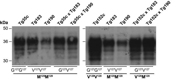
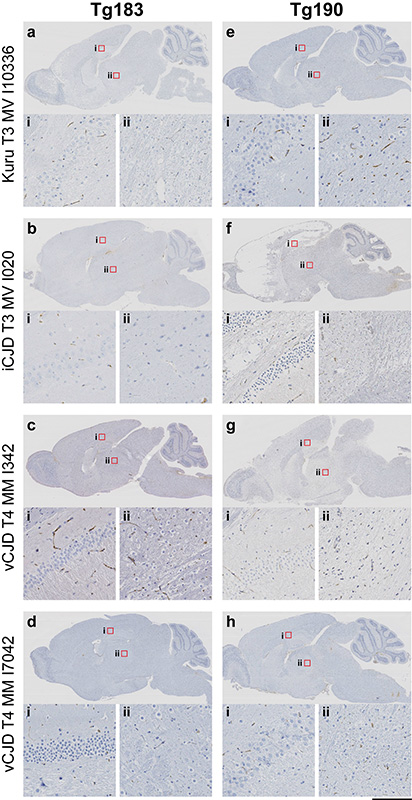
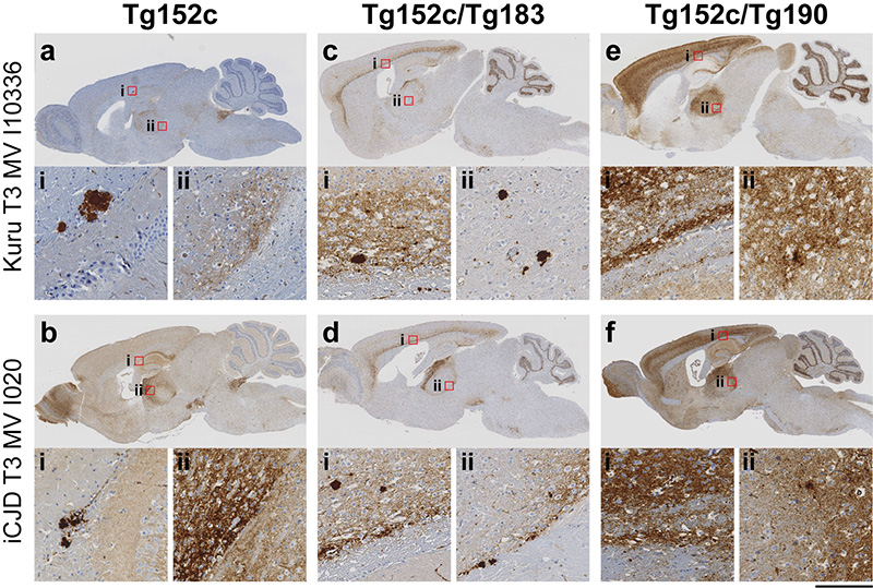
Mammalian prions, transmissible agents causing lethal neurodegenerative diseases, are composed of assemblies of misfolded cellular prion protein (PrP). A novel PrP variant, G127V, was under positive evolutionary selection during the epidemic of kuru--an acquired prion disease epidemic of the Fore population in Papua New Guinea--and appeared to provide strong protection against disease in the heterozygous state. Here we have investigated the protective role of this variant and its interaction with the common, worldwide M129V PrP polymorphism. V127 was seen exclusively on a M129 PRNP allele. We demonstrate that transgenic mice expressing both variant and wild-type human PrP are completely resistant to both kuru and classical Creutzfeldt-Jakob disease (CJD) prions (which are closely similar) but can be infected with variant CJD prions, a human prion strain resulting from exposure to bovine spongiform encephalopathy prions to which the Fore were not exposed. Notably, mice expressing only PrP V127 were completely resistant to all prion strains, demonstrating a different molecular mechanism to M129V, which provides its relative protection against classical CJD and kuru in the heterozygous state. Indeed, this single amino acid substitution (G→V) at a residue invariant in vertebrate evolution is as protective as deletion of the protein. Further study in transgenic mice expressing different ratios of variant and wild-type PrP indicates that not only is PrP V127 completely refractory to prion conversion but acts as a potent dose-dependent inhibitor of wild-type prion propagation.


Figures

Extended Data Figure 1
Extended Data Figure 1. Immunoblots showing relative PrPC expression levels in transgenic mice
The provenance of each brain sample is designated above each lane and molecular markers are indicated on the left. The PRNP codon 127 (G, glycine, V, valine) or codon 129 (M, methionine, V, valine) genotypes of the transgenic mice are designated below. Transgenic mouse brains were analysed by enhanced chemiluminescence without proteinase-K digestion and equal amounts of total protein loaded in each well and probed with anti-PrP monoclonal antibody ICSM 35. Wild type human PrP expression levels of G127M129/G127M129 Tg35c and G127V129/G127V129 Tg152c mice are 2- and 6-fold higher respectively, than seen in 10 % (w/v) pooled human brain homogenate. Homozygous V127M129 Tg183 and Tg190 mice have 2-fold higher or equivalent PrP expression levels respectively compared to 10 % (w/v) pooled human brain homogenate.
Extended Data Figure 2
Extended Data Figure 2. Immunohistochemical analysis of transgenic mouse brain following challenge with vCJD prions
All mice were intracerebrally challenged with the same vCJD prion isolates (I342 and I7042). Abnormal PrP deposition in fixed post-mortem brain from affected mice was detected using anti-PrP monoclonal antibody ICSM 35. (a-b) Homozygous Tg35c mice (expressing G127M129/G127M129 wild type PrP only) show intense and widespread PrP plaque deposits. Magnified areas show (ai) hippocampus, (bi) frontal cortex and (aii and bii) thalamus. In contrast, heterozygous G127M129/V127M129 Tg35c/Tg183 mice expressing equivalent levels of G127M129 and V127M129 PrP (c-d) show only weak PrP deposition in the corpus callosum (ci and di) with no abnormal PrP deposition detected in other brain areas, for example, in the thalamus (cii and dii). Heterozygous G127M129/V127M129 Tg35c/Tg190 mice which express a lower level of V127M129 PrP relative to wild type G127M129 PrP (e-f) show greater levels of PrP deposition than seen in Tg35c/Tg183 mice following challenge with the same vCJD prion isolates, (ei, eii and fi) corpus callosum; (fii) pons. Scale bar, upper panels (a-f) 2 mm; magnified panels, 100 μm.
Extended Data Figure 3
Extended Data Figure 3. Immunohistochemical analysis of homozygous PRNP V127M129/V127M129 transgenic mouse brain following challenge with human prions
Mice were intracerebrally challenged with kuru, classical and variant CJD prions. Following prolonged (>600 days) post inoculation periods, abnormal PrP deposition in fixed post-mortem brain was examined using anti-PrP monoclonal antibody ICSM 35. (a-d) V127M129/V127M129 Tg183 mice with 2-fold overexpression of V127M129 PrP. (E-H) V127M129/V127M129 Tg190 mice expressing endogenous levels of V127M129 PrP. Red square boxes in the main panels (a-h) define the area of magnified images of hippocampus (i) and thalamus (ii) shown in the lower panels. Scale bar, a-h, 2 mm; magnified panels i and ii, 100 μm. The lack of detection of abnormal PrP deposition in brain indicates that the mice are not subclinically infected with prions.
Extended Data Figure 4
Extended Data Figure 4. Schematic diagram summarising transmissions of kuru and classical CJD prions to transgenic mice expressing human PrP
G127M129/G127M129 Tg35c mice or G127V129/G127V129 Tg152c mice are homozygous for wild type human PrP alleles and are fully susceptible to kuru and classical CJD prions. V127M129/V127M129 Tg183 or V127M129/V127M129 Tg190 transgenic mice are homozygous for the variant V127M129 allele found only in humans from the kuru-exposed population of Papua New Guinea and are entirely resistant to infection with kuru and classical CJD prions. The levels of PrP expression in the brain of these homozygous transgenic mice relative to a pooled human brain homogenate are 1× (Tg190 mice) 2× (Tg35c and Tg183 mice) and 6× (Tg152c mice). Generation of F1 mice through inter-breeding the various homozygous lines produces different combinations of the various human PrP alleles leading to differences in the relative expression levels of the various prion proteins in brain. The PrP expression ratios from the two PrP alleles in the crosses are shown in parentheses above the cartoon mice. Full, intermediate or low susceptibility of the mice to infection with kuru and classical CJD prions is indicated by three, two or one diagonal red bar, respectively, drawn across the mice. Mice with no red bar are entirely resistant to infection with kuru and classical CJD prions.
Extended Data Figure 5
Extended Data Figure 5. Immunohistochemical analysis of homozygous PRNP G127V129/G127V129 and heterozygous G127V129/V127M129 transgenic mouse brain following challenge with human prions
Mice were intracerebrally challenged with kuru and classical CJD prions and abnormal PrP deposition in fixed post-mortem brain from affected mice was examined using anti-PrP monoclonal antibody ICSM 35. Red square boxes labelled i and ii in panels a-f mark brain areas that are magnified and displayed below; (i) cortex and (ii) thalamus. (a-b) Wild type G127V129/G127V129 Tg152c mice. (c-d) Heterozygous G127V129/V127M129 Tg152c/Tg183 mice. (e-f) Heterozygous G127V129/V127M129 Tg152c/190 mice Scale bar, a-f, 2 mm; magnified panels i and ii, 100 μm. The detection of abnormal PrP deposition in brain indicates that the mice are infected with prions.
Figure 1
Figure 1. Transmission rates of human prions to transgenic mice homo- or heterozygous for human PrP V127
Transgenic mice were intracerebrally inoculated with brain homogenate from patients with kuru (a) or classical CJD (b). Codon 127 and 129 PRNP genotypes of the recipient mice are shown (G, glycine, M, methionine, V, valine). G127M129 is a wild type human allele, and the V127M129 allele is seen only in humans from the kuru-exposed population of Papua New Guinea. Attack rate reports the total of clinically affected and sub-clinically infected mice as a proportion of the number of inoculated mice after prolonged (>600 days) post-inoculation periods. Primary prion transmission data are reported in tables 2 and 3.

Comment in

References

    1. Collinge J. Prion diseases of humans and animals: their causes and molecular basis. Annu. Rev. Neurosci. 2001;24:519–550. - PubMed
    1. Mead S, et al. A novel protective prion protein variant that colocalizes with kuru exposure. N Engl J Med. 2009;361:2056–2065. - PubMed
    1. Jucker M, Walker LC. Self-propagation of pathogenic protein aggregates in neurodegenerative diseases. Nature. 2013;501:45–51. - PMC - PubMed
    1. Collinge J, Palmer MS, Dryden AJ. Genetic predisposition to iatrogenic Creutzfeldt-Jakob disease. Lancet. 1991;337:1441–1442. - PubMed
    1. Palmer MS, Dryden AJ, Hughes JT, Collinge J. Homozygous prion protein genotype predisposes to sporadic Creutzfeldt-Jakob disease. Nature. 1991;352:340–342. - PubMed

Publication types

MeSH terms