

Review
doi: 10.4155/fmc.15.42.
SecA: a potential antimicrobial target
Affiliations
- PMID: 26062397
- PMCID: PMC4479503
- DOI: 10.4155/fmc.15.42
Item in Clipboard
Review
SecA: a potential antimicrobial target
Future Med Chem.
2015.
Abstract
There is a consensus in the medical profession of the pressing need for novel antimicrobial agents due to issues related to drug resistance. In practice, solutions to this problem to a large degree lie with the identification of new and vital targets in bacteria and subsequently designing their inhibitors. We consider SecA a very promising antimicrobial target. In this review, we compile and analyze information available on SecA to show that inhibition of SecA has a multitude of consequences. Furthermore, we discuss issues critical to the design and evaluation of SecA inhibitors.
Figures
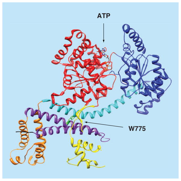
Color coding of SecA domains: NBD, red; IRA2, dark blue; SD, cyan; HWD, orange; PBD, yellow; IRA1, purple; and CTL (first 4 residues), black. Also shown are the bound ATP molecule (in ball and stick, colored by elements) and C34 regulating W775 residue (green). The figure was created using the UCSF Chimera package, using the coordinates of the EcSecA (PDB code 2FSG) [51]. For color images please see online www.future-science.com/doi/full/10.4155/FMC.15.42
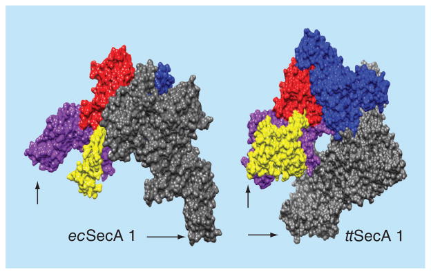
Dimeric SecA proteins were structurally aligned on one of their protomers (the nucleotide binding domain NBD, red; the intramolecular regulator of ATPase 2 IRA2, dark blue; the protein binding domain PBD, yellow and the C-domain, purple) so as to demonstrate the variable position that the second (grey) protomer occupies; arrows indicate C-terminus. The figure was created using the UCSF Chimera package [67,68]. The structures used are: Escherichia coli (ecSecA1; 2FSF) [51]. Thermus thermophilus (ttSecA; 2IPC) [61]. For color images please see online www.future-science.com/doi/full/10.4155/FMC.15.42
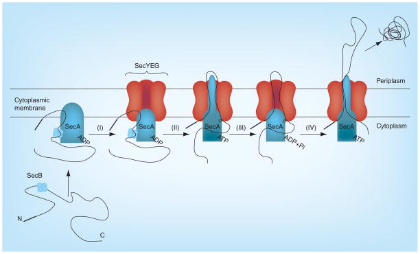
(I) Membrane SecA complexed with the SecB and preprotein binds to SecYEG; (II) SecA acquires the translocation ATPase activity and inserts itself into SecYEG channel; (III) SecYEG channel loosens and gets ready to push the pre-proteins; and (IV) the preprotein is pushed into the periplasm across the cytoplasmic membrane. Adapted with permission from [91] © Elsevier (2011).
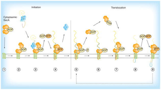
Steps 1–4: Initiation of protein translocation is commenced by SecA cytoplasmic homodimer (1) and ends with the release of SecB chaperone (4). Steps 5–9: The translocation cycle is initiated with the release of SecA protomer (5) which is recycled back into the translocation cycle (6–7), finally culminating in the release of preprotein into the periplasm (9). Reproduced with permission from [46] © Springer (2011).
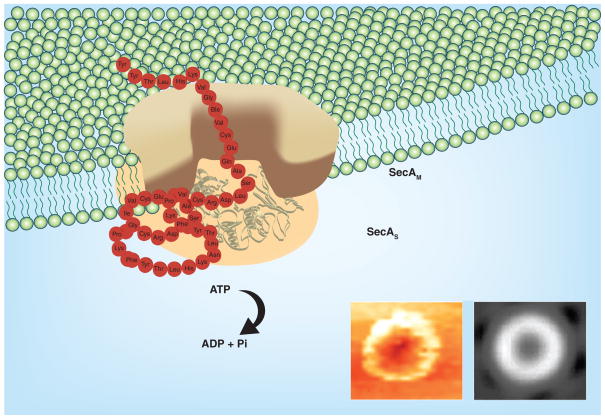
The Tai model [114]: SecA dimer [54,118] alone acts as the protein conducting-channel to promote protein translocation and ion-channel activity [
,–122]. There exist two forms of integral SecA in the membranes [115,116]. SecAS has the same conformation as in soluble form, and SecAM is specific for lipids, acting as channels [–55]. Reproduced from [114].
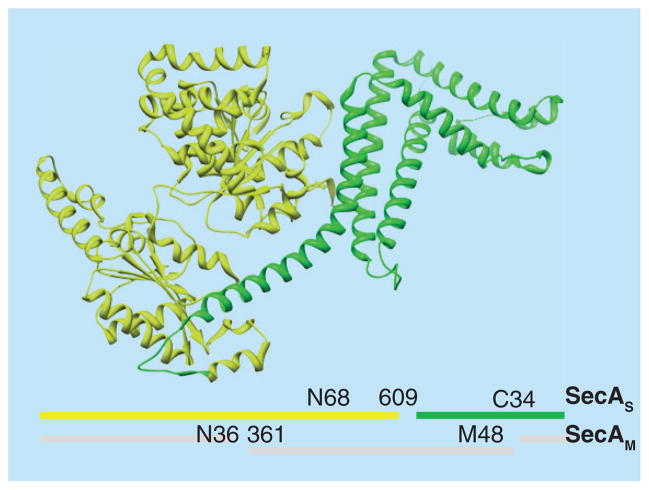
Two forms of SecA exist in the membrane: SecAS, which is similar to the soluble form with two separable domains: N68 and C34, and the other SecAM with the N36 and M48 domains spanning the lipid membrane [114]. X-ray ribbon structure of EcSecA with N68 (yellow) and C34 (green) is shown. For color images please see online www.future-science.com/doi/full/10.4155/FMC.15.42
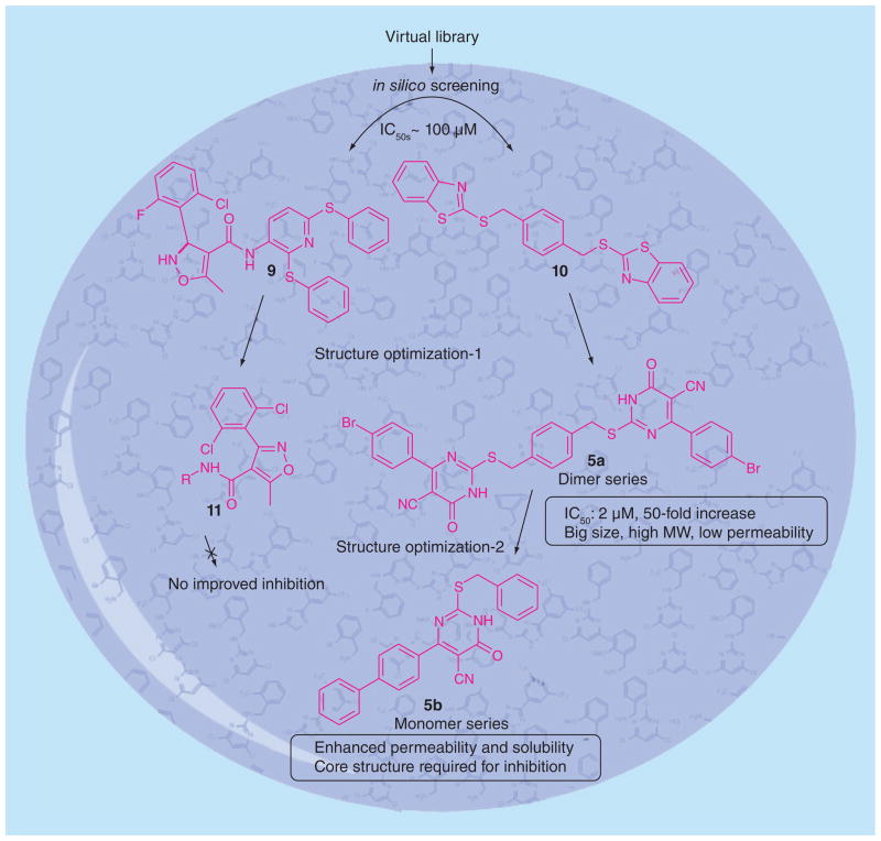
Structure optimization of 5-cyano-6-aryl-2-thiouracils derivatives.
References
-
- Falagas ME, Karageorgopoulos DE. Pandrug resistance (PDR), extensive drug resistance (XDR), and multidrug resistance (MDR) among Gram-negative bacilli: need for international harmonization in terminology. Clin Infect Dis. 2008;46(7):1121–1122. - PubMed
-
- Falagas ME, Koletsi PK, Bliziotis IA. The diversity of definitions of multidrug-resistant (MDR) and pandrugresistant (PDR) Acinetobacter baumannii and Pseudomonas aeruginosa. J Med Microbiol. 2006;55(12):1619–1629. - PubMed
Publication types
MeSH terms
Substances
Grants and funding
LinkOut - more resources
Full Text Sources
Other Literature Sources
Medical
Molecular Biology Databases