

Dietary supplementation with omega-3 fatty acid attenuates 5-fluorouracil induced mucositis in mice
- PMID: 26063053
- PMCID: PMC4473827
- DOI: 10.1186/s12944-015-0052-z
Dietary supplementation with omega-3 fatty acid attenuates 5-fluorouracil induced mucositis in mice
Abstract
Background: Studies showed the positive effects of omega-3 fatty acid (n-3 FA) for the treatment of inflammatory bowel disease as it alleviated the symptoms and promoted better mucosal integrity. The objective of this study was to determine whether a diet with the addition of n-3 FA helps control the inflammation observed in 5-fluorouracil (5-FU) induced mucositis.
Methods: BALB/c mice were randomly divided into four groups as follows: 1: control (CTL), fed a standard chow diet; 2: CTL + n-3 FA - n-3 FA, fed a diet with n-3; 3: mucositis (MUC), fed a standard chow diet and subjected to mucositis; and 4: MUC+ n-3 FA, fed a diet with n-3 FA and subjected to mucositis. On the 8th day, the animals of the MUC and MUC + n-3 FA groups received an intraperitoneal injection of 300 mg/kg 5-FU for mucositis induction. After 24 h or 72 h, all mice were euthanized and evaluated for intestinal permeability, bacterial translocation, intestinal histology and apoptosis.
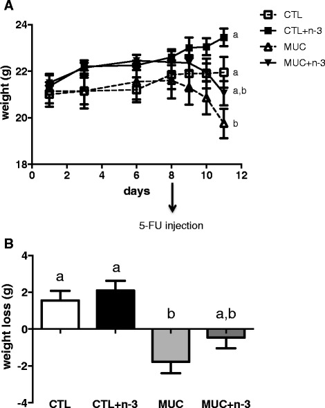
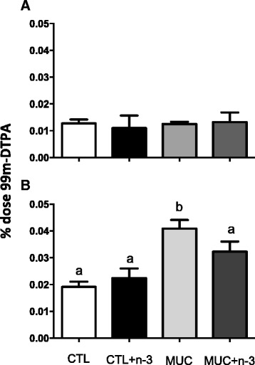
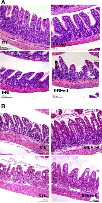
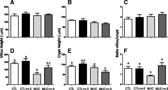
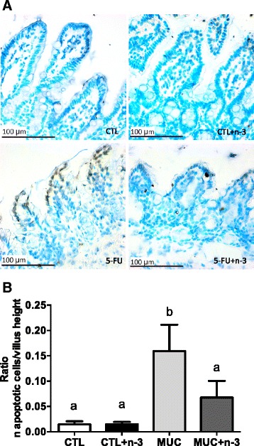
Results: Mice that received the diet with n-3 FA and a 5-FU injection showed less weight loss compared to the animals of the MUC group (p < 0.005). Decreased intestinal permeability and bacterial translocation were also observed in animals fed n-3 FA, and these mice underwent mucositis compared to the MUC group (p < 0.005). These data were associated with mucosal integrity and a reduced number of apoptotic cells in the ileum mucosa compared to the mice that received the control diet and 5-FU injection.
Conclusion: Together, these results show that omega-3 fatty acid decreases the mucosal damage caused by 5-FU-induced mucositis.
Introdução: Estudos têm demonstrado efeitos positivos da utilização do ácido graxo ômega-3 (n-3 AG) no tratamento de doenças inflamatórias do intestino, aliviando os sintomas e promovendo melhora da integridade da mucosa. Assim, no presente estudo, foi avaliado o potencial de uma dieta adicionada n-3 AG poderia ajudar a controlar a inflamação observada na mucosite intestinal induzida pelo 5-fluoracil (5-FU).
Métodos: Camundongos BALB/c foram divididos aleatoriamente em quatro grupos: 1. Controle (CTL) - alimentados com ração padrão; 2. CTL + n-3 AG - alimentados com uma dieta adicionada de n-3 AG; 3. Mucosite (MUC) - alimentados com a dieta ração padrão e submetidos à indução da mucosite; 4. MUC + n-3 FA – alimentados com dieta adicionada de n-3 AG e submetidos à indução da mucosite. No oitavo dia, os animais dos grupos MUC e MUC + n-3 AG receberam uma injeção intraperitoneal de 300 mg/kg de 5-FU para a indução da mucosite. Após 24 h ou 72 h, todos os camundongos foram eutanaziados para a avaliação da permeabilidade intestinal, translocação bacteriana, histologia intestinal e ensaio de apoptose.
Resultados: Os animais que receberam a dieta adicionada de n-3 AG e injeção de 5-FU mostraram menor perda de peso comparado com os animais do grupo MUC (p <0,005). Foi observado diminuição da permeabilidade intestinal e translocação bacteriana nos animais alimentados com n-3 AG e submetidos a mucosite. Estes dados foram associadas com melhor integridade da mucosa e uma redução do número de células apoptóticas na mucosa do íleo em comparação com os camundongos que receberam ração controle dieta e injeção com 5-FU.
Conclusão: Estes resultados mostram que o ácido graxo ômega-3 pode diminuir o dano da mucosa causada pela mucosite induzida pelo 5-FU.
Figures
References
-
- Wall R, Ross RP, Fitzgerald GF, Stanton C. Fatty acids from fish: The anti-inflammatory potential of long-chain omega-3 fatty acids. Nutrition Reviews. 2010;280–289. - PubMed
Publication types
MeSH terms
Substances
LinkOut - more resources
Full Text Sources
Other Literature Sources
Medical
