. 2015 Aug;89(16):8580-9.
doi: 10.1128/JVI.01043-15.

Characterization of Ribosomal Frameshifting in Theiler's Murine Encephalomyelitis Virus

Characterization of Ribosomal Frameshifting in Theiler's Murine Encephalomyelitis Virus

Leanne K Finch et al. J Virol. 2015 Aug.

Abstract

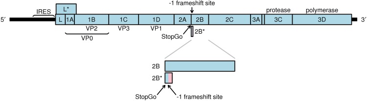
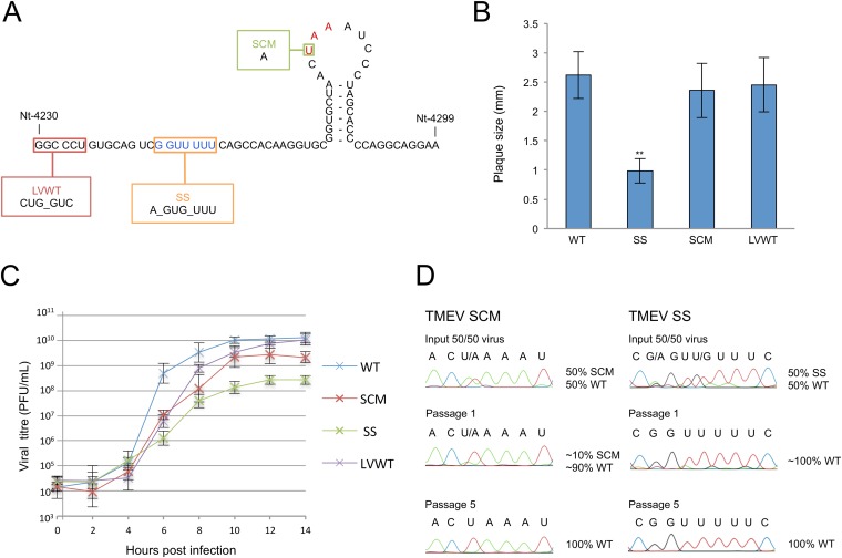
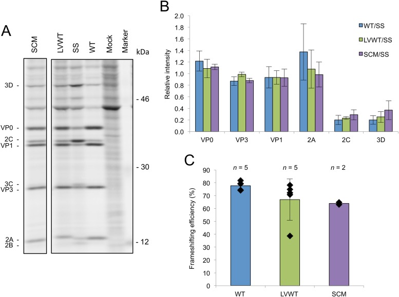
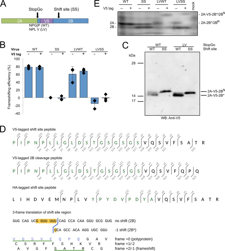
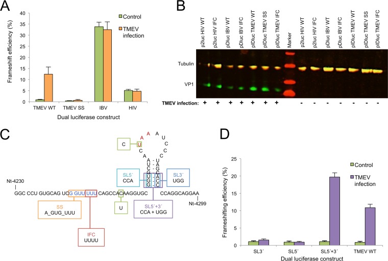
Theiler's murine encephalomyelitis virus (TMEV) is a member of the genus Cardiovirus in the Picornaviridae, a family of positive-sense single-stranded RNA viruses. Previously, we demonstrated that in the related cardiovirus, Encephalomyocarditis virus, a programmed-1 ribosomal frameshift (1 PRF) occurs at a conserved G_GUU_UUU sequence within the 2B-encoding region of the polyprotein open reading frame (ORF). Here we show that-1 PRF occurs at a similar site during translation of the TMEV genome. In addition, we demonstrate that a predicted 3= RNA stem-loop structure at a noncanonical spacing downstream of the shift site is required for efficient frameshifting in TMEV and that frameshifting also requires virus infection. Mutating the G_GUU_UUU shift site to inhibit frameshifting results in an attenuated virus with reduced growth kinetics and a small-plaque phenotype. Frameshifting in the virus context was found to be extremely efficient at 74 to 82%, which, to our knowledge, is the highest frameshifting efficiency recorded to date for any virus. We propose that highly efficient-1 PRF in TMEV provides a mechanism to escape the confines of equimolar expression normally inherent in the single-polyprotein expression strategy of picornaviruses.


Figures

FIG 1
FIG 1
Schematic representation of the TMEV genome. The predicted −1 PRF site is situated at codons 5 and 6 downstream of the junction between the 2A and 2B coding sequences. Frameshift translation would yield a 14-aa transframe protein termed 2B*, whose N terminus would be encoded in the polyprotein frame (blue) and whose C terminus would be encoded in the −1 frame (pink).
FIG 2
FIG 2
Analysis of TMEV mutants. (A) Schematic representation of mutations. SS, shift site mutant; SCM, 2B* extension mutant; LVWT, StopGo mutant. The shift site sequence is in blue, and the 2B* stop codon is in red. (B) Mean plaque sizes for TMEV WT and mutant viruses. Mean plaque sizes are the averages for 100 representative plaques; error bars indicate standard deviations. **, SS is significantly different from WT (two-tailed t test, P < 0.001). (C) One-step growth curves. BHK-21 cells were infected with WT, SS, SCM, or LVWT viruses at an MOI of 10 and harvested at the indicated time points. Titers were measured by plaque assay. At least two biological repeats were performed for each virus; error bars indicate standard deviations. (D) Competition assay. BHK-21 cells were infected with a mixture of an MOI of 0.1 of either SS plus WT or SCM plus WT viruses. At 24 h p.i., virus was harvested and used to reinfect BHK-21 cells. Passaging was repeated five times. RNA was extracted from passages 0, 1, and 5, and a region of ∼1,000 nt encompassing the mutated region was sequenced.
FIG 3
FIG 3
Analysis of frameshifting in the viral context. (A) Radiolabeled TMEV translation products. BHK-21 cells were infected with either WT, SCM, SS, or LVWT viruses at an MOI of 8 or mock infected. Cells were labeled from 6 to 7 h p.i. and harvested at 7 h p.i., and proteins were separated by SDS-PAGE. Note that the more slowly migrating 2A band for LVWT may contain both 2A-2B* and a 2A-2B cleavage product. All samples were run on the same gel; an irrelevant lane has been excised. (B) Relative amounts of TMEV proteins. Individual band intensities for WT, LVWT, and SCM were normalized first by methionine content, then by the means of these values for VP0, VP1, and VP3 (to control for lane loading), and then by the corresponding similarly normalized band for SS. Each bar represents the mean (± standard deviation) from all biological repeats in which the corresponding band could be resolved and quantified (see the text). (C) Frameshifting efficiency. The intensity in each of the VP0, VP3, VP1, and 2C bands for WT, LVWT, and SCM viruses was normalized first by methionine content, then by the means of these values for VP0, VP1, and VP3 (to control for lane loading), and then by the corresponding similarly normalized values for SS. Then the value for 2C (downstream product) was divided by the average of the values for VP0, VP3, and VP1 (upstream products). Subtracting this value from 1 and multiplying the result by 100 gives the percent frameshifting efficiency. Each bar represents the mean value (± standard deviation) from five, five, and two biological repeats for WT, LVWT, and SCM viruses, respectively.
FIG 4
FIG 4
Characterization of tagged viruses. (A) Schematic representation of the tagged viruses. A V5 tag was inserted just after the first proline of 2B in the WT sequence (after valine in the GP-to-LV StopGo-mutated viruses), to tag products containing 2B or 2B*. (B) Frameshifting efficiencies of tagged and untagged viruses. BHK-21 cells were infected with WT, SS, LVWT, or LVSS viruses or their tagged equivalents, and frameshifting efficiencies were calculated from radiolabeled products as described in Fig. 3C. All viruses were normalized by SS; thus, the frameshifting efficiency for SS is zero by definition. Negative frameshifting efficiencies are likely an artifact of measurement errors and/or biological variability. Each bar represents the mean value (± standard deviation) from three biological repeats (the three untagged WT and LVWT data points are also used in Fig. 3C). (C) Western blot of virus-infected cell lysates. BHK-21 cells were infected with V5-tagged viruses at MOI of 1 for LVSS, 5 for SS, and 10 for WT and LVWT, and lysates were prepared when cytopathic effect was extensive. Fivefold-smaller amounts of the LVWT and LVSS samples were loaded to give band intensities similar to those of the WT and SS samples, where only proteins generated by StopGo failure are detected. The proteins with the GP-to-LV mutation may migrate slightly faster than the wild-type ones. Samples were run on bis-Tris gels with 6 M urea and MOPS buffer, which results in altered mobility of the prestained 14-kDa and 17-kDa markers. (D) Mass spectrometric analysis of tagged products. Lysates from BHK-21 cells infected with V5- or HA-tagged TMEV LVWT were immunoprecipitated with V5 or HA antibodies, respectively, and immunoprecipitates were separated by SDS-PAGE. Products migrating at the expected size for 2A-tag-2B* were subjected to in-gel trypsin digest, and peptides were analyzed by LC-MS/MS. Fragmentation ions are shown for the shift site peptide derived from V5-tagged 2A-2B* (top), a peptide consistent with 3C-Pro cleavage of tagged 2A-2B at the conserved Q|G encoded just downstream of the frameshift site (middle), and the shift site peptide derived from HA-tagged 2A-2B* (bottom). Amino acids derived from the V5 or HA tag are in green. The b- and y-series ions correspond to N- and C-terminal fragments. See Fig. S1 in the supplemental material for the fragmentation spectra. Below, the nucleotide sequence in the vicinity of the shift site G_GUU_UUU is shown, with conceptual amino acid translations in all three reading frames. The C-terminal end of the frameshift tryptic peptide is underlined in green, and the C-terminal end of the 2B cleavage peptide is in blue. (E) Low-molecular-mass radiolabeled TMEV translation products. BHK-21 cells were infected with WT, SS, LVWT, or LVSS viruses or their tagged equivalents at an MOI of 10 or mock infected. Cells were labeled from 8 to 9 h p.i. and harvested at 9 h p.i., and proteins were separated by SDS-PAGE.
FIG 5
FIG 5
Analysis of frameshift stimulators. (A) Frameshifting efficiencies measured using dual-luciferase constructs. BHK-21 cells were transfected with frameshift reporter constructs and 18 h later were either infected with WT virus at an MOI of 10 or mock infected. Lysates were harvested at 7 h p.i. and assayed for Renilla and firefly luciferase activity. Frameshift efficiencies were determined by comparing luciferase activities to an in-frame control (IFC) construct. Mean values and standard deviations are shown, each based on nine separate transfections. (B) Western blot verifying infection of infected samples. Aliquots of each of the cell lysates were separated on a 10 to 20% Tris-Tricine gradient gel and probed with rat monoclonal anti-tubulin (red, IRDye 700-labeled secondary) and mouse monoclonal anti-VP1 (TMEV capsid protein) (green, IRDye 800-labeled secondary) antibodies. Note that the rat monoclonal primary cross-reacts with both the secondary antibodies. (C) Schematic representation of the fragments cloned into the pIDluc vector. All constructs contain the U-to-C mutation removing the −1 frame UAA stop codon (red) to allow expression of the downstream luciferase and the C-to-U mutation to introduce a zero-frame UAA stop codon just 3′ of the shift site (see the text). pIDluc IFC contains an extra U in the G_GUU_UUU shift site sequence (blue). (D) Frameshifting efficiencies of dual-luciferase constructs containing stem-loop mutants. See the description of panel A for details. Mean values and standard deviations are shown, each based on nine separate transfections.

References

    1. Palmenberg AC, Kirby EM, Janda MR, Drake NL, Duke GM, Potratz KF, Collett MS. 1984. The nucleotide and deduced amino acid sequences of the encephalomyocarditis viral polyprotein coding region. Nucleic Acids Res 12:2969–2985. doi:10.1093/nar/12.6.2969. - DOI - PMC - PubMed
    1. Jackson RJ. 1986. A detailed kinetic analysis of the in vitro synthesis and processing of encephalomyocarditis virus products. Virology 149:114–127. doi:10.1016/0042-6822(86)90092-9. - DOI - PubMed
    1. Palmenberg AC. 1990. Proteolytic processing of picornaviral polyprotein. Annu Rev Microbiol 44:603–623. doi:10.1146/annurev.mi.44.100190.003131. - DOI - PubMed
    1. Batson S, Rundell K. 1991. Proteolysis at the 2A/2B junction in Theiler's murine encephalomyelitis virus. Virology 181:764–767. doi:10.1016/0042-6822(91)90914-W. - DOI - PubMed
    1. Palmenberg AC, Parks GD, Hall DJ, Ingraham RH, Seng TW, Pallai PV. 1992. Proteolytic processing of the cardioviral P2 region: primary 2A/2B cleavage in clone-derived precursors. Virology 190:754–762. doi:10.1016/0042-6822(92)90913-A. - DOI - PubMed

Publication types

LinkOut - more resources