. 2015 Sep;134(9):941-50.
doi: 10.1007/s00439-015-1571-4. Epub 2015 Jun 11.

Mutation of ATF6 causes autosomal recessive achromatopsia

Collaborators, Affiliations

Mutation of ATF6 causes autosomal recessive achromatopsia

Muhammad Ansar et al. Hum Genet. 2015 Sep.

Abstract

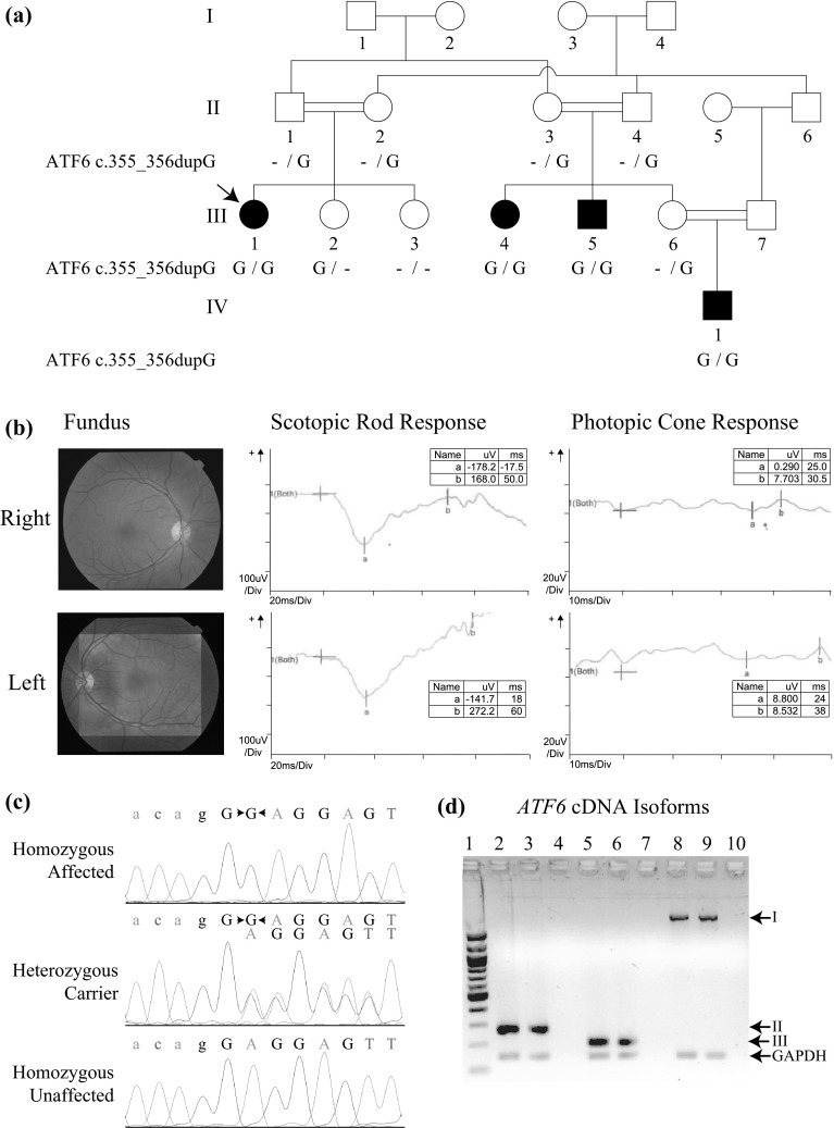
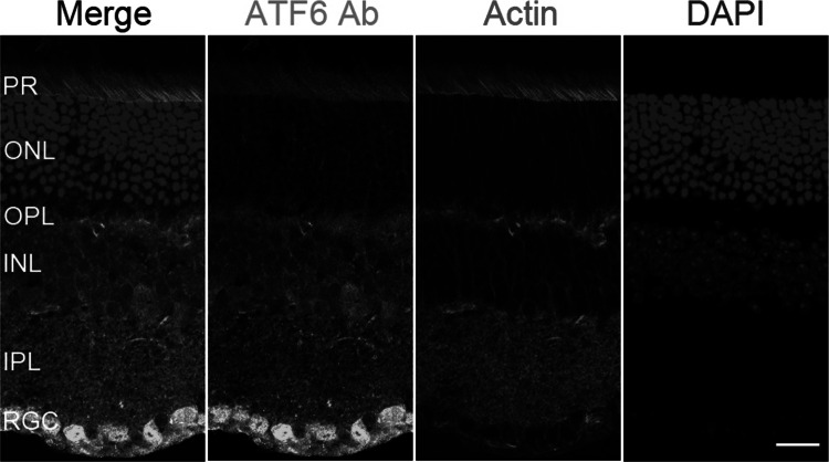
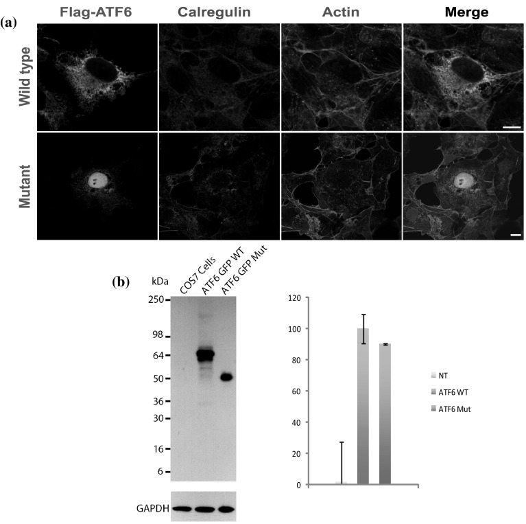
Achromatopsia (ACHM) is an early-onset retinal dystrophy characterized by photophobia, nystagmus, color blindness and severely reduced visual acuity. Currently mutations in five genes CNGA3, CNGB3, GNAT2, PDE6C and PDE6H have been implicated in ACHM. We performed homozygosity mapping and linkage analysis in a consanguineous Pakistani ACHM family and mapped the locus to a 15.12-Mb region on chromosome 1q23.1-q24.3 with a maximum LOD score of 3.6. A DNA sample from an affected family member underwent exome sequencing. Within the ATF6 gene, a single-base insertion variant c.355_356dupG (p.Glu119Glyfs*8) was identified, which completely segregates with the ACHM phenotype within the family. The frameshift variant was absent in public variant databases, in 130 exomes from unrelated Pakistani individuals, and in 235 ethnically matched controls. The variant is predicted to result in a truncated protein that lacks the DNA binding and transmembrane domains and therefore affects the function of ATF6 as a transcription factor that initiates the unfolded protein response during endoplasmic reticulum (ER) stress. Immunolabeling with anti-ATF6 antibodies showed localization throughout the mouse neuronal retina, including retinal pigment epithelium, photoreceptor cells, inner nuclear layer, inner and outer plexiform layers, with a more prominent signal in retinal ganglion cells. In contrast to cytoplasmic expression of wild-type protein, in heterologous cells ATF6 protein with the p.Glu119Glyfs*8 variant is mainly confined to the nucleus. Our results imply that response to ER stress as mediated by the ATF6 pathway is essential for color vision in humans.


Figures

Fig. 1
Fig. 1
Pedigree drawing, clinical findings, chromatograms and RT-PCR expression results for ATF6. a Pedigree drawing of MA28 family with autosomal recessive ACHM. Genotypes for the ATF6 c.355_356dupG variant are shown below each symbol of corresponding family members with available DNA samples. b Fundoscopy images and ERG data for affected proband III-1 (arrow in panel a) which shows bilateral loss of the foveal reflex and reduced cone response, respectively. Other retinal features are normal, including a pale-centered optic disc with clear borders, 0.3–0.4 cup-to-disc ratio, and no arteriovenous nicking, hemorrhages, emboli or infarcts. c Chromatograms of family members who are homozygous, heterozygous and wild-type for the ATF6 variant. d RT-PCR results showed the presence of multiple ATF6 isoforms in human eye and retinal pigment epithelium cells. I NM_007348.3 exons 1–16, 2028 bp; II NM_007348.3 exons 15–16, 265 bp; III AB208929 exon 14, 190 bp. Table S1 has additional details on isoforms and exons tested. GAPDH (143 bp) was used as internal control. Lanes: 1 100-bp leader; 2, 5, 8 whole eye; 3, 6, 9 retinal pigment epithelium; 4, 7, 10 negative control
Fig. 2
Fig. 2
Localization of ATF6 in wild-type mouse retina. Immunostaining in CD1 mouse retina with antibodies for DAPI (blue), rhodamine phalloidin (red) for actin-based structures and ATF6 (green). ATF6 strongly localized to the retinal ganglion cells (RGC), and had moderate staining of the outer and inner segments of photoreceptor cells (PR) and the inner plexiform (IPL), inner nuclear (INL) and outer plexiform (OPL) layers. ATF6 is weakly localized to the outer nuclear layer (ONL) which contains photoreceptor cell bodies (color figure online)
Fig. 3
Fig. 3
Expression of wild-type and mutant ATF6 in transfected COS-7 cells. a Flag-tagged wild-type ATF6 protein is expressed throughout the cytoplasm and localized in the endoplasmic reticulum. In contrast, the p.Glu119Glyfs*8 variant affected the stability, targeting and localization of ATF6, with expression confined to the nucleus and significantly reduced cytoplasmic localization. Scale bars 10 μm. b COS-7 cells were transiently transfected with the same quantity of wild-type or mutant ATF6 constructs. Protein extracts from the cell lysates were analyzed by western blot using an anti-ATF6 antibody. Flag-tagged wild-type ATF6 protein is demonstrated at the expected size ~64 kDa and with dimer formation. In contrast, mutant ATF6 had reduced protein size. GAPDH antibody was used as loading control. Quantification of the steady-state expression did not reveal any significant difference between the wild-type and mutant ATF6

References

    1. Abecasis GR, Cherny SS, Cookson WO, Cardon LR. Merlin-rapid analysis of dense genetic maps using sparse gene flow trees. Nat Genet. 2002;30:97–101. doi: 10.1038/ng786. - DOI - PubMed
    1. Aboshiha J, Dubis AM, Cowing J, Fahy RT, Sundaram V, Bainbridge JW, Ali RR, Dubra A, Nardini M, Webster AR, Moore AT, Rubin G, Carroll J, Michaelides M. A prospective longitudinal study of retinal structure and function in achromatopsia. Investig Ophthalmol Vis Sci. 2014;55:5733–5743. doi: 10.1167/iovs.14-14937. - DOI - PMC - PubMed
    1. Chang B, Grau T, Dangel S, Hurd R, Jurklies B, Sener EC, Andreasson S, Dollfus H, Baumann B, Bolz S, Artemyev N, Kohl S, Heckenlively J, Wissinger B. A homologous genetic basis of the murine cpfl1 mutant and human achromatopsia linked to mutations in the PDE6C gene. Proc Natl Acad Sci USA. 2009;106:19581–19586. doi: 10.1073/pnas.0907720106. - DOI - PMC - PubMed
    1. Chiang WC, Hiramatsu N, Messah C, Kroeger H, Lin JH. Selective activation of ATF6 and PERK endoplasmic reticulum stress signaling pathways prevent mutant rhodopsin accumulation. Investig Ophthalmol Vis Sci. 2012;53:7159–7166. doi: 10.1167/iovs.12-10222. - DOI - PMC - PubMed
    1. Cinaroglu A, Gao C, Imrie D, Sadler KC. Activating transcription factor 6 plays protective and pathological roles in steatosis due to endoplasmic reticulum stress in zebrafish. Hepatology. 2011;54:495–508. doi: 10.1002/hep.24396. - DOI - PMC - PubMed
Web resources
    1. Burrows–Wheeler Aligner, http://bio-bwa.sourceforge.net/
    1. Clustal Omega, http://www.ebi.ac.uk/Tools/msa/clustalo/
    1. dbSNP, http://www.ncbi.nlm.nih.gov/SNP/
    1. Exome Aggregation Consortium (ExAC), http://exac.broadinstitute.org/
    1. Genome Analysis Toolkit (GATK), https://www.broadinstitute.org/gatk/

Publication types

MeSH terms

Substances