. 2015 Aug 1;26(15):2742-54.
doi: 10.1091/mbc.E15-02-0094. Epub 2015 Jun 10.

HJURP is involved in the expansion of centromeric chromatin

Affiliations

HJURP is involved in the expansion of centromeric chromatin

Marinela Perpelescu et al. Mol Biol Cell. .

Abstract

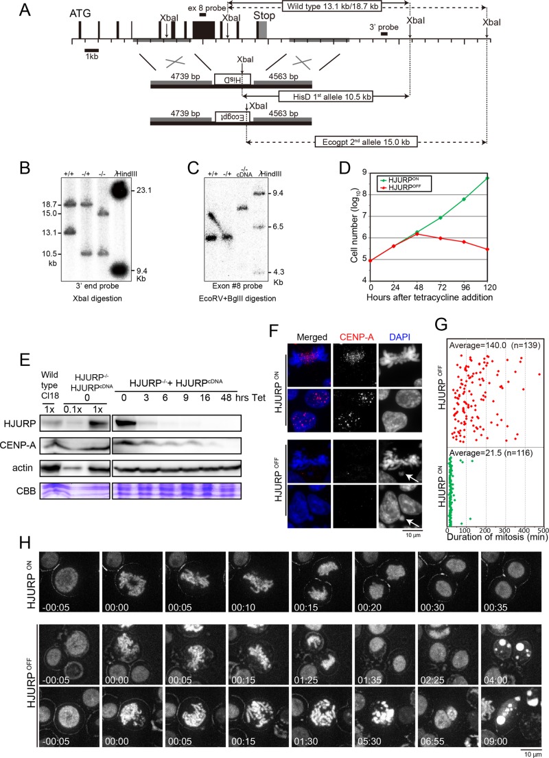
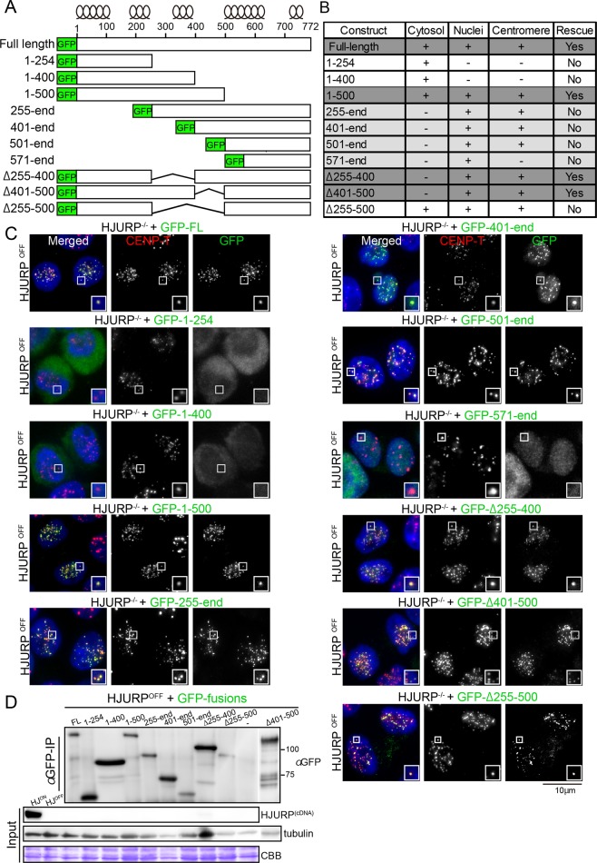
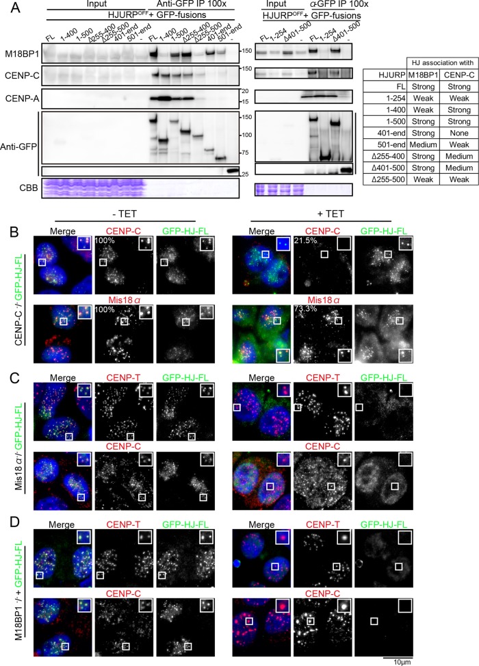
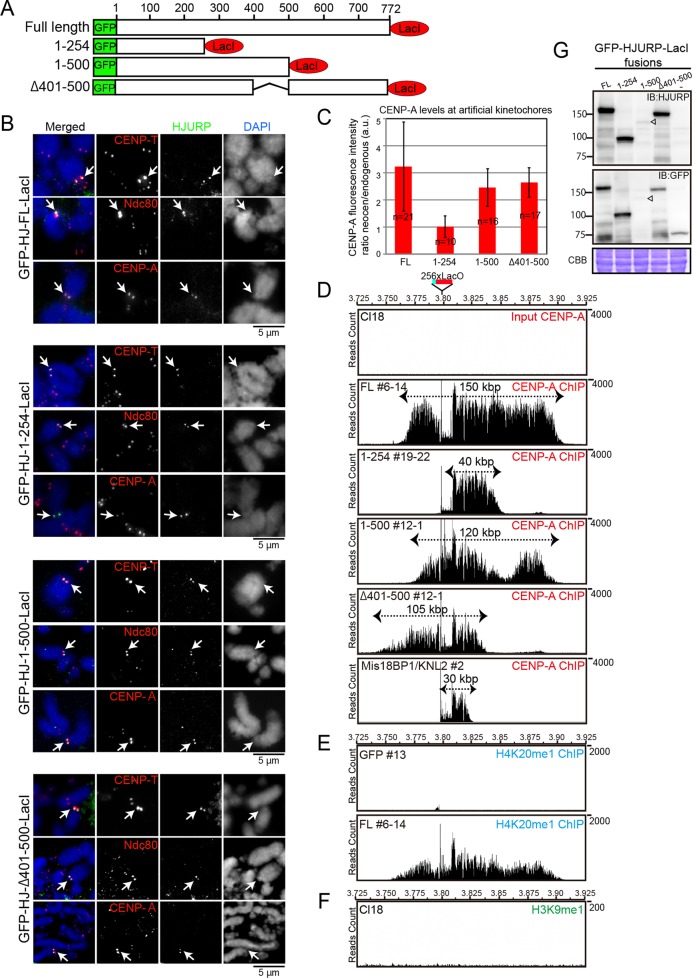
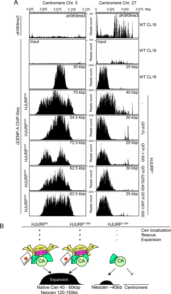
The CENP-A-specific chaperone HJURP mediates CENP-A deposition at centromeres. The N-terminal region of HJURP is responsible for binding to soluble CENP-A. However, it is unclear whether other regions of HJURP have additional functions for centromere formation and maintenance. In this study, we generated chicken DT40 knockout cell lines and gene replacement constructs for HJURP to assess the additional functions of HJURP in vivo. Our analysis revealed that the middle region of HJURP associates with the Mis18 complex protein M18BP1/KNL2 and that the HJURP-M18BP1 association is required for HJURP function. In addition, on the basis of the analysis of artificial centromeres induced by ectopic HJURP localization, we demonstrate that HJURP exhibits a centromere expansion activity that is separable from its CENP-A-binding activity. We also observed centromere expansion surrounding natural centromeres after HJURP overexpression. We propose that this centromere expansion activity reflects the functional properties of HJURP, which uses this activity to contribute to the plastic establishment of a centromeric chromatin structure.


Figures

FIGURE 1:
FIGURE 1:
HJURP is essential for mitotic progression in chicken DT40 cells. (A) Genome map around the chicken HJURP locus and targeting constructs. Restriction sites for XbaI are shown. The left arm of the knockout construct contains the genomic region 19090–23829 of chromosome 1 and the right arm, 30513–34990. There is a polymorphism for XbaI sites. Solid and dashed lines show the respective alleles. After the first targeting, the 13.1-kb fragment was reduced to 10.5 kb (solid line). After the second targeting, the 18.7-kb fragment was reduced to 15.0 kb (dashed line). (B) Southern blot analysis for confirmation of gene disruption. The probe is shown in A. (C) Additional confirmation of gene disruption based on Southern blot analysis using a probe for exon 8 of the HJURP gene. Exon 8 hybridized with the HJURP gene locus before the second allele knockout, but it hybridized with a rescue construct (HJURP cDNA) integrated at a different locus after the second allele knockout. (D) Growth curve of HJURP conditional knockout cells in the presence (HJURPOFF) or absence (HJURPON) of tetracycline. Numbers indicate hours (h) after addition of tetracycline. (E) Western blot analysis of HJURP conditional knockout cell lines with anti-HJURP or anti–CENP-A antibodies. Numbers indicate hours (h) after addition of tetracycline. Lysate from Cl18 (clone 18: wild-type DT40 cells) and conditional knockout cells (HJURP/, HJURPcDNA) were prepared. CBB, protein samples stained by Coomassie brilliant blue. (F) Typical images of HJURP-deficient cells (HJURPOFF). Cells were stained with anti–CENP-A antibody (red). Chromosomes and nuclei were counterstained with DAPI (blue). Arrows shows micronuclei (HJURPOFF bottom) and misaligned chromosomes (HJURPOFF top). Scale bar, 10 μm. (G) Average time span necessary for HJURPON (green diamonds) or HJURPOFF (red diamonds) cells to complete mitosis, starting from nuclear envelope breakdown (NEBD) to anaphase onset. Each diamond represents one cell. This measurement was performed by live-cell imaging observation. n, number of analyzed cells. (H) Typical images from live-cell imaging of HJURP conditional knockout cells stably expressing H2B-RFP in the presence (HJURPOFF) or absence (HJURPON) of tetracycline. Time 0 indicates entry of mitosis, recognized as nuclear envelope breakdown and start of nuclear condensation. The live-cell observation was started at 48 h after tetracycline addition, and time-lapse images were taken every 5 min for the next 16 h. Scale bar, 10 μm.
FIGURE 2:
FIGURE 2:
Most centromere proteins are gradually lost in HJURP-deficient cells. (A) Representative images of immunofluorescence staining for indicated centromere proteins in HJURP conditional knockout cells in the absence (HJURPON) or presence (48hrs, HJURPOFF) of tetracycline. Antibodies against CENP-A, CENP-C, CENP-T, CENP-H, and Ndc80 were used. (B) Quantification of signal intensities for each centromere protein in HJURP conditional knockout cells in the presence (48 h, HJURPOFF) or absence (HJURPON) of tetracycline.
FIGURE 3:
FIGURE 3:
Middle region of HJURP is essential for cell viability. (A) Scheme of different HJURP GFP-tagged expression constructs. The predicted position of α-helixes obtained with PSIPRED version 3.3 software. (B) Summary of results for localization and complementation assay of indicated GFP-tagged HJURP mutant proteins. Supplemental Figure S2 shows the results of cell viability assay. (C) Representative images showing cellular localization of different GFP-HJURP mutant proteins (green). CENP-T was used as a centromere marker (red). DAPI was used for DNA staining (blue). HJURPON refers to cells grown in the absence of tetracycline and HJURPOFF to those grown for 48 h in the presence of tetracycline. Because cells expressing C-terminal truncation of HJURPs (HJURP1-254 and HJURP1-400) restrictively localize to the cytoplasm, it is possible that the middle region may be related to the nuclear localization of HJURP. (D) Western blot analysis with anti-GFP antibody showing the expression levels for each of the GFP-mutant proteins. Although GFP-tagged HJURP1-500 shows unexpectedly slower migration, similar to GFP-tagged HJURPFL, expression of GFP-tagged HJURP1-500 was confirmed by genomic PCR.
FIGURE 4:
FIGURE 4:
The Mis18 complex is associated with HJURP. (A) Immunoprecipitation/Western blot analysis showing preferential associations of HJURP with M18BP1/KNL2, CENP-C, and CENP-A. Coprecipitation was also confirmed by mass spectrometry analysis. Whole-cell lysates of HJURP conditional knockout cell lines in which expression of HJURP was replaced with GFP-tagged-HJURPFL, -HJURP1-400, -HJURP1-500, -HJURPΔ255-400, -HJURPΔ255-400, and -HJURP401-end (left gel) and -HJURP1-254 and -HJURPΔ401-500 (right gel) were immunoprecipitated with anti-GFP antibody, and the eluates were applied to SDS–PAGE and subjected to Western blot analysis with the indicated antibodies. Because the majority of M18BP1/KNL2 localizes in nuclei and HJURP mainly exists in the cytoplasm (Supplemental Figure S3B), cytoplasmic and nuclear soluble fractions were prepared, and IP experiments were also done using these two mixed fractions (Supplemental Figure S3C). (B) Immunofluorescence with anti-Mis18α and anti–CENP-C antibodies in CENP-C–deficient cells (+TET). CENP-C was expressed in the absence of tetracycline (–TET). GFP-HJURP-FL was stably expressed in these cells. Mis18α and CENP-C are visible in CENP-C–deficient cells (+TET). (C) Immunofluorescence with anti–CENP-T and –CENP-C antibodies in Mis18α-deficient cells (+TET). Mis18α was expressed in the absence of tetracycline (–TET). GFP–HJURP-FL was stably expressed in these cells. HJURP signals were not detected in Mis18α-deficient cells (+TET). (D) Immunofluorescence with anti–CENP-T and –CENP-C antibodies in M18BP1/KNL2-deficient cells (+TET). M18BP1/KNL2 was expressed in the absence of tetracycline (–TET). GFP–HJURP-FL was stably expressed in these cells. HJURP signals were not detected in M18BP1/KNL2-deficient cells (+TET).
FIGURE 5:
FIGURE 5:
Middle region of HJURP contributes to centromere expansion. (A) Schematic representation of different HJURP fusion constructs with GFP and LacI used for induction of artificial kinetochores at the LacO site on chromosome Z. (B) Immunofluorescence images with anti–CENP-T, -Ndc80, and -CENP-A antibodies on artificial kinetochores induced by HJURPFL, HJURP1-254, HJURP1-500, and -HJURPΔ401-500 tethering. Mitotic spreads were used for immunofluorescence experiments. Arrows shows artificial kinetochores. (C) Quantification of signal intensity of CENP-A from B for each type of HJURP-induced artificial kinetochore. The intensity at induced kinetochores was calculated relative to that of centromeres of chromosome 1 or 2. Error bars are SDs. Cell numbers (n) are shown. (D) ChIP-seq profiles with anti–CENP-A antibody around LacO sites in indicated artificial centromeres induced by HJURPFL (#6-14), HJURP1-254 (#19-22), HJURP1-500 (#12-1), HJURPΔ401-500 (#2-1), and M18BP1 (#2). DNA purified from wild-type DT40 cells (Cl18) before ChIP was used as input DNA sample. Raw reads of sequence were mapped, as CENP-A peaks around centromere regions were highly enriched and normalization was not necessary. The 256 copies of LacO sequences were inserted into the chicken galGAL4 genome database (UCSC Genome Browser), which was further used as a reference for mapping. Note that CENP-A incorporation at the LacO site is not high and is expanded in adjacent regions, but some clones shows still high incorporation of CENP-A at the LacO site. (E) ChIP-seq profile with anti-H4K20me1 antibody around LacO sites in the artificial centromeres induced by HJURPFL (#6-14). Cells in which LacI-GFP was tethered at the LacO site were used as control. (F) ChIP-seq profile with anti-H3K9me3 antibody around LacO sites in DT40 cells (Cl18). (G) Western blot analysis showing expression level of each LacI-fused HJURP protein. Anti-HJURP and anti-GFP antibodies were used.
FIGURE 6:
FIGURE 6:
Centromere expansion around native centromeres upon HJURP overexpression. (A) Distribution of CENP-A and heterochromatic marker H3K9me3 around native centromere of chromosomes 5 and 27 in wild-type DT40 cells (Cl18, top). CENP-A distribution was examined around native centromere of chromosomes 5 and 27 in cells overexpressing HJURPFL, HJURP1-500, and HJURPΔ255-400 and HJURPΔ401-500, which could rescue HJURP deficiency. Centromere expansion was observed around centromere 5 upon overexpression of HJURPs but was not around centromere 27, where heterochromatin was enriched around centromeres. (B) Schematic summary emphasizing the importance of the middle region for HJURP function in centromere formation. HJURPFL or HJURP1-500 containing the middle region (aa 255–500), which efficiently binds the Mis18 complex, shows chromatin expansion activity, whereas HJURP1-254 cannot establish a native centromere or form an expanded centromere even if HJURP1-254 is tethered to the LacO site.

References

    1. Barnhart MC, Kuich PH, Stellfox ME, Ward JA, Bassett EA, Black BE, Foltz DR. HJURP is a CENP-A chromatin assembly factor sufficient to form a functional de novo kinetochore. J Cell Biol. 2011;194:229–243. - PMC - PubMed
    1. Black BE, Cleveland DW. Epigenetic centromere propagation and the nature of CENP-a nucleosomes. Cell. 2011;144:471–479. - PMC - PubMed
    1. Dambacher S, Deng W, Hahn M, Sadic D, Frohlich J, Nuber A, Hoischen C, Diekmann S, Leonhardt H, Schotta G. CENP-C facilitates the recruitment of M18BP1 to centromeric chromatin. Nucleus. 2012;3:101–110. - PMC - PubMed
    1. Dignam JD, Martin PL, Shastry BS, Roeder RG. Eukaryotic gene transcription with purified components. Methods Enzymol. 1983;101:582–598. - PubMed
    1. Dunleavy EM, Roche D, Tagami H, Lacoste N, Ray-Gallet D, Nakamura Y, Daigo Y, Nakatani Y, Almouzni-Pettinotti G. HJURP is a cell-cycle-dependent maintenance and deposition factor of CENP-A at centromeres. Cell. 2009;137:485–497. - PubMed

Publication types