. 2015 Aug;114(2):857-68.
doi: 10.1152/jn.00238.2015. Epub 2015 Jun 10.

Abnormal tuning of saccade-related cells in pontine reticular formation of strabismic monkeys

Affiliations

Abnormal tuning of saccade-related cells in pontine reticular formation of strabismic monkeys

Mark M G Walton et al. J Neurophysiol. 2015 Aug.

Abstract

Strabismus is a common disorder, characterized by a chronic misalignment of the eyes and numerous visual and oculomotor abnormalities. For example, saccades are often highly disconjugate. For humans with pattern strabismus, the horizontal and vertical disconjugacies vary with eye position. In monkeys, manipulations that disturb binocular vision during the first several weeks of life result in a chronic strabismus with characteristics that closely match those in human patients. Early onset strabismus is associated with altered binocular sensitivity of neurons in visual cortex. Here we test the hypothesis that brain stem circuits specific to saccadic eye movements are abnormal. We targeted the pontine paramedian reticular formation, a structure that directly projects to the ipsilateral abducens nucleus. In normal animals, neurons in this structure are characterized by a high-frequency burst of spikes associated with ipsiversive saccades. We recorded single-unit activity from 84 neurons from four monkeys (two normal, one exotrope, and one esotrope), while they made saccades to a visual target on a tangent screen. All 24 neurons recorded from the normal animals had preferred directions within 30° of pure horizontal. For the strabismic animals, the distribution of preferred directions was normal on one side of the brain, but highly variable on the other. In fact, 12/60 neurons recorded from the strabismic animals preferred vertical saccades. Many also had unusually weak or strong bursts. These data suggest that the loss of corresponding binocular vision during infancy impairs the development of normal tuning characteristics for saccade-related neurons in brain stem.

Keywords: PPRF; esotropia; exotropia; monkey; strabismus.


Figures

Fig. 1.
Fig. 1.
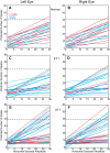
Linear regression fits relating the number of spikes in each burst to horizontal saccade amplitude. Each line represents data from a single neuron. A, C, and E: left eye. B, D, and F: right eye. A and B: for almost every long-lead burst neuron (LLBN; red) and medium-lead burst neuron (MLB; blue) recorded from the normal animals, the number of spikes increased monotonically with horizontal amplitude. For many neurons recorded from subjects ET1 (C and D) and XT1 (E and F), however, the slopes were near 0. On the other hand, there were eight neurons recorded from monkey XT1 for which the number of spikes exceeded 40 for 30° saccades (horizontal dashed lines). This was the case for only one neuron recorded from the normal animals.
Fig. 2.
Fig. 2.
Slopes for regression analysis relating the number of spikes in the burst to horizontal saccade amplitude. For the normal animals (black diamonds), there is a clear preponderance of neurons with slopes between 0.5 and 1.0 (large gray circles). For the strabismic animals (cyan = monkey XT1; red = monkey ET1), a strong majority had slopes outside of this range. In particular, note the large number of neurons with slopes near 0. For the strabismic animals (particularly XT1), there were also many neurons with unusually large slopes.
Fig. 3.
Fig. 3.
Raster plots for one predominantly vertical neuron, recorded from right pontine paramedian reticular formation (PPRF) of monkey XT1. Note that the neuron consistently burst for predominantly upward saccades (A), but there were no spikes for predominantly downward (B), rightward (C), or leftward (D) saccades. Note that all of the saccades in D have a small upward component for the left eye, but not for the right eye. The cumulative spike histogram was created by placing the spike data into 5-ms bins and then summing all of the spikes for each bin across the depicted trials.
Fig. 4.
Fig. 4.
Relationship between the number of spikes in the burst and the horizontal (A and B) and vertical (C and D) amplitude of the left (A and C) and right (B and D) eyes for a neuron recorded from left PPRF of monkey ET1. The neuron clearly has a downward preferred direction and quite strong bursts.
Fig. 5.
Fig. 5.
Example Gaussian fits, relating the number of spikes in the burst to polar saccade direction for the left eye (A) and right eye (B). This example neuron was recorded from monkey ET1. The inset in A illustrates the convention: 0° = right, 180° = left, 90° = up, 270° = down.
Fig. 6.
Fig. 6.
Summary of preferred directions of PPRF neurons, estimated from Gaussian fits (see Fig. 5). Red arrows depict neurons recorded from right PPRF; blue arrows depict neurons recorded from left PPRF. The length of each arrow corresponds to the height of the Gaussian function. Solid lines represent MLBs; dashed lines with double arrowheads represent LLBNs. A, C, and E: left eye. B, D, and F: right eye. A and B: all PPRF neurons recorded from the normal animals had preferred directions within 30° of horizontal. C and D: in contrast, neurons recorded from left PPRF in subject ET1 showed highly variable preferred directions, including some MLBs that were almost pure vertical. For right PPRF, however, the distribution of preferred directions was mostly normal. E and F: a similar asymmetry was found for subject XT1, except that the highly variable data were from right PPRF. For left PPRF in this animal, there appears to be a downward bias for the right eye, and a slight upward bias for the left eye. Note that all neurons with predominantly vertical preferred directions were MLBs, with the exception of one LLBN in monkey XT1, when the data were plotted with respect to the left eye (E).
Fig. 7.
Fig. 7.
Microstimulation of an example site at which a vertically-tuned burst neuron was recorded from monkey XT1. The gray shaded area indicates the period of stimulation (200-ms train, 400 Hz, 30 μA). Three traces are plotted, aligned with respect to the first pulse in the stimulation train. A and B: horizontal eye position and velocity traces, respectively. C and D: vertical position and velocity traces, respectively. Note that stimulation evoked constant velocity ramp eye movements that persisted for the duration of each train. These characteristics are typical of PPRF stimulation. Insets compare superimposed example spike waveforms from all vertically-tuned neurons to that of a burst-tonic neuron recorded more dorsally.
Fig. 8.
Fig. 8.
Eye velocity sensitivities, derived from a dynamic analysis that predicts the instantaneous firing rate based on horizontal and vertical velocity terms for each eye: A, left eye; B, right eye. Red, subject XT1; blue, subject ET1. For the sake of comparison, A also shows data from subject N2 (black), derived from a simplified version of the model that included terms for horizontal and vertical cyclopean velocity. A: the horizontal gray bar indicates the range of vertical sensitivities found in this normal subject. Triangles represent neurons recorded from left PPRF; circles represent right PPRF. For neurons recorded on both sides of the brain, the horizontal velocity coefficients were usually positive for the left eye (A) and negative for the right (B). Note that this does not mean that cells in left PPRF discharged for rightward saccades. Rather, it means that, for a given (leftward) velocity of the right eye, these neurons discharged at a higher rate if the left eye's (leftward) velocity was lower. Most neurons in right PPRF, however, discharged at a higher rate if the (rightward) velocity of the left eye was higher (independent of the velocity of the right eye). Mathematically, this is equivalent to saying that most PPRF neurons in the strabismic animals were sensitive to dynamic changes in strabismus angle, independent of cyclopean horizontal velocity. Note also that many neurons had vertical velocity sensitivities outside the range obtained from the normal subject.
Fig. 9.
Fig. 9.
Comparison of R2 values for the reduced (only including terms for one eye) monocular versions of Eq. 1. Red, monkey ET1; blue, monkey XT1. Overall, for both monkeys, the firing rates were better correlated with the velocity of the left eye.

References

    1. Bucci MP, Kapoula Z, Yang Q, Roussat B, Bremond-Gignac D. Binocular coordination of saccades in children with strabismus before and after surgery. Invest Ophthalmol Vis Sci 43: 1040–1047, 2002. - PubMed
    1. Busettini C, Mays LE. Saccade-vergence interactions in macaques. II. Vergence enhancement as the product of a local feedback vergence motor error and a weighted saccadic burst. J Neurophysiol 94: 2312–2330, 2005. - PubMed
    1. Carpenter J, Bithell J. Bootstrap confidence intervals: when, which, what? A practical guide for medical statisticians. Stat Med 19: 1141–1164, 2000. - PubMed
    1. Chu L, Kaneko CR. Do the saccadic system and the vestibulo-ocular reflex of monkeys share a common neural integrator? Neurosci Lett 187: 193–196, 1995. - PubMed
    1. Cohen B, Komatsuzaki A. Eye movements induced by stimulation of the pontine reticular formation: evidence for integration in oculomotor pathways. Exp Neurol 36: 101–117, 1972. - PubMed

Publication types

LinkOut - more resources