. 2015:2015:574676.
doi: 10.1155/2015/574676. Epub 2015 May 12.

Surgical Treatment of Peri-Implantitis: A 17-Year Follow-Up Clinical Case Report

Affiliations

Surgical Treatment of Peri-Implantitis: A 17-Year Follow-Up Clinical Case Report

Fabrizio Bassi et al. Case Rep Dent. 2015.

Abstract

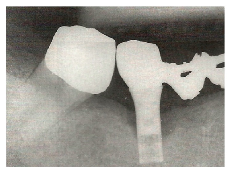
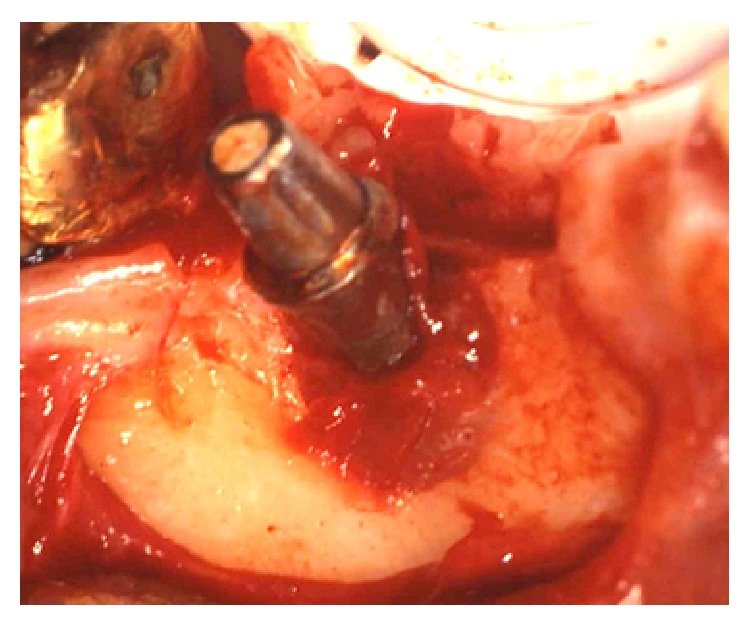
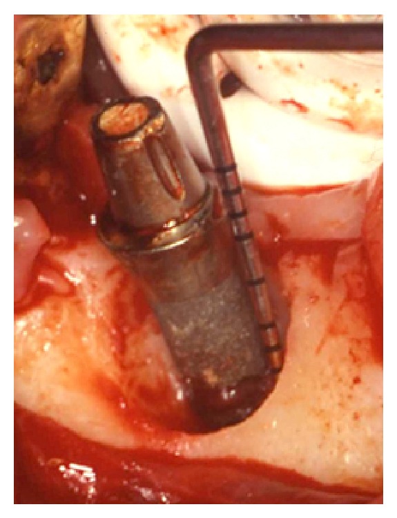
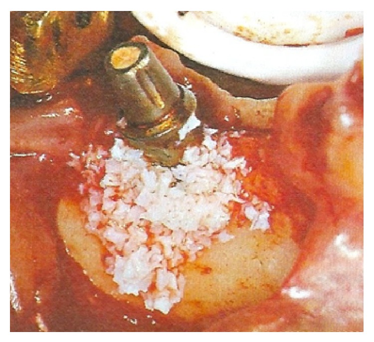
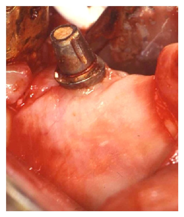
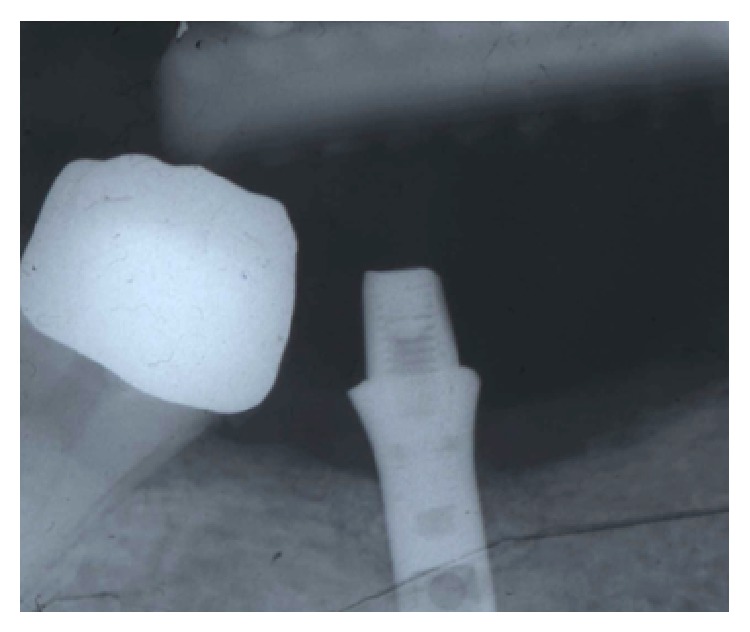
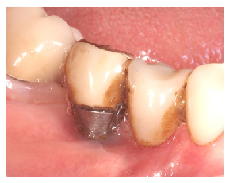
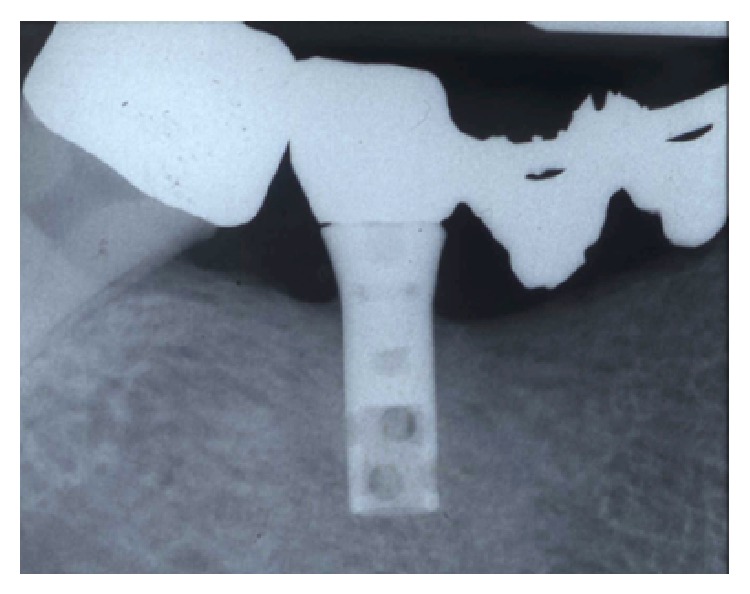
The purpose of the present case report was to describe the surgical treatment of a peri-implantitis lesion associated with a regenerative approach. A 48-year-old patient came to authors' attention 36 months after the placement of a dental implant (ITI-Bonefit Straumann, Waldenburg, Switzerland) in position 46. A swelling of the peri-implant soft tissues was observed, associated with bleeding on probing and probing depth > 10 mm. A significant peri-implant bone loss was clearly visible on the periapical radiograph. A nonsurgical periodontal supportive therapy was firstly conducted to reduce the inflammation, followed by the surgical treatment of the defect. After mechanical and chemical decontamination with tetracycline solution, a regenerative approach consisting in the application of deproteinized bovine bone mineral (Bio-Oss, Geistlich Pharma AG, Wolhusen, Switzerland) and a collagen membrane (Bio-Gide, Geistlich Pharma AG, Wolhusen, Switzerland) was performed. An antibiotic therapy was associated with the treatment. The 17-year follow-up showed a physiological probing depth with no clinical signs of peri-implant inflammation and bleeding on probing. No further radiographic bone loss was observed. The treatment described in the present case report seemed to show improved clinical results up to a relevant follow-up period.


Figures

Figure 1
Figure 1
Clinical situation of the peri-implantitis affected implant, before treatment procedures. A mesial PD of 11 mm and a distal PD of 12 mm were recorded.
Figure 2
Figure 2
Preoperative intraoral radiography. A typical peri-implantitis crater-like defect was evident.
Figure 3
Figure 3
Clinical situation after the elevation of a mucoperiosteal flap. Granulation tissue delimitating the peri-implantitis defect was clearly visible around the implant.
Figure 4
Figure 4
Clinical situation after the careful removal of the granulation tissue. The typical crater-like peri-implantitis bone defect was present circumferentially around the implant.
Figure 5
Figure 5
The bone defect was packed with heterologous DBBM graft.
Figure 6
Figure 6
A bioabsorbable collagen membrane was placed in order to create a secluded space over the DBBM graft.
Figure 7
Figure 7
Postoperative intraoral radiography. The graft filled into the bone defect was observable in close contact with the implant surfaces. During the healing period the implant was left unloaded.
Figure 8
Figure 8
Clinical situation during the follow-up recall. Peri-implant soft tissues appeared healthy, with no sign of inflammation and suppuration.
Figure 9
Figure 9
17-year follow-up intraoral radiography. The peri-implant marginal bone levels appeared radiographically stable, without any sign of bone resorption mesially and distally to the implant.

References

    1. Lindhe J., Meyle J. Peri-implant diseases: consensus report of the sixth European workshop on periodontology. Journal of Clinical Periodontology. 2008;35(8):282–285. doi: 10.1111/j.1600-051x.2008.01283.x. - DOI - PubMed
    1. Lang N. P., Berglundh T. Periimplant diseases: where are we now?—consensus of the Seventh European Workshop on Periodontology. Journal of Clinical Periodontology. 2011;38(supplement 11):178–181. doi: 10.1111/j.1600-051x.2010.01674.x. - DOI - PubMed
    1. de Boever A. L., de Boever J. A. Early colonization of non-submerged dental implants in patients with a history of advanced aggressive periodontitis. Clinical Oral Implants Research. 2006;17(1):8–17. doi: 10.1111/j.1600-0501.2005.01175.x. - DOI - PubMed
    1. Fürst M. M., Salvi G. E., Lang N. P., Persson G. R. Bacterial colonization immediately after installation on oral titanium implants. Clinical Oral Implants Research. 2007;18(4):501–508. doi: 10.1111/j.1600-0501.2007.01381.x. - DOI - PubMed
    1. Leonhardt Å., Renvert S., Dahlén G. Microbial findings at failing implants. Clinical Oral Implants Research. 1999;10(5):339–345. doi: 10.1034/j.1600-0501.1999.100501.x. - DOI - PubMed