

Ectopic Opening of the Common Bile Duct into the Duodenal Bulb Accompanied with Cholangitis and Gallbladder Cancer: A Report of Two Cases
- PMID: 26064829
- PMCID: PMC4461673
- DOI: 10.5946/ce.2015.48.3.260
Ectopic Opening of the Common Bile Duct into the Duodenal Bulb Accompanied with Cholangitis and Gallbladder Cancer: A Report of Two Cases
Abstract
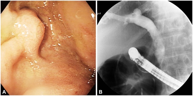
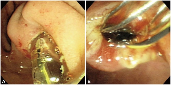
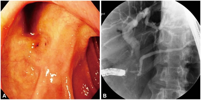
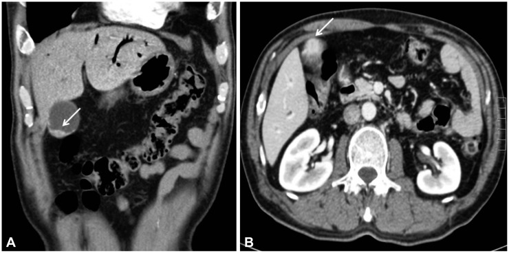
An ectopic opening of the common bile duct (CBD) into the duodenal bulb is a very rare congenital anomaly of the biliary system, which may cause recurrent duodenal ulcer or biliary diseases such as choledocholithiasis and cholangitis. Endoscopic retrograde cholangiopancreatography (ERCP) plays a major role in the diagnosis of this anomaly. We report two such cases: one in a 61-year-old man and the other in a 57-year-old man. In the first case, this anomaly caused acute cholangitis with multiple CBD stones, which were successfully treated by ERCP. In the second case, abdominal computed tomography showed pneumobilia, which was further evaluated using ERCP. Besides, this patient was diagnosed with an ectopic opening of the CBD associated with gallbladder cancer. We report these unusual cases and review the relevant medical literature.
Keywords: Acute cholangitis; Common bile duct; Duodenal bulb; Ectopic opening; Gallbladder neoplasms.
Conflict of interest statement
Figures
References
-
- Kanematsu M, Imaeda T, Seki M, Goto H, Doi H, Shimokawa K. Accessory bile duct draining into the stomach: case report and review. Gastrointest Radiol. 1992;17:27–30. - PubMed
-
- Pereira-Lima J, Pereira-Lima LM, Nestrowski M, Cuervo C. Anomalaous location of the papilla of vater. Am J Surg. 1974;128:71–74. - PubMed
-
- Moosman DA. The surgical significance of six anomalies of the biliary duct system. Surg Gynecol Obstet. 1970;131:655–660. - PubMed
-
- Kubota T, Fujioka T, Honda S, et al. The papilla of Vater emptying into the duodenal bulb. Report of two cases. Jpn J Med. 1988;27:79–82. - PubMed
-
- Lee SS, Kim MH, Lee SK, et al. Ectopic opening of the common bile duct in the duodenal bulb: clinical implications. Gastrointest Endosc. 2003;57:679–682. - PubMed
LinkOut - more resources
Full Text Sources
Other Literature Sources
