. 2015:2015:319454.
doi: 10.1155/2015/319454. Epub 2015 May 10.

Prohibitin: A Novel Molecular Player in KDEL Receptor Signalling

Affiliations

Prohibitin: A Novel Molecular Player in KDEL Receptor Signalling

Monica Giannotta et al. Biomed Res Int. 2015.

Abstract

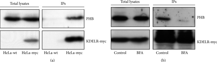
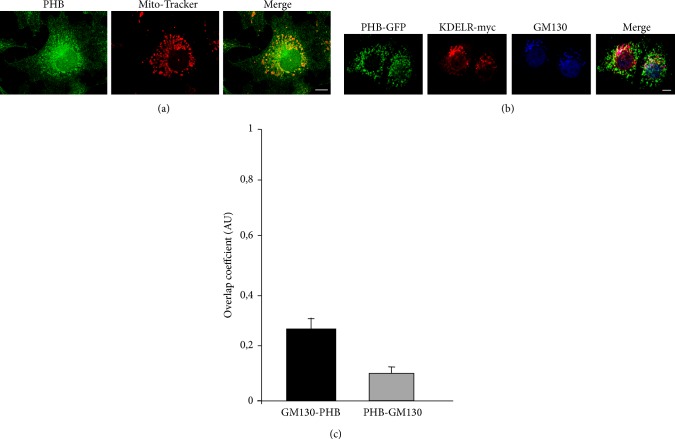
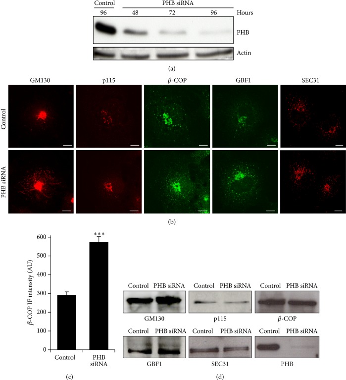
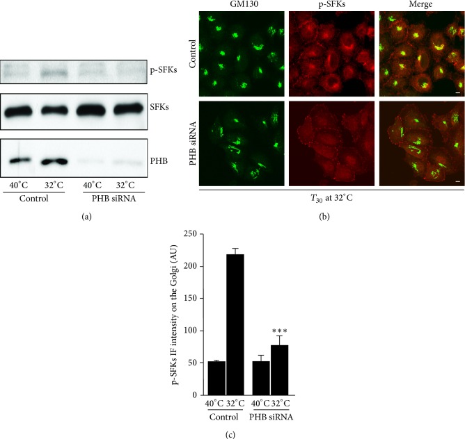
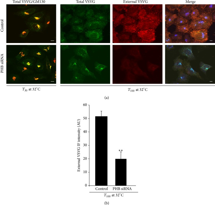
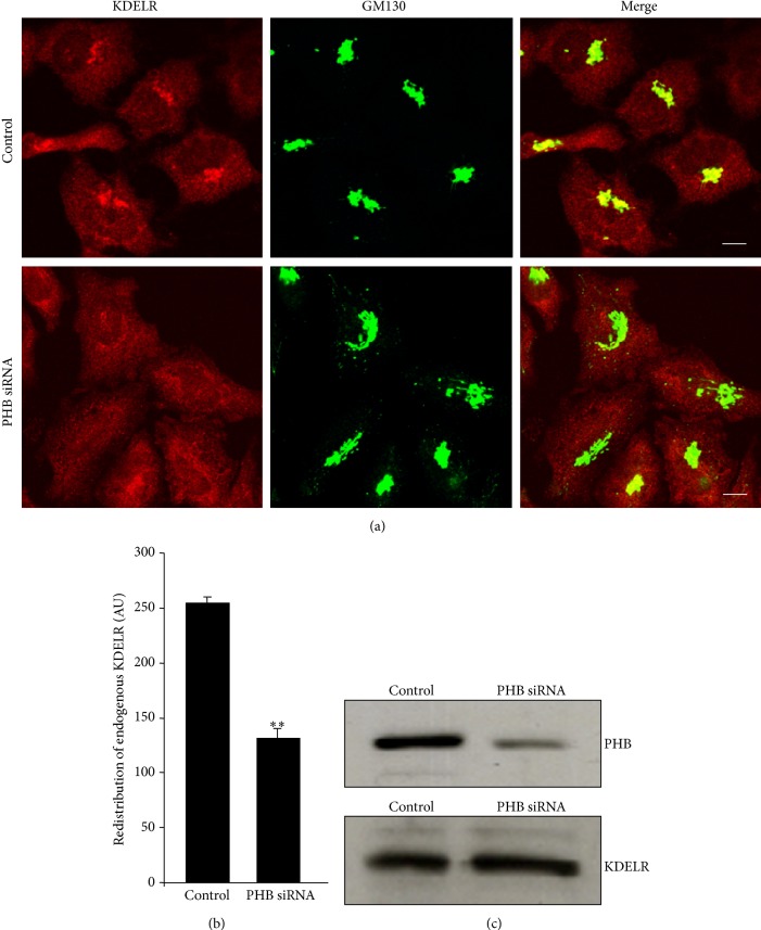
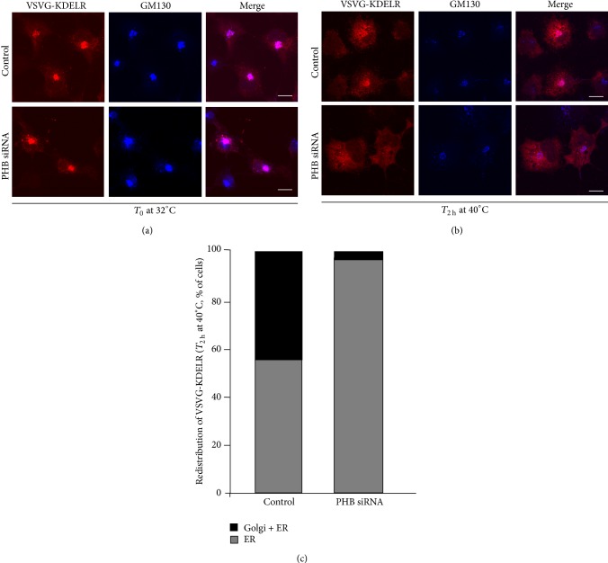
The KDEL receptor (KDELR) is a seven-transmembrane-domain protein involved in retrograde transport of protein chaperones from the Golgi complex to the endoplasmic reticulum. Our recent findings have shown that the Golgi-localised KDELR acts as a functional G-protein-coupled receptor by binding to and activating Gs and Gq. These G proteins induce activation of PKA and Src and regulate retrograde and anterograde Golgi trafficking. Here we used an integrated coimmunoprecipitation and mass spectrometry approach to identify prohibitin-1 (PHB) as a KDELR interactor. PHB is a multifunctional protein that is involved in signal transduction, cell-cycle control, and stabilisation of mitochondrial proteins. We provide evidence that depletion of PHB induces intense membrane-trafficking activity at the ER-Golgi interface, as revealed by formation of GM130-positive Golgi tubules, and recruitment of p115, β-COP, and GBF1 to the Golgi complex. There is also massive recruitment of SEC31 to endoplasmic-reticulum exit sites. Furthermore, absence of PHB decreases the levels of the Golgi-localised KDELR, thus preventing KDELR-dependent activation of Golgi-Src and inhibiting Golgi-to-plasma-membrane transport of VSVG. We propose a model whereby in analogy to previous findings (e.g., the RAS-RAF signalling pathway), PHB can act as a signalling scaffold protein to assist in KDELR-dependent Src activation.


Figures

Figure 1
Figure 1
The KDELR–PHB interaction. (a) PHB coimmunoprecipitates with the KDELR. Protein from wild-type (HeLa-wt, control) and stably transfected KDELR-myc (HeLa-myc) HeLa cells were immunoprecipitated using anti-myc antibodies. The proteins from cell lysates (Total lysates) and immunoprecipitated (IPs) were separated by polyacrylamide gel electrophoresis and analysed by Western blotting for PHB and KDELR-myc. The images shown are representative of three independent experiments. (b) BFA treatment dissociates the KDELR–PHB complex. HeLa-myc cells were treated with vehicle (Control) or for 5 min with 5 μg/mL BFA and the proteins were immunoprecipitated with anti-myc antibodies. The proteins from cell lysates (Total lysates) and immunoprecipitated (IPs) were separated by polyacrylamide gel electrophoresis and analysed by Western blotting for PHB and KDELR-myc, as indicated. The images shown are representative of two independent experiments.
Figure 2
Figure 2
Intracellular distribution of PHB. (a) PHB mainly localises to the mitochondria. COS-7 cells were fixed, permeabilised, and stained for PHB (green) and Mito-Tracker (red). The merged image of green and red signals is also shown. (b) Colocalisation analysis of PHB and Golgi proteins. COS-7 cells were transiently transfected with GFP-tagged PHB and myc-tagged KDELR. The day after, the cells were fixed and stained for myc (red), and GM130 (blue). The merged image of the green (PHB-GFP), red and blue signals is also shown. (a, b) The images shown are representative of three independent experiments. Scale bars, 10 μm. (c) Quantification of PHB-GFP fluorescence colocalizing with GM130. The black bar indicates the extent of GM130 overlapping with PHB. The gray bar indicates the extent of PHB overlapping with GM130. Data are means of overlapping coefficient ± SEM, representative of two independent experiments assessing at least 25 cells each. AU: arbitrary units.
Figure 3
Figure 3
Effects of PHB knock-down on five proteins localised at the ER–Golgi interface. (a) Analysis of PHB knockdown efficiency. HeLa-myc cells were treated with siRNAs against PHB (PHB siRNA) for the indicated times. Scrambled interfered cells (Control) are shown as reference. The cells were lysed, and the proteins were analysed by Western blotting for PHB expression levels. Actin was used as the loading control. (b) PHB knockdown affects the intracellular distribution of ER and Golgi proteins. Mock-interfered (Control) and PHB-interfered (PHB siRNA) COS-7 cells were fixed and stained for GM130, p115, β subunit of the COPI coatomer complex (β-COP), GBF1 and SEC31, as indicated. The images are representative of two independent experiments. Scale bars, 10 μm. (c) Quantification of β-COP immunofluorescence levels on the Golgi complex. Data are means ± SEM of β-COP immunofluorescence from two independent experiments, with at least 25 cells quantified per experiment. ∗∗∗ p < 0.001 compared to control cells (t-test). AU: arbitrary units. (d) PHB knockdown does not affect the expression levels of ER and Golgi proteins. COS-7 cells treated as in B were lysed and their proteins analysed by Western blotting using antibodies to GM130, p115, β-COP, GBF1 and SEC31. The levels of PHB knockdown were investigated as a control.
Figure 4
Figure 4
PHB knock-down hinders KDELR-dependent SFKs activation. (a) Depletion of PHB inhibits SFKs activation. HeLa-myc cells were treated with scrambled siRNAs (Control) or siRNAs against PHB (PHB siRNA) for 96 h. After infection with VSV for 45 min, the cells were incubated at 40°C for 3 h (temperature block) and then shifted to 32°C for 30 min (block release). The cells were lysed and analysed by Western blotting for active phosphorylated SFKs (p-SFKs). The total SFKs (SFKs) was used as the loading control, while the knock-down levels were assessed with a PHB antibody. The images shown are representative of two independent experiments. (b) Depletion of PHB inhibits SFKs activation on the Golgi complex. HeLa-myc cells were treated as in (a). Following the 30 min of temperature-block release, the control cells and siRNA-treated cells were fixed and stained for GM130 (marker for Golgi area definition; green) and for active SFKs (p-SFKs; red); the merged images are also shown. Scale bars, 10 μm. (c) Quantification of p-SFKs immunofluorescence intensity on the Golgi complex of HeLa-myc cells treated as in (a). Data are means ± SEM of three independent experiments, with at least 50 cells quantified per experiment. ∗∗∗ p < 0.001 compared to control cells at 32°C (t-test).
Figure 5
Figure 5
Trafficking of VSVG in PHB knock-down cells. (a) Depletion of PHB does not interfere with transport of VSVG to the Golgi complex, but it impairs VSVG arrival at the plasma membrane. HeLa-myc cells were treated with scrambled siRNAs (Control) or with siRNAs against PHB (PHB siRNA,) for 96 h. After infection with VSV for 45 min, the cells were incubated at 40°C for 3 h (temperature block) and then shifted to 32°C for the indicated times (temperature-block release). Left panels: The cells were fixed and stained for VSVG (green) and GM130 (marker for Golgi area definition, red); the merged images are also shown (Total VSVG/GM130). Right panels: Immunostaining for total VSVG (green), VSVG at the plasma membrane (revealed by an antibody against the extracellular domain of VSVG; External VSVG, red). The merged images shown include immunostaining for GM130 (blue). Scale bars, 10 μm. (b) Quantification of VSVG localised at the plasma membrane of HeLa-myc cells treated as in (a). Data are means ± SEM of VSVG immunofluorescence on the plasma membrane, from three independent experiments, with at least 50 cells quantified per experiment. ∗∗ p < 0.05 compared to control cells (t-test).
Figure 6
Figure 6
Intracellular distribution of endogenous KDELR in PHB knock-down cells. (a) KDELR redistributes from the Golgi in PHB-depleted cells. HeLa cells were treated with scrambled siRNAs (Control) or with siRNAs against PHB (PHB siRNA,) for 96 h. The cells were fixed and stained for KDELR (red) and GM130 (marker for Golgi area definition, green); the merged images are also shown. Scale bars, 10 μm. (b) Quantification of KDELR in the Golgi area of HeLa cells treated as in (a). Data are means ± SEM for KDELR immunofluorescence on the Golgi, from three independent experiments, with at least 50 cells quantified per experiment. ∗∗∗ p < 0.001 compared to control cells (t-test). (c) PHB knockdown does not affect the expression levels of KDELR. HeLa cells treated as in (a) were lysed, and their proteins analysed by Western blotting using antibodies to KDELR. The levels of PHB knockdown were investigated as a control.
Figure 7
Figure 7
The intracellular dynamics of VSVG-KDELR chimera are affected by PHB knock-down. (a, b) The VSVG–KDELR chimera redistributes towards the ER in PHB-depleted cells. COS-7 cells were treated with scrambled siRNAs (Control) and with siRNAs against PHB (PHB siRNA,) for 48 h, transfected for the VSVG–KDELR chimera, incubated overnight at 32°C, and fixed (a), or following the incubation at 32°C, the cells were further incubated at 40°C for 2 h, and fixed (b). The cells were stained for the VSVG–KDELR chimera (red) and GM130 (marker for Golgi area definition, blue); the merged images are also shown. Scale bars, 10 μm. (c) Quantification of the KDELR in the Golgi and ER, or exclusively in the ER of COS-7 cells treated as in (b). Data are means ± SEM of three independent experiments, with at least 50 cells quantified per experiment. ∗∗ p < 0.05 compared to control cells (t-test).

References

    1. Lewis M. J., Pelham H. R. B. A human homologue of the yeast HDEL receptor. Nature. 1990;348(6297):162–163. doi: 10.1038/348162a0. - DOI - PubMed
    1. Lewis M. J., Pelham H. R. B. Sequence of a second human KDEL receptor. Journal of Molecular Biology. 1992;226(4):913–916. doi: 10.1016/0022-2836(92)91039-R. - DOI - PubMed
    1. Hsu V. W., Shah N., Klausner R. D. A brefeldin A-like phenotype is induced by the overexpression of a human ERD-2-like protein, ELP-1. Cell. 1992;69(4):625–635. doi: 10.1016/0092-8674(92)90226-3. - DOI - PMC - PubMed
    1. Collins J. E., Wright C. L., Edwards C. A., et al. A genome annotation-driven approach to cloning the human ORFeome. Genome Biology. 2004;5(10):p. R84. doi: 10.1186/gb-2004-5-10-r84. - DOI - PMC - PubMed
    1. Semenza J. C., Hardwick K. G., Dean N., Pelham H. R. B. ERD2, a yeast gene required for the receptor-mediated retrieval of luminal ER proteins from the secretory pathway. Cell. 1990;61(7):1349–1357. doi: 10.1016/0092-8674(90)90698-E. - DOI - PubMed

Publication types

LinkOut - more resources