. 2015;6(4):385-94.
doi: 10.1080/21505594.2015.1027479.

Micafungin triggers caspase-dependent apoptosis in Candida albicans and Candida parapsilosis biofilms, including caspofungin non-susceptible isolates

Affiliations

Micafungin triggers caspase-dependent apoptosis in Candida albicans and Candida parapsilosis biofilms, including caspofungin non-susceptible isolates

F Shirazi et al. Virulence. 2015.

Abstract

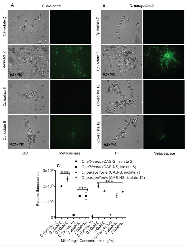
Candida biofilms play an important role in infections associated with medical devices and are resistant to antifungals. We hypothesized that the echinocandin micafungin (MICA) exerts an enhanced antifungal activity against caspofungin (CAS)-susceptible (CAS-S) and CAS-non-susceptible (CAS-NS) Candida albicans and Candida parapsilosis which is at least in part through apoptosis, even in the biofilm environment. Apoptosis was characterized by detecting reactive oxygen species (ROS) accumulation, depolarization of mitochondrial membrane potential (MMP), DNA fragmentation, lack of plasma membrane integrity, and metacaspase activation following exposure of Candida biofilm to MICA for 3h at 37°C in RPMI 1640 medium. The minimum inhibitory concentration was higher for CAS (2.0-16.0 μg/mL) than for MICA (1.0-8.0 μg/mL) for Candida biofilms. Elevated intracellular ROS levels and depolarization of MMP was evident in CAS-S C. albicans (3.0-4.2 fold) and C. parapsilosis (4.8-5.4 fold) biofilms compared with CAS-NS (1.2 fold) after exposure to MICA (0.25x-1xMIC). Elevated intracellular ROS levels and depolarization of MMP was evident in CAS-S C. albicans (3.0-4.2 fold) and C. parapsilosis (4.8-5.4 fold) biofilms compared with CAS-NS (1.2 fold) after exposure to MICA (0.25x-1xMIC). Finally higher ß-1, 3 glucan levels were seen in sessile cells compared to planktonic cells, especially in CAS-NS strains. MICA treatment might induce a metacaspase-dependent apoptotic process in biofilms of both CAS-S C. albicans and C. parapsilosis, and to some degree in CAS-NS strains.

Keywords: Apoptosis; biofilms; caspofungin; micafungin; reactive oxygen species.


Figures

Figure 1.
Figure 1.
Fungicidal action of MICA against C. albicans (A–C) and C. parapsilosis (D–F) biofilms as shown by the morbidity stain DiBAC. (A) Fluorescence images of C. albicans stained with DiBAC. Relative fluorescence of DiBAC staining of CAS-S C. albicans (isolate 2) (B) and CAS-NS C. albicans (isolate 6) (C) treated with MICA. (D) Fluorescence images of C. parapsilosis stained with DiBAC. Relative fluorescence of DiBAC staining of CAS-S C. parapsilosis (isolate 7) (E) and CAS-NS C. parapsilosis (isolate 12) (F) treated with MICA. DIC: differential interference contrast; Ca: C. albicans; Cp: C. parapsilosis; C: untreated control. *P < 0.05; **P < 0.001; ***P < 0.0001 (compared with untreated control). The experiments were performed in triplicate and repeated 3 times.
Figure 2.
Figure 2.
Extracellular β-1,3 glucan concentrations from C. albicans (A) and C. parapsilosis (B) planktonic and sessile cells. Each bar represents the mean and standard deviation from 2 replicates. *P < 0.05; **P < 0.001; ***P < 0.0001 (compared with untreated control).
Figure 3.
Figure 3.
Intracellular ROS accumulation detected using fluorescence microscopy and fluorescence spectrophotometry in C. albicans (CAS-S isolate 2 and CAS-NS isolate 6) and C. parapsilosis (CAS-S isolate 7 and CAS-NS isolate 12) cells treated with MICA. Fluorescence images and relative fluorescence of CAS-S and CAS-NS strains of C. albicans (A, B) stained with DHR-123. Fluorescence images and relative fluorescence of CAS-S and CAS-NS strains of C. parapsilosis (C, D) stained with DHR-123. DIC: differential interference contrast; Ca: C. albicans; Cp: C. parapsilosis; C: untreated control. *P < 0.05; ***P < 0.0001 (compared with untreated controls).
Figure 4.
Figure 4.
Measurement of depolarization of mitochondrial membrane potential by Rh-123 in C. albicans and C. parapsilosis sessile cells treated with MICA. Fluorescence images and relative fluorescence of CAS-S and CAS-NS strains of C. albicans (A, B) stained with Rh-123. Fluorescence images and relative fluorescence of CAS-S and CAS-NS strains of C. parapsilosis (C, D) stained with Rh -123. DIC: differential interference contrast; Ca: C. albicans; Cp: C. parapsilosis; C: untreated control. *P < 0.05; **P < 0.001; ***P < 0.0001 (compared with untreated control).
Figure 5.
Figure 5.
Detection of DNA fragmentation by TUNEL assay in CAS-S and CAS-NS C. albicans (isolates 2 and 6, respectively) and C. parapsilosis (isolates 7 and 12, respectively) sessile cells treated with MICA. Fluorescence images and relative fluorescence of CAS-S and CAS-NS strains of C. albicans (A, B) as detected by TUNEL assay. Fluorescence images and relative fluorescence of CAS-S and CAS-NS strains of C. parapsilosis (C, D) as detected by TUNEL. DIC: differential interference contrast; Ca: C. albicans; Cp: C. parapsilosis; C: untreated control. *P < 0.05; ***P < 0.0001 (compared with untreated control).
Figure 6.
Figure 6.
Caspase-like activity in CAS-S and CAS-NS C. albicans and C. parapsilosis strains treated with MICA. Fluorescence images of CAS-S and CAS-NS strains of C. albicans (A) and C. parapsilosis (B) treated with MICA and stained with CaspACE FITC-VAD-FMK. (C) Relative fluorescence of CAS-S and CAS-NS C. albicans and C. parapsilosis sessile cells stained with CaspACE FITC-VAD-FMK. DIC: differential interference contrast; Ca: C. albicans; Cp: C. parapsilosis; C: untreated control. ***P < 0.0001 (compared with untreated control).

References

    1. Pfaller MA, Diekema DJ. Epidemiology of invasive candidiasis: a persistent public health problem. Clin Microbiol Rev 2007; 20:133-63; PMID:17223626; http://dx.doi.org/10.1128/CMR.00029-06 - DOI - PMC - PubMed
    1. Pappas PG, Kauffman CA, Andes D, Benjamin DK, Jr, Calandra TF, Edwards JE, Jr, Filler SG, Fisher JF, Kullberg BJ, Ostrosky-Zeichner L, et al. . Clinical practice guidelines for the management of candidiasis: 2009 update by the Infectious Diseases Society of America. Clin Infect Dis 2009; 48:503-35; PMID:19191635; http://dx.doi.org/10.1086/596757 - DOI - PMC - PubMed
    1. Kojic EM, Darouiche RO. Candida infections of medical devices. Clin Microbiol Rev 2004; 17:255-67; PMID:15084500; http://dx.doi.org/10.1128/CMR.17.2.255-267.2004 - DOI - PMC - PubMed
    1. Douglas LJ. Candida biofilms and their role in infection. Trends Microbiol 2003; 11:30-36; PMID:12526852; http://dx.doi.org/10.1016/S0966-842X(02)00002-1 - DOI - PubMed
    1. ten Cate JM, Klis FM, Pereira-Cenci T, Crielaard W, de Groot PW. Molecular and cellular mechanisms that lead to Candida biofilm formation. J Dent Res 2009; 88:105-15; PMID:19278980; http://dx.doi.org/10.1177/0022034508329273 - DOI - PubMed

Publication types

MeSH terms

LinkOut - more resources