. 2015 Jun 11;10(6):e0130126.
doi: 10.1371/journal.pone.0130126. eCollection 2015.

B7-H3 Promotes Pathogenesis of Autoimmune Disease and Inflammation by Regulating the Activity of Different T Cell Subsets

Affiliations

B7-H3 Promotes Pathogenesis of Autoimmune Disease and Inflammation by Regulating the Activity of Different T Cell Subsets

Liqun Luo et al. PLoS One. .

Abstract

B7-H3 is a cell surface molecule in the immunoglobulin superfamily that is frequently upregulated in response to autoantigens and pathogens during host T cell immune responses. However, B7-H3's role in the differential regulation of T cell subsets remains largely unknown. Therefore, we constructed a new B7-H3 deficient mouse strain (B7-H3 KO) and evaluated the functions of B7-H3 in the regulation of Th1, Th2, and Th17 subsets in experimental autoimmune encephalomyelitis (EAE), experimental asthma, and collagen-induced arthritis (CIA); these mouse models were used to predict human immune responses in multiple sclerosis, asthma, and rheumatoid arthritis, respectively. Here, we demonstrate that B7-H3 KO mice have significantly less inflammation, decreased pathogenesis, and limited disease progression in both EAE and CIA mouse models when compared with littermates; these results were accompanied by a decrease in IFN-γ and IL-17 production. In sharp contrast, B7-H3 KO mice developed severe ovalbumin (OVA)-induced asthma with characteristic infiltrations of eosinophils in the lung, increased IL-5 and IL-13 in lavage fluid, and elevated IgE anti-OVA antibodies in the blood. Our results suggest B7-H3 has a costimulatory function on Th1/Th17 but a coinhibitory function on Th2 responses. Our studies reveal that B7-H3 could affect different T cell subsets which have important implications for regulating pathogenesis and disease progression in human autoimmune disease.


Conflict of interest statement

Competing Interests: The authors have declared that no competing interests exist.

Figures

Fig 1
Fig 1. Generation and characterization of B7-H3 KO mice.
(A) Mapping of the B7-H3 genomic locus, targeting vector, and the replaced allele. (B) Southern blot analysis of SpeI-digested DNA from targeted embryonic stem (ES) cell clones. The wild-type allele generated a 9.6-kb fragment, and the targeted allele yielded an 8.0-kb fragment. (C) PCR identification of genomic DNA isolated from tails of wild type (+/+), heterozygous (+/−) or homozygous (−/−) B7-H3 mutant mice. The wild type allele generated an 894-bp fragment and the targeted allele generated a 632-bp fragment. (D) Examination of B7-H3 mRNA by RT-PCR. The cDNA was generated from the spleens of wild type (+/+), heterozygous (+/−) or homozygous (−/−) B7-H3 mutant mice using primers specific for B7-H3 or β-actin (control).
Fig 2
Fig 2. B7-H3 KO mice are resistant to EAE.
EAE incidence and severity in wild type and B7-H3 KO mice. Data are presented as an average clinical score from four independent experiments (5–10 mice each group). There were significant differences between two groups starting on day 16. Data are shown as mean ± SEM,*p<0.05, **p<0.01, ***p<0.001, versus control.
Fig 3
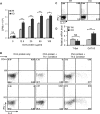
Fig 3. Decreased MOG-specific T cell responses in B7-H3 KO mice.
The brachial and axillary draining-lymph nodes (DLN) were removed from wild type and B7-H3 KO mice 10 days after MOG/CFA immunization. (A) Comparing DLN size and number. Representative image of wt and B7-H3 KO mice DLNs (top), which taken from one mouse per group. DLNs number counting is from five mice per group (bottom). Data are representative of three independent experiments. (B) Proliferative response of DLN cells to MOG peptide, as determined by incorporation of 3H-TdR. DLNs were collected from five mice per group. Data are representative of four independent experiments with similar results. (C) Cytokine IL-2, IL-12, IL-17, and IFN-γ concentrations in cultured supernatants of DLN cells were measured by ELISA 2 days after restimulation with MOG peptide (40 μg/ml). Data are representative of three independent experiments with similar results. (D) DLN cells (mixed 5 individual cells per group) were cultured with MOG peptide (40 μg/ml) for 5 days under Th1 or Th2 polarizing conditions. Then, Th1 and Th2 cell subpopulations were analyzed by intracellular cytokine staining. Data are representative of 3 independent experiments with similar results. (E) Real-time PCR analysis of T-bet and GATA3 were performed on cDNA from wt and B7-H3 KO mice DLN cells, 5 individual cells per group in triplicates. Data are representative of 2 independent experiments. Data are shown as mean ± SEM, *p<0.05, **p<0.01, ***p<0.001, versus control.
Fig 4
Fig 4. Enhanced airway allergic responses in B7-H3 KO mice.
(A) Total and eosinophil cell counts in BALF from OVA protein-immunized and challenged wild type or B7-H3 KO mice. Data were from 5 individual mice per group each experiment and data are representative of four independent experiments. Eos, eosinophils; Macs, macrophage; Lymphs, lymphocytes; PMNs, polymorphonuclear neutrophils. (B) Characterization of inflammatory cells in BALF. The cells were spun to slides, and then stained with HEMA3 (Original magnification: ×60). (C) Lung paraffin sections of naïve, allergic wild type, and B7-H3 KO mice were stained with H & E and PAS (Original magnification: ×20). Peribronchial and perivas cular inflammation and mucus-secreting cells in the airways were scored (right plots). (D) Th2 cytokine levels in BALF of wild type or B7-H3 KO mice assessed by ELISA. (E) Levels of serum OVA-specific IgE were determined by ELISA in wild type and B7-H3 KO mice after OVA protein challenge. Data are shown as mean ± SEM, *p<0.05, **p<0.01, ***p<0.001, versus control.
Fig 5
Fig 5. Predominant Th2 response in B7-H3 KO mice.
Spleens were removed from OVA protein-immunized wild type and B7-H3 KO mice on day 12. (A) Splenocytes were restimulated with varying concentrations OVA protein. Cell proliferation was determined by 3H-TdR incorporation. 5 mice per group and data are representative of 3 independent experiments with similar results. (B) Splenocytes (mixed 5 individual cells per group) were cultured with OVA protein (50 μg/ml) for 5 days under Th-1 or Th-2 polarizing conditions. Th1 and Th2 cell subpopulations were analyzed by intracellular cytokine staining. Data are representative of three independent experiments with similar results. (C) Splenocytes from wt or B7-H3 KO mice were restimulated with PMA (5ng/ml) + ionomycin (250 ng/ml) and GolgiPlug (1 μl/ml) in vitro for 5 hrs. IL-17 producing cells were analyzed by flow cytometry. 5 mice per group and data are representative of 2 independent experiments. (D) Real-time PCR analysis of T-bet and GATA3 were performed on the cDNA from 5 individual splenocytes per group in triplicates. Data are representative of 2 independent experiments. Data are shown as mean ± SEM, *p<0.05, **p<0.01, ***p<0.001, versus control.
Fig 6
Fig 6. B7-H3 KO mice are resistant to CIA.
(A) The arthritis score in CIA mice. 5–10 mice per group per experiment and data shown are representative of 3 independent experiments with similar results. (B) Representative images of hind paws from wild type (left) and B7-H3 KO (right) CIA mice on 40 days after primary immunization. (C) Representative hematoxylin and eosin—stained sections of toe and talus joints (original magnification ×4 and ×10, respectively) from wild type and B7-H3 KO CIA mice. (D) Serum anti-type II collagen Ab (IgG) levels (100 units is approximately 0.1 mg IgG antibody/ml). Data shown are representative of 3 independent experiments. (E) Staining for B7-H3 in normal and arthritic joints. Hind toe joints from normal, CIA-wild type, and B7-H3 KO mice (Original magnification ×10). (F) Splenocytes from wild type and B7-H3 KO CIA mice were restimulated with chicken CII protein in various concentrations. 5 mice per group each experiment. Cell proliferation was determined by 3H-TdR incorporation. Data shown are representative of 3 independent experiments. (G) Th1/Th2/Th17 Cytokine concentrations of cultured supernatants from chicken CII protein (20 μg/ml)-restimulated splenocytes (mixed 5 individual cells per group) after 1 and 2 days were assessed using mouse Th1/Th2/Th17 Cytokine kits (CBA); data were analyzed using FCAP Array V2.0. Data shown are representative of two independent experiments. Data are shown as mean ± SEM, *p<0.05, **p<0.01, ***p<0.001, versus control.

References

    1. Zhu Y, Yao S, Chen L. Cell surface signaling molecules in the control of immune responses: a tide model. Immunity. 2011. April 22;34(4):466–478. 10.1016/j.immuni.2011.04.008 - DOI - PMC - PubMed
    1. Chen L, Flies DB. Molecular mechanisms of T cell co-stimulation and co-inhibition. Nat Rev Immunol. 2013. April;13(4):227–242. 10.1038/nri3405 - DOI - PMC - PubMed
    1. Chapoval AI, Ni J, Lau JS, Wilcox RA, Flies DB, Liu D, et al. B7-H3: a costimulatory molecule for T cell activation and IFN-gamma production. Nat Immunol. 2001. March;2(3):269–274. - PubMed
    1. Hashiguchi M, Kobori H, Ritprajak P, Kamimura Y, Kozono H, Azuma M. Triggering receptor expressed on myeloid cell-like transcript 2 (TLT-2) is a counter-receptor for B7-H3 and enhances T cell responses. Proc Natl Acad Sci U S A. 2008. July 29;105(30):10495–10500. 10.1073/pnas.0802423105 - DOI - PMC - PubMed
    1. Leitner J, Klauser C, Pickl WF, Stockl J, Majdic O, Bardet AF, et al. B7-H3 is a potent inhibitor of human T-cell activation: No evidence for B7-H3 and TREML2 interaction. Eur J Immunol. 2009. July;39(7):1754–1764. 10.1002/eji.200839028 - DOI - PMC - PubMed

Publication types

MeSH terms