Review
. 2015 Jun 11;10(3):034006.
doi: 10.1088/1748-6041/10/3/034006.

From cardiac tissue engineering to heart-on-a-chip: beating challenges

Affiliations
Review

From cardiac tissue engineering to heart-on-a-chip: beating challenges

Yu Shrike Zhang et al. Biomed Mater. .

Abstract

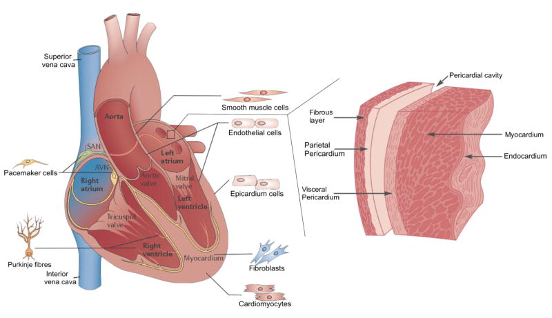
The heart is one of the most vital organs in the human body, which actively pumps the blood through the vascular network to supply nutrients to as well as to extract wastes from all other organs, maintaining the homeostasis of the biological system. Over the past few decades, tremendous efforts have been exerted in engineering functional cardiac tissues for heart regeneration via biomimetic approaches. More recently, progress has been made toward the transformation of knowledge obtained from cardiac tissue engineering to building physiologically relevant microfluidic human heart models (i.e. heart-on-chips) for applications in drug discovery. The advancement in stem cell technologies further provides the opportunity to create personalized in vitro models from cells derived from patients. Here, starting from heart biology, we review recent advances in engineering cardiac tissues and heart-on-a-chip platforms for their use in heart regeneration and cardiotoxic/cardiotherapeutic drug screening, and then briefly conclude with characterization techniques and personalization potential of the cardiac models.


Figures

Figure 1
Figure 1
Schematics showing mature heart cell populations with geographical anatomy, and general histological representation of the heart wall. Adapted with permission from Ashley et al., 2004 and Xin et al., 2013 [29, 41].
Figure 2
Figure 2
(A) (i) Fabrication of multi-layered cardiac cell sheets; (ii) Fluorescence image on the right shows Troponin T staining of the cardiac muscle in a construct containing 6 layers of cardiac cell sheets. Green: Troponin T; blue, nuclei. Scale bar: 20 μm. Adapted with permission from Masuda et al., 2008 and Sakaguchi et al., 2013 [44, 93]; (B) Fabrication of PEG microwells and formation of EBs of uniform sizes. Cardiogenesis was maximized in EBs of 450 μm in diameter. Adapted with permission from Karp et al., 2007 and Hwang et al., 2009 [48, 52]; (C) (i) Schematic diagram showing the fabrication of micropatterned hydrogels using a micromolding technique for cardiomyocytes alignment. (ii) Bright-field images of micropatterned MeTro with 20 × 20 μm (width × spacing) channels; (iii) Fluorescence images of aligned cardiomyocytes cultured on the micropatterned MeTro; scale bars: 200 μm. Adapted with permission from Annabi et al., 2013 [73]; (D) (i) The biowire approach where a surgical suture was used to induce compaction and alignment of cardiomyocytes in the surrounding hydrogel. (ii, iii) Cultured biowires under electrical stimulation improved the phenotype of cardiomyocytes. Adapted with permission from Nunes et al., 2013 [74]; (E) (i) Preparation of scaffolds with suspended electrospun nanofibers. (ii) A superimposed confocal image of (iii–v) showing cardiomycytes on aligned nanofibers where the nanofibers were stained red, f-actin green, and nuclei blue; (vi) image showing parallel alignment of sarcomeres, where nanofibers were stained bright pink and α-actin stained red. Scale bars: (ii) 100 μm, (iii) 50 μm, (iv) 50 μm, (v) 100 μm, and (vi) 10 μm. Adapted with permission from Orlova et al., 2011 [75]; (F) (i) Scaffolds with an accordion-like honeycomb structure resulted in anisotropic mechanical properties possessed by the native myocardium. (ii) Fluorescence image showing alignment of cardiomyocytes cultured on an accordion-like honeycomb scaffold. Green indicates F-actin. Scale bars: (i) 1 mm, and (ii) 200 μm. Adapted with permission from Engelmayr et al., 2008[76].
Figure 3
Figure 3
Schematics illustrating methods of conducting electrical stimulation and enhancing the electrical properties of the matrices for cardiomyocytes.
Figure 4
Figure 4
Heart-on-a-chip models. (A) Schematics illustrating the fabrication process of contracting heart stripes; (B) Time-lapse bright-field images showing contracting myocardium stripes. Scale bar: 5 mm. Adapted with permimssion from Grosberg et al., 2011 [20]; (C) Schematic cross-sectional view of a heart-on-a-chip pump made of a PDMS elastomer hollow sphere covered with a sheet of pulsating cardiomyocytes. The contraction of the cell monolayer squeezes the sphere and pumps the fluid through the connected microchannel. (D) Displacement over time of a particle caused by the pulsation. The light grey and black plots indicate the movement of the particle before and after transplantation of the cardiomyocytes monolayer, respectively. Adapted with permission from Tanaka et al., 2007 [147]; (E) Schematic showing the heart-in-a-channel model, where the inner surface of a microfluidic channel was coated with MeTro and seeded with a monolayer of beating cardiomyocytes; (F) Confocal images showing cardiomyocytes stained with troponin I (red) and nuclei (blue) in the left panel, and α-actinin (green), connexin-43 (red), and nuclei (blue) in the right panel. Scale bar: 50 μm. Adapted with permission from Annabi et al., 2013 [150].
Figure 5
Figure 5
(A) Experimental setup showing on-chip monitoring of cardiomyocytes beating using a webcam-based lens-less microscope; (B) Relative beating rate of cardiomyocytes treated with doxorubicin (1, 5, 10, 100 nM); (C) Representative pattern of the beating signal recorded from cardiomyocytes showing a clear increase in the beating frequency following isoprenaline (10 nM) treatment; (D) Relative beating rate of cardiomyocytes treated with doxorubicin (10, 100, 200, and 300 μM); (E) Representative pattern of the beating signal recorded from cardiomyocytes showing a clear decrease in the beating frequency following doxorubicin (100 μM) treatment. Adapted with permision from Kim et al., 2011 [106].

References

    1. van Berlo JH, Molkentin JD. Nature medicine. 2014;20:1386–1393. - PMC - PubMed
    1. Chiu LL, Iyer RK, Reis LA, Nunes SS, Radisic M. Frontiers in bioscience (Landmark edition) 2011;17:1533–1550. - PubMed
    1. Radisic M, Christman KL. Mayo Clinic Proceedings. Vol. 88. Elsevier; 2013. Materials science and tissue engineering: repairing the heart; pp. 884–898. - PMC - PubMed
    1. Chiu L, Radisic M. Current Opinion in Chemical Engineering. 2013;2:41–52.
    1. Zhang B, Xiao Y, Hsieh A, Thavandiran N, Radisic M. Nanotechnology. 2011;22:494003. - PubMed

Publication types