. 2015 Jun 12:6:7331.
doi: 10.1038/ncomms8331.

Molecular snapshots of the Pex1/6 AAA+ complex in action

Affiliations

Molecular snapshots of the Pex1/6 AAA+ complex in action

Susanne Ciniawsky et al. Nat Commun. .

Abstract

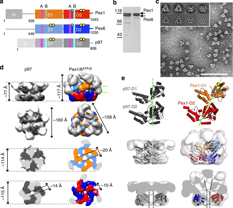
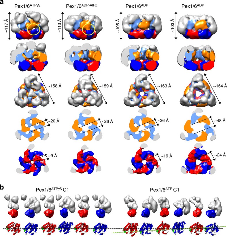
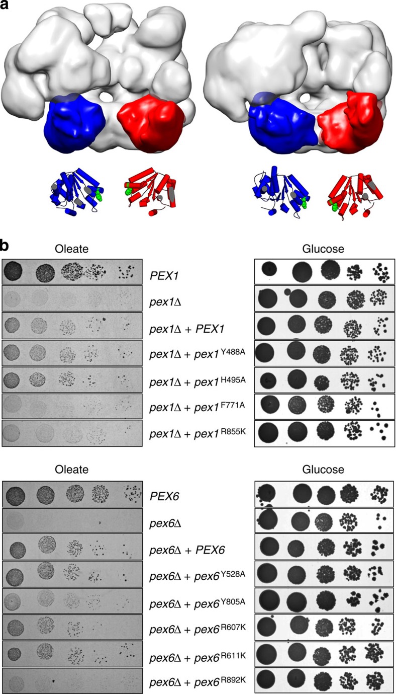
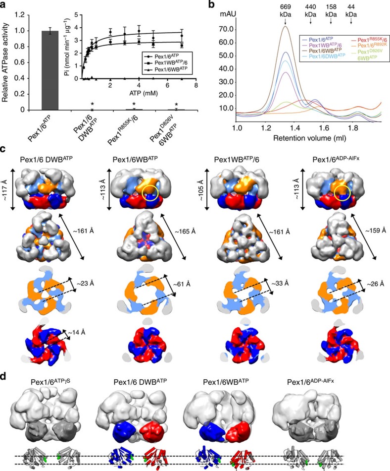
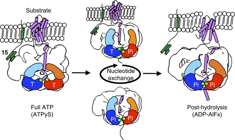
The peroxisomal proteins Pex1 and Pex6 form a heterohexameric type II AAA+ ATPase complex, which fuels essential protein transport across peroxisomal membranes. Mutations in either ATPase in humans can lead to severe peroxisomal disorders and early death. We present an extensive structural and biochemical analysis of the yeast Pex1/6 complex. The heterohexamer forms a trimer of Pex1/6 dimers with a triangular geometry that is atypical for AAA+ complexes. While the C-terminal nucleotide-binding domains (D2) of Pex6 constitute the main ATPase activity of the complex, both D2 harbour essential substrate-binding motifs. ATP hydrolysis results in a pumping motion of the complex, suggesting that Pex1/6 function involves substrate translocation through its central channel. Mutation of the Walker B motif in one D2 domain leads to ATP hydrolysis in the neighbouring domain, giving structural insights into inter-domain communication of these unique heterohexameric AAA+ assemblies.


Figures

Figure 1
Figure 1. Pex1/6 hexamers are trimers of dimers.
(a) Schematic domain representation of Pex1/Pex6 protomers compared with p97 (N domain, D1/D2 domain). Conserved motifs and residues of each AAA+ domain are indicated: Walker A (A, magenta bars), Walker B (B, turquoise bars), substrate-binding loops (green dots) and arginine finger residues (yellow dots). Non-canonical Walker A and B motifs are indicated as dotted lines. (b) Coomassie-stained SDS–polyacrylamide gel electrophoresis of purified Pex1/6ATP (5 μg, lane 1) or Pex1/6 DWBATP (5 μg, lane 2) overexpressed in E. coli or Saccharomyces cerevisiae. (c) Raw negative stain electron micrograph showing Pex1/6 complexes (40 μg ml−1) incubated with ATPγS. Representative class averages derived from multivariate statistical analysis show top and side views of the Pex1/6ATPγS complex (inset, upper row) and corresponding reprojections of the final 3D reconstruction in the Euler angle directions assigned to the class averages (lower row). Each class contains an average of 5–10 images. Scale bar, 100 nm. (d) Pex1/6ATPγS EM density map as side, top and cross-section views of D1 and D2 rings. Colour code: Pex1 D1 (orange), Pex1 D2 (red), Pex6 D1 (pale blue), Pex6 D2 (blue) and Pex1/6N domains (grey). Equivalent views of p97 (pdb-ID: 3CF3) filtered to 20 Å are shown for comparison. p97 single subunits are coloured alternately light and dark grey. Cross-section viewing planes are indicated by green lines. (e) Cartoon representation of a p97 protomer without N domains and of a Pex1 protomer homology model, seen from the side of the complex. Domain offset between Pex1 D1 and Pex1 D2 is indicated by green dotted lines. Walker A and Walker B motifs are shown as spheres and coloured as in a (upper row). Side view of a p97 dimer fitted as a rigid body into low-pass filtered p97 crystal structure and Pex1/6 heterodimer docked to Pex1/6ATPγS 3D map (middle row). Cut-open side views of the low-pass filtered p97 crystal structure with p97 D2 placed into the EM density map and of Pex1/6ATPγS map with fitted Pex1 D2 and Pex6 D2 homology models. Black dotted lines indicate the central channel (lower row).
Figure 2
Figure 2. Symmetric and asymmetric wild-type Pex1/6 complexes in the presence of different nucleotides.
(a) EM reconstructions of Pex1/6 complexes in the presence of ATPγS (Pex1/6ATPγS), ADP-AlFx (Pex1/6ADP-AlFx), ATP (Pex1/6ATP) and ADP (Pex1/6ADP) seen as side views (upper row), followed by cut-open side views, top views and cross-sections of the D1 and D2 layers (lower rows). Domain colours correspond to Fig. 1d. (b) Surface representation of each protomer of the asymmetric Pex1/6ATPγS and Pex1/6ATP complex. Pex1 (red) or Pex6 (blue) D2 domains are highlighted. Underneath, a cartoon representation based on rigid body fits of homology models into negative stain EM maps is shown. Pex1F771 and Pex6Y805 are shown as green spheres. The black dotted line indicates the position of pore-facing loops in the D2 domains of asymmetric Pex1/6ATPγS. A green dotted line indicates the asymmetric arrangement of D2 domains in Pex1/6ATP.
Figure 3
Figure 3. ATP hydrolysis translocates tyrosine loops through movements of D2 domains.
(a) Side-view surface representation of Pex1/6ATPγS and Pex1/6ADP-AlFx. One heterodimer is omitted from the hexamer. One Pex1 D2 and Pex6 D2 domain of each complex is highlighted in red and blue, respectively. Underneath, a cartoon representation based on rigid body fits of homology models into negative stain EM maps is shown. Conserved aromatic residues Pex1F771 and Pex6Y805 are shown as green spheres. (b) Growth of strains expressing either wild-type (PEX1, PEX6), no (pex1Δ, pex6Δ) or mutated pex1, pex6 alleles with a modified pore loop (pex1Y488A, pex1H495A, pex1F771A, pex6Y528A, pex6Y805A) or arginine finger residues (pex1R855K, pex6R607K, pex6R611K, pex6R892K) on either glucose or oleate as a single carbon source.
Figure 4
Figure 4. ATPase activity of Pex6 D2 domains drive conformational changes.
(a) ATPase activities of wild-type Pex1/6 (Pex1/6ATP), single Walker B (Pex1WBATP/6, Pex1/6WBATP) or double-Walker B mutants (Pex1/6 DWBATP) or Pex1 arginine finger (Pex1R855K/6) or ISS motif (Pex1D826V/6WBATP) mutants. Assays were performed in the presence of 1 mM ATP and 5 mM Mg2+ at 37 °C. Error bars represent s.d. of two independent experiments with five technical replicates each. *P<0.0001 in comparison with wild-type Pex1/6ATP (one-way ANOVA). Michaelis–Menten plot of enzyme activity of Pex1/6ATP, Pex1WBATP/6 or Pex1/6WBATP at various ATP concentrations (inset). Error bars represent s.d. of two independent experiments including three technical replicates. (b) Size-exclusion chromatography A290 nm profiles of purified Pex1/6 complexes used for ATPase activity assays. (c) EM reconstructions of double- (Pex1/6 DWBATP) or single- (Pex1/6WBATP, Pex1WBATP/6) Walker B complexes in the presence of ATP as side views (upper row), followed by top views and cross-sections of the D1 and D2 rings (lower rows). (d) Side-view surface representation as in Fig. 3a. Underneath, a cartoon representation based on rigid body fits of homology models into negative stain EM maps is shown. Pex1/6ATPγS and Pex1/6ADP-AlFx are depicted in grey for comparison. Domains are coloured according to Fig. 1d. Green spheres depict residues Pex1F771 and Pex6Y805.
Figure 5
Figure 5. Model for Pex1/6 movements during ATP binding and hydrolysis.
The Pex1/6 complex anchors to the peroxisomal membrane via binding of Pex6 N domains to Pex15. Pex1 N domains establish interactions with the substrate. ATP binding to Pex1 D2 and Pex6 D2 (full ATP, ATPγS) elevates substrate-binding loops in the D2 domain, ready to grab the substrate. ATP turnover creates a power stroke that pulls the substrate along the central pore (post hydrolysis, ADP-AlFx). Nucleotide exchange in Pex6 D2 or Pex1 D2 translocates the substrate along the central pore (Pex1/6WBATP, Pex1WBATP/6). One Pex1 and Pex6 protomer are denoted as a simple cartoon representation. Conserved aromatic residues of substrate-binding loops are shown as green dots. Representative tertiary structures of substrate protein (purple) and membrane anchor Pex15 (green) are depicted as cartoon representations. Nucleotide occupancy of each D2 domain is indicated by T for ATP or Pi for the transition state.

References

    1. van den Bosch H., Schutgens R. B., Wanders R. J. & Tager J. M. Biochemistry of peroxisomes. Annu. Rev. Biochem. 61, 157–197 (1992). - PubMed
    1. Voorn-Brouwer T., van der Leij I., Hemrika W., Distel B. & Tabak H. F. Sequence of the PAS8 gene, the product of which is essential for biogenesis of peroxisomes in Saccharomyces cerevisiae. Biochim. Biophys. Act 1216, 325–328 (1993). - PubMed
    1. Erdmann R., Veenhuis M., Mertens D. & Kunau W. H. Isolation of peroxisome-deficient mutants of Saccharomyces cerevisiae. Proc. Natl. Acad. Sci. USA 86, 5419–5423 (1989). - PMC - PubMed
    1. Platta H. W., Grunau S., Rosenkranz K., Girzalsky W. & Erdmann R. Functional role of the AAA peroxins in dislocation of the cycling PTS1 receptor back to the cytosol. Nat. Cell Biol. 7, 817–822 (2005). - PubMed
    1. Reuber B. E. et al.. Mutations in PEX1 are the most common cause of peroxisome biogenesis disorders. Nat. Genet. 17, 445–448 (1997). - PubMed

Publication types

MeSH terms

Substances