

Current MRI techniques for the assessment of renal disease
- PMID: 26066472
- PMCID: PMC4467464
- DOI: 10.1097/MNH.0000000000000122
Current MRI techniques for the assessment of renal disease
Abstract
Purpose of review: Over the past decade, a variety of MRI methods have been developed and applied to many kidney diseases. These MRI techniques show great promise, enabling the noninvasive assessment of renal structure, function and injury in individuals. This review will highlight the current applications of functional MRI techniques for the assessment of renal disease and discuss future directions.
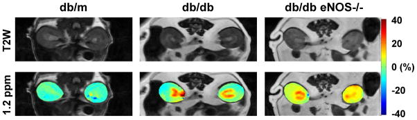
Recent findings: Many pathological (functional and structural) changes or factors in renal disease can be assessed by advanced MRI techniques. These include renal vascular structure and function (contrast-enhanced MRI, arterial spin labelling), tissue oxygenation (blood oxygen level dependent MRI), renal tissue injury and fibrosis (diffusion or magnetization transfer imaging, magnetic resonance elastography), renal metabolism (chemical exchange saturation transfer, spectroscopic imaging), nephron endowment (cationic-contrast imaging), sodium concentration (23Na-MRI) and molecular events (targeted-contrast imaging).
Summary: Current advances in MRI techniques have enabled the noninvasive investigation of renal disease. Further development, evaluation and application of the MRI techniques should facilitate better understanding and assessment of renal disease, and the development of new imaging biomarkers, enabling the intensified treatment of high-risk populations and a more rapid interrogation of novel therapeutic agents and protocols.
Conflict of interest statement
None
Figures
References
Publication types
MeSH terms
Grants and funding
LinkOut - more resources
Full Text Sources
Medical
Research Materials
Miscellaneous
