. 2015 Sep;20(9):1200-10.
doi: 10.1007/s10495-015-1144-y.

Critical role of the α1-Na(+), K(+)-ATPase subunit in insensitivity of rodent cells to cytotoxic action of ouabain

Affiliations

Critical role of the α1-Na(+), K(+)-ATPase subunit in insensitivity of rodent cells to cytotoxic action of ouabain

Olga A Akimova et al. Apoptosis. 2015 Sep.

Abstract

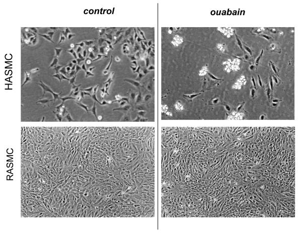
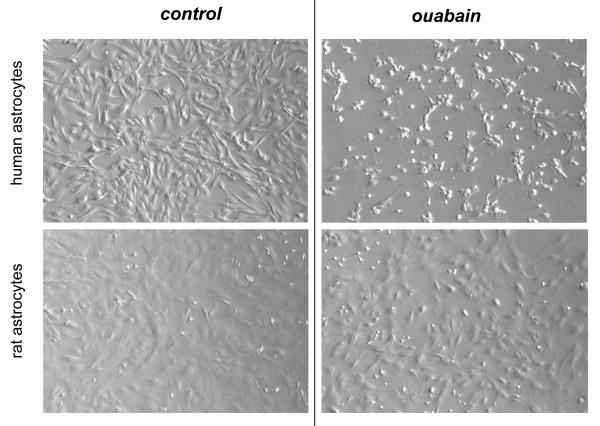
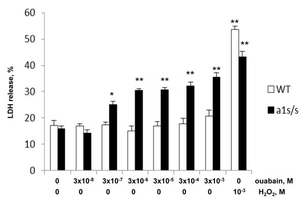
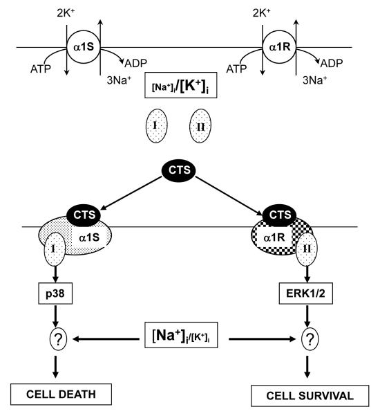
In rodents, ubiquitous α1-Na(+), K(+)-ATPase is inhibited by ouabain and other cardiotonic steroids (CTS) at ~10(3)-fold higher concentrations than those effective in other mammals. To examine the specific roles of the CTS-sensitive α1S- and CTS-resistant α1R-Na(+), K(+)-ATPase isoforms, we compared the effects of ouabain on intracellular Na(+) and K(+) content, cell survival, and mitogen-activated protein kinases (MAPK) in human and rat vascular smooth muscle cells (HASMC and RASMC), human and rat endothelial cells (HUVEC and RAEC), and human and rat brain astrocytes. 6-h exposure of HASMC and HUVEC to 3 μM ouabain dramatically increased the intracellular [Na(+)]/[K(+)] ratio to the same extend as in RASMC and RAEC treated with 3000 μM ouabain. In 24, 3 μM ouabain triggered the death of all types of human cells used in this study. Unlike human cells, we did not detect any effect of 3000-5000 μM ouabain on the survival of rat cells, or smooth muscle cells from mouse aorta (MASMC). Unlike in the wild-type α1(R/R) mouse, ouabain triggered death of MASMC from α1(S/S) mouse expressing human α1-Na(+), K(+)-ATPase. Furthermore, transfection of HUVEC with rat α1R-Na(+), K(+)-ATPase protected them from the ouabain-induced death. In HUVEC, ouabain led to phosphorylation of p38 MAPK, whereas in RAEC it stimulated phosphorylation of ERK1/2. Overall, our results, demonstrate that the drastic differences in cytotoxic action of ouabain on human and rodent cells are caused by unique features of α1S/α1R-Na(+), K(+)-ATPase, rather than by any downstream CTS-sensitive/resistant components of the cell death machinery.


Figures

Fig. 1
Fig. 1
Phase-contrast microscopy of smooth muscle cells from human (HASMC) and rat (RASMC) aorta exposed to ouabain. HASMC and RASMC were incubated for 24 h in DMEM containing 3 and 3,000 μM ouabain, respectively. These representative images were captured using a Nikon phase-contrast microscope with 100× magnification.
Fig. 2
Fig. 2
Phase-contrast microscopy demonstrating the effect of ouabain on endothelial cells isolated from human umbilical vein (HUVEC) and rat aorta (RAEC). HUVEC and RAEC were incubated for 24 h in EMB-2 and DMEM containing 3 and 3,000 μM ouabain, respectively. These representative images were captured using a Nikon phase-contrast microscope with 100× magnification
Fig. 3
Fig. 3
Hoffman modulation contrast microscopy images demonstrating the effects of ouabain on human and rat astrocytes. Human and rat astrocytes were incubated for 24 h in DMEM containing 10% fetal bovine serum and 20 and 5,000 μM ouabain for human and rat cells, respectively. These representative images were captured using an Olympus IX-71 setup with 100× magnification.
Fig. 4
Fig. 4
Effects of ouabain and H2O2 on LDH release from smooth muscle cells isolated from aorta of wild-type (WT) mice (open bars) and α1S/S mice, expressing human α1S-Na+,K+-ATPase (solid bars). Cells were serum-starved for 24 h and then treated for additional 24 h with drugs at concentrations indicated at the bottom. The total content of LDH was taken as 100%. Means ± S.E. from 3 experiments performed in triplicates are shown. *, ** p<0.005 and 0.001 as compared to untreated cells.
Fig. 5
Fig. 5
Dose-dependent effects of ouabain on the intracellular Na+ content (A) and LDH release (B) from mock- and α1R-transfected HUVEC. Cells were serum-starved for 24 h and then treated for additional 6 (A) or 24 (B) h with ouabain at concentrations indicated at the X-axes. The total content of LDH was taken as 100%. Treatment with 1000 mM H2O2 was used as a positive control for the cell death. Means ± S.E. from 3 experiments performed in triplicates (A) or quadruplicate (B) are shown.
Fig. 6
Fig. 6
Effects of ouabain on phosphorylation of the mitogen-activated protein kinases in HUVEC and RAEC. A, Representative Western blots showing the effects of ouabain on the content of phosphorylated ERK1/2, JNK1/2 and p38 MAPK. HUVEC and RAEC were treated during 6 hr with 3 and 3,000 μM ouabain, respectively. B, Normalized changes in the immunoreactivity of phosphorylated ERK1/2, JNK1/2 and MAPK in ouabain-treated HUVEC and RAEC. The immunoreactivity of phosphorylated MAPKs in control cells, which were not treated with ouabain, was taken as 1.00. Means ± S.E. from 3 experiments are shown.
Fig. 7
Fig. 7
Hypothetical mechanisms underlying the distinct impact of cardiotonic steroids (CTS) on survival of cells, expressing the CTS-sensitive (α1S) and the CTS-resistant (α1R) Na+,K+-ATPase subunits. In both cases, saturating levels of CTS strongly increase the [Na+]i/[K+]i ratio. In addition, CTS trigger distinct conformational changes in α1S and α1R isoforms that, in turn, affect their interactions with unknown protein partner(s) I and II. These subsequent signaling events lead to activation of p38 and ERK1/2 MAPK and result in cell death and survival, respectively. ? – steps showing possible additional effects of the transcriptomic changes, which can be directly induced by elevation of the [Na+]i/[K+]i ratio. For more details, see text.

References

    1. Therien AG, Blostien R. Mechanisms of sodium pump regulation. Am.J.Physiol. 2000;279:C541–C566. - PubMed
    1. Scheiner-Bobis G. The sodium pump. Its molecular properties and mechanisc of ion transport. Eur.J.Biochem. 2002;269:2424–2433. - PubMed
    1. Bagrov AY, Shapiro JI, Fedorova OV. Endogenous cardiotonic steroids: physiology, pharmacology, and novel therapeutic targets. Pharmacol.Rev. 2009;61:9–38. - PMC - PubMed
    1. Schoner W, Scheiner-Bobis G. Endogenous and exogenous cardiac glycosides: their role in hypertension, salt metabolism, and cell growth. Am.J.Physiol.Cell Physiol. 2007;293:C509–C536. - PubMed
    1. Lingrel JB, Argьello JM, Van Huysse JW, Kuntzweiler TA. Cation and cardiac glycoside binding sites of the Na,K-ATPase. Ann.N.Y.Acad.Sci. 1997;843:194–206. - PubMed

Publication types

LinkOut - more resources