. 2015 Jul 24;290(30):18333-42.
doi: 10.1074/jbc.M114.619494. Epub 2015 Jun 11.

A Novel Interaction between Complement Inhibitor C4b-binding Protein and Plasminogen That Enhances Plasminogen Activation

Affiliations

A Novel Interaction between Complement Inhibitor C4b-binding Protein and Plasminogen That Enhances Plasminogen Activation

Vaibhav Agarwal et al. J Biol Chem. .

Abstract

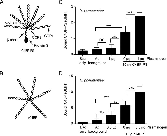
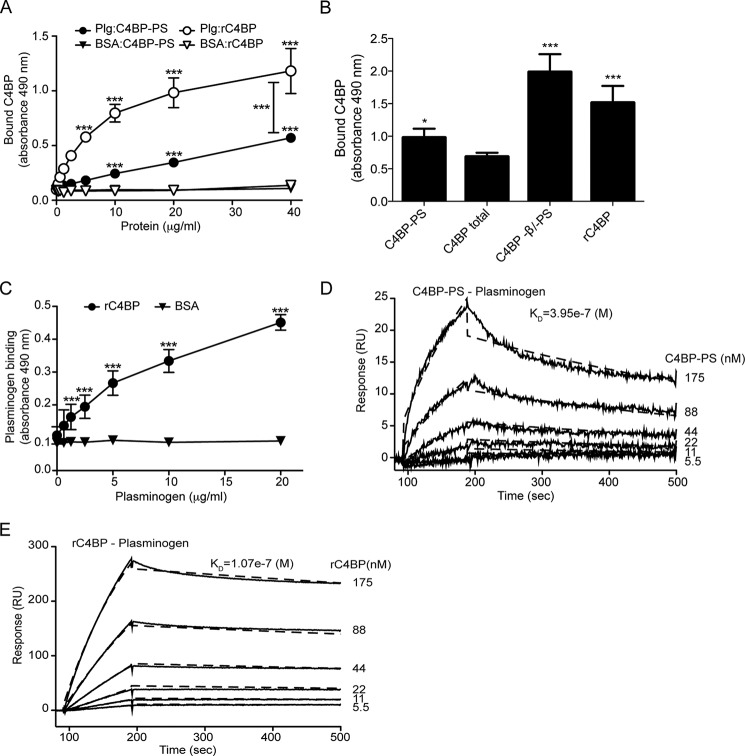
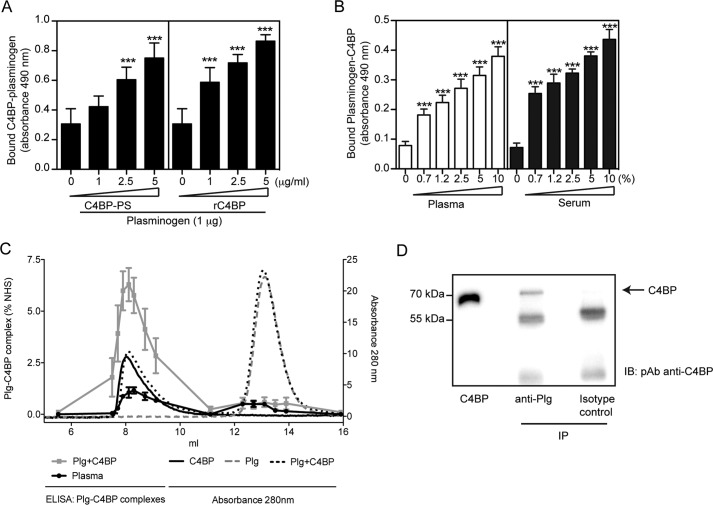
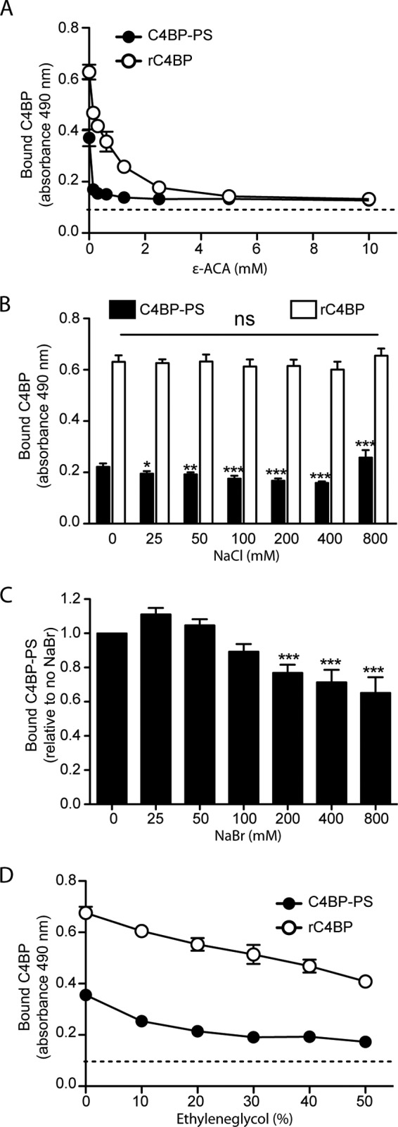
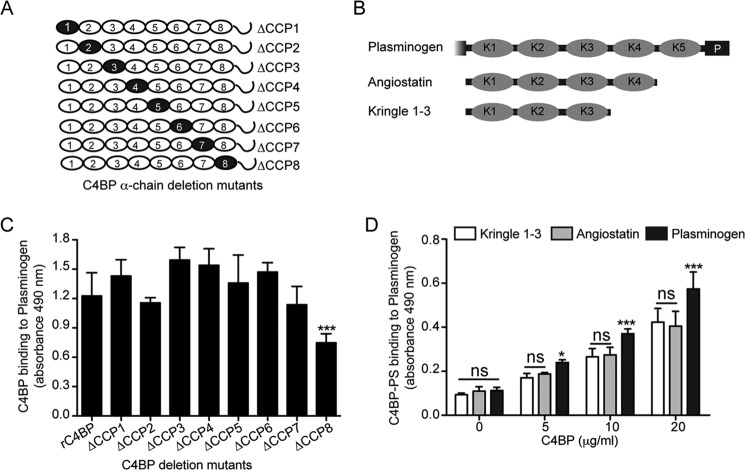
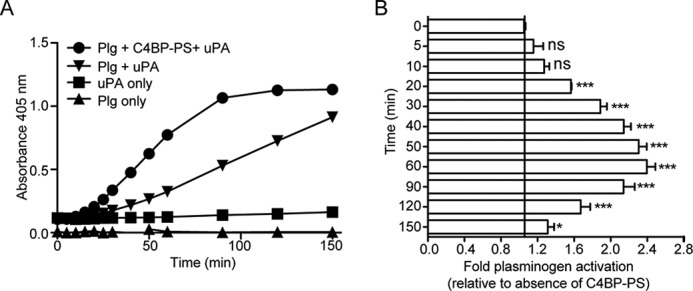
The complement, coagulation, and fibrinolytic systems are crucial for the maintenance of tissue homeostasis. To date numerous interactions and cross-talks have been identified between these cascades. In line with this, here we propose a novel, hitherto unknown interaction between the complement inhibitor C4b-binding protein (C4BP) and plasminogen of the fibrinolytic pathway. Binding of C4BP to Streptococcus pneumoniae is a known virulence mechanism of this pathogen and it was increased in the presence of plasminogen. Interestingly, the acute phase variant of C4BP lacking the β-chain and protein S binds plasminogen much stronger than the main isoform containing the β-chain and protein S. Indeed, the complement control protein (CCP) 8 domain of C4BP, which would otherwise be sterically hindered by the β-chain, primarily mediates this interaction. Moreover, the lysine-binding sites in plasminogen kringle domains facilitate the C4BP-plasminogen interaction. Furthermore, C4BP readily forms complexes with plasminogen in fluid phase and such complexes are present in human serum and plasma. Importantly, whereas the presence of plasminogen did not affect the factor I cofactor activity of C4BP, the activation of plasminogen by urokinase-type plasminogen activator to active plasmin was significantly augmented in the presence of C4BP. Taken together, our data demonstrate a novel interaction between two proteins of the complement and fibrinolytic system. Most complexes might be formed during the acute phase of inflammation and have an effect on the homeostasis at the site of injury or acute inflammation.

Keywords: C4b-binding protein; complement; plasmin; plasminogen; protein-protein interaction; surface plasmon resonance (SPR).


Figures

FIGURE 1.
FIGURE 1.
Additive binding of C4BP and plasminogen to S. pneumoniae. A and B, schematic representation of the different variants of C4BP used: C4BP-PS (A) and rC4BP (B). The main isoform of C4BP contains seven identical α-chains and one unique β-chain. The β-chain-containing C4BP in circulation is bound to vitamin K-dependent anticoagulant Protein S (PS), forming a C4BP-PS complex. The rC4BP contains six α-chains and lacks the β-chain and the associated PS. C and D, increased binding of C4BP to pneumococci in the presence of plasminogen. Binding of C4BP-PS (C) and rC4BP (D) to pneumococci was analyzed in the presence or absence of plasminogen, by flow cytometry. The geometric mean fluorescence intensity (GMFI) and S.D. calculated from three independent experiments are shown. Bac only represents bacteria that were neither incubated with protein nor Abs, whereas the Ab background represents bacteria incubated with Abs only. A one-way analysis of variance test was used to calculate statistical significance. ns, not significant; **, p < 0.01; ***, p < 0.001.
FIGURE 2.
FIGURE 2.
Binding of C4BP to plasminogen. Microtiter plates were coated with plasminogen (10 μg/ml) and (A) increasing amounts of plasma-purified C4BP-PS, or rC4BP, were added. Binding was detected using specific polyclonal Abs. BSA was used as negative control. B, binding of C4BP-PS complex, C4BP total (all forms found in blood, predominantly C4BP-PS complex), C4BP without β-chain and protein S (−β/−PS), and rC4BP (exclusively α-chains), all at 25 μg/ml, was measured. C, microplates were coated with rC4BP, and increasing concentrations of plasminogen were added. BSA was used as control. Mean ± S.D. of three independent experiments performed in duplicates are presented. Statistical significance was calculated using two-way analysis of variance test; ***, p < 0.001; *, p < 0.01. Binding of C4BP-PS (D) and rC4BP (E) to immobilized plasminogen as analyzed by surface plasmon resonance. Increasing concentrations of C4BP (5.5–175 nm) were injected onto a plasminogen-coated CM5 sensor chip. The amount of C4BP associating with plasminogen was measured in response units (RU). Representative sensorgrams are presented.
FIGURE 3.
FIGURE 3.
C4BP forms complexes with plasminogen. A and B, interaction between C4BP and plasminogen. Polyclonal sheep anti-plasminogen (10 μg/ml, A) or polyclonal anti-C4BP Ab (10 μg/ml, B) were coated on 96-well microtiter plates. A mixture of plasminogen with plasma-purified C4BP-PS or rC4BP (A), or normal plasma or serum (B) were added. Bound C4BP-plasminogen complexes were detected with a polyclonal rabbit anti-C4BP Ab (A) and sheep anti-plasminogen Ab (B) and peroxidase-conjugated respective secondary Abs and substrate. A one-way analysis of variance test was used to calculate statistical significance. ns, not significant; ***, p < 0.001. C, rC4BP (0.2 mg/ml) and plasminogen (0.2 mg/ml) were incubated separately or together overnight at 37 °C and analyzed on a Superose 12 gel filtration column. The A280 nm for rC4BP (black solid line), plasminogen (gray dotted line), and rC4BP with plasminogen (black dotted line) are shown. Complexes of rC4BP and plasminogen were measured using a sandwich ELISA and plotted as percentage of complex present in normal human serum (gray squares). Furthermore, ELISA was used to detect C4BP-plasminogen complexes in pooled human plasma (black dots). ELISA data represent mean ± S.D. of three independent experiments performed in duplicates. D, protein G-coated-Sepharose beads where incubated with a monoclonal anti-plasminogen Ab, and then incubated with IgG-depleted normal serum. C4BP was detected by Western blotting under reduced conditions using a polyclonal anti-C4BP Ab. Isotype control Ab was used as negative control.
FIGURE 4.
FIGURE 4.
The C4BP-plasminogen interaction is mediated by lysine residues and is partially ionic and hydrophobic in nature. A, microtiter plates were coated with plasminogen (10 μg/ml) and a constant amount of C4BP-PS or rC4BP was added in the presence of increasing concentrations of the lysine analog ϵ-ACA. Binding of C4BP-PS (B–D) or rC4BP (B and D) was analyzed under increasing amounts of NaCl (B), NaBr (C), or ethylene glycol (D). Bound C4BP was detected using a specific polyclonal Abs. Dotted line represents the Ab background. Data represent mean ± S.D. of three independent experiments conducted in duplicates. Statistical significance was calculated using one-way analysis of variance and Dunnett's post-test to compare the binding in the absence of inhibitor; ns, not significant; *, p < 0.05; **, p < 0.01; ***, p < 0.001.
FIGURE 5.
FIGURE 5.
Localization of binding domains within C4BP and plasminogen. A and B, schematic representation of different C4BP α-chain deletion mutants (A) and various plasminogen derivatives (B) used. Black circles within each α-chain represent the C4BP α-chain deletion mutants lacking single CCP domains. Although for plasminogen, the kringle domains are depicted by K1 to K5 and the serine protease domain is depicted by P. C, the C4BP variants were allowed to bind to plasminogen immobilized on a plate. Bound C4BP was detected with polyclonal Abs. The graph represents data from three independent experiments done in duplicates ± S.D. Statistical significance was calculated using a one-way analysis of variance test. **, p = 0.01; ***, p = 0.001. D, microtiter plates were coated with variants of plasminogen (10 μg/ml) and incubated with plasma-purified C4BP-PS. Bound C4BP was detected using specific Abs. The data represent mean ± S.D. of three independent experiments performed in duplicate. Statistical significance was calculated using a two-way analysis of variance test. ns, not significant; *, p < 0.05; ***, p < 0.001.
FIGURE 6.
FIGURE 6.
C4BP enhances plasminogen activation in fluid phase. A and B, activation of plasminogen (Plg) to active serine protease plasmin was determined using the chromogenic substrate S-2251. Plasminogen incubated with or without plasma-purified C4BP-PS was mixed together with uPA and S-2251 and the plate was read at 405 nm in 10-min intervals up to 150 min. uPA or plasminogen alone incubated with S-2251 were used as negative control. A representative graph of three independent experiments is shown (A) and the fold-activation of plasminogen in the presence of C4BP-PS compared with the absence is presented as mean ± S.D. of three independent experiments conducted in duplicate (B). Statistical significance was calculated using a one-way analysis of variance and Dunnett's post test to compare the binding in the absence of inhibitor; ns, not significant; *, p < 0.05; ***, p < 0.001.

References

    1. Holers V. M. (2014) Complement and its receptors: new insights into human disease. Annu. Rev. Immunol. 32, 433–459 - PubMed
    1. Ricklin D., Hajishengallis G., Yang K., Lambris J. D. (2010) Complement: a key system for immune surveillance and homeostasis. Nat. Immunol. 11, 785–797 - PMC - PubMed
    1. Sjöberg A. P., Trouw L. A., Blom A. M. (2009) Complement activation and inhibition: a delicate balance. Trends Immunol. 30, 83–90 - PubMed
    1. Zipfel P. F., Skerka C. (2009) Complement regulators and inhibitory proteins. Nat. Rev. Immunol. 9, 729–740 - PubMed
    1. Fujita T., Gigli I., Nussenzweig V. (1978) Human C4-binding protein: II. role in proteolysis of C4b by C3b-inactivator. J. Exp. Med. 148, 1044–1051 - PMC - PubMed

Publication types

MeSH terms