. 2015 Jul 24;290(30):18806-16.
doi: 10.1074/jbc.M115.660191. Epub 2015 Jun 11.

Interplay between Ku and Replication Protein A in the Restriction of Exo1-mediated DNA Break End Resection

Affiliations

Interplay between Ku and Replication Protein A in the Restriction of Exo1-mediated DNA Break End Resection

Danielle S Krasner et al. J Biol Chem. .

Abstract

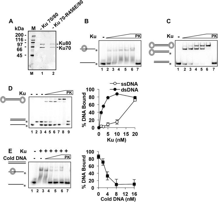
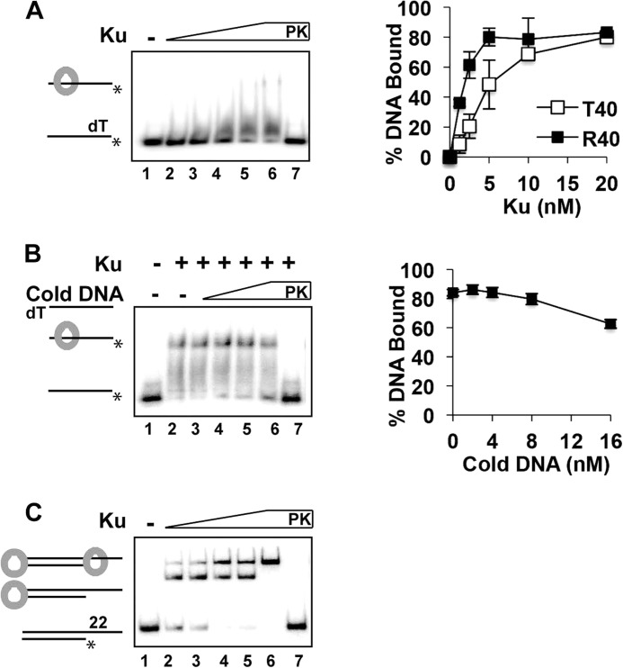
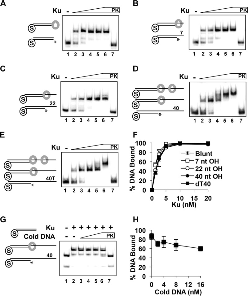
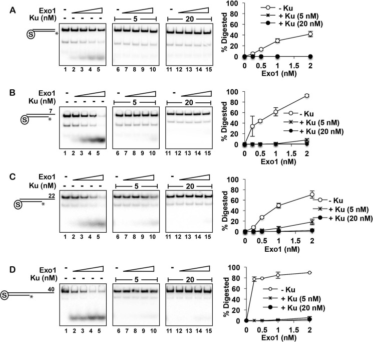
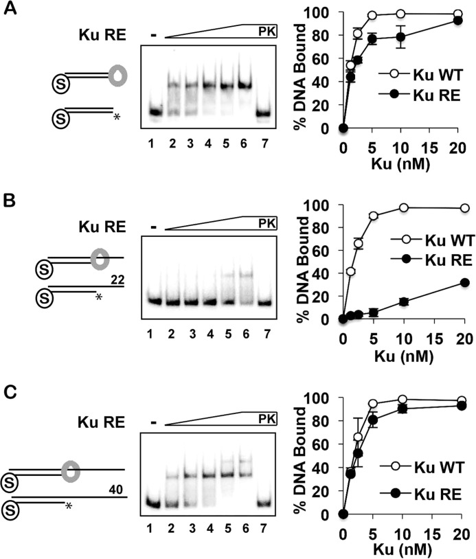
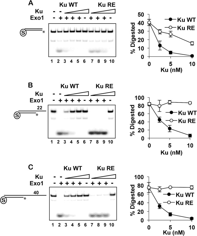
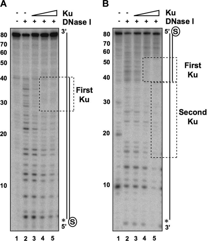
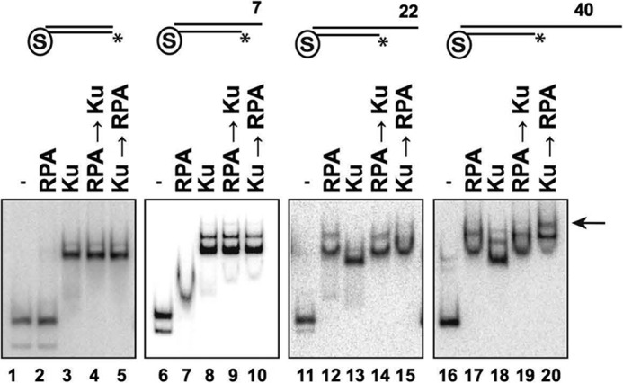
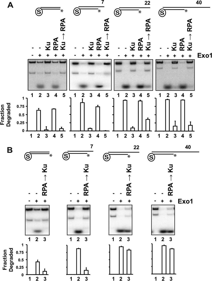
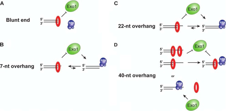
DNA double-strand breaks can be eliminated via non-homologous end joining or homologous recombination. Non-homologous end joining is initiated by the association of Ku with DNA ends. In contrast, homologous recombination entails nucleolytic resection of the 5'-strands, forming 3'-ssDNA tails that become coated with replication protein A (RPA). Ku restricts end access by the resection nuclease Exo1. It is unclear how partial resection might affect Ku engagement and Exo1 restriction. Here, we addressed these questions in a reconstituted system with yeast proteins. With blunt-ended DNA, Ku protected against Exo1 in a manner that required its DNA end-binding activity. Despite binding poorly to ssDNA, Ku could nonetheless engage a 5'-recessed DNA end with a 40-nucleotide (nt) ssDNA overhang, where it localized to the ssDNA-dsDNA junction and efficiently blocked resection by Exo1. Interestingly, RPA could exclude Ku from a partially resected structure with a 22-nt ssDNA tail and thus restored processing by Exo1. However, at a 40-nt tail, Ku remained stably associated at the ssDNA-dsDNA junction, and RPA simultaneously engaged the ssDNA region. We discuss a model in which the dynamic equilibrium between Ku and RPA binding to a partially resected DNA end influences the timing and efficiency of the resection process.

Keywords: DNA damage; DNA damage response; DNA-binding protein; Exo1; Ku; RPA; Saccharomyces cerevisiae; homologous recombination; non-homologous DNA end joining.


Figures

FIGURE 1.
FIGURE 1.
Binding of ssDNA by Ku. A, SDS-PAGE analysis of purified Ku and the Ku-RE mutant. Molecular masses are indicated in kilodaltons (M lane). B, Ku (1.25, 2.5, 5, 10, and 20 nm) was incubated with the 40-mer ssDNA (4 nm) for 10 min. C, same conditions, except that the 40-bp dsDNA was tested. D, same conditions, except that both the 40-mer ssDNA and 40-bp dsDNA substrates were included in the same reactions. Quantification of the results is shown to the right. E, Ku (5 nm) was incubated with the radiolabeled 40-mer ssDNA (4 nm) for 10 min, and unlabeled 40-bp dsDNA (0, 2, 4, 8, and 16 nm) was added, followed by a further 10 min of incubation. Quantification of the results is shown to the right. In B–E, PK lanes represent a reaction that was treated with SDS and proteinase K prior to analysis, and asterisks denote the 32P label. Error bars in D and E represent 1 S.D. based on results from three independent experiments.
FIGURE 2.
FIGURE 2.
Binding of unstructured ssDNA by Ku. A, Ku (1.25, 2.5, 5, 10, and 20 nm) was incubated with oligo(dT)40 (dT; 4 nm) for 10 min. Quantification of results in Fig. 1B (R40) and those pertaining to oligo(dT)40 (T40) binding is shown to the right. B, Ku (5 nm) was incubated with the radiolabeled 40-mer ssDNA (4 nm) for 10 min, and unlabeled oligo(dT)40 (0, 2, 4, 8, and 16 nm) was added, followed by a 10-min incubation. Quantification of the results is shown to the right. C, the substrate was 40-bp dsDNA with a blunt end and a 22-nt 5′-recessed end. PK lanes represent a reaction that was treated with SDS and proteinase K prior to analysis, and asterisks denote the 32P label. Error bars in A and B represent 1 S.D. based on results from three independent experiments.
FIGURE 3.
FIGURE 3.
Engagement of 5′-recessed DNA ends by Ku. A–E, Ku (1.25, 2.5, 5, 10, and 20 nm) was incubated with DNA substrate (4 nm each) with one of its ends blocked by a biotin-streptavidin complex and also with a free blunt end (A) or a 7-nt (B), 22-nt (C), 40-nt (D), or oligo(dT)40 (40T or dT40; E) 5′-recessed end for 10 min. F, quantification of the results in A–E. G, Ku (5 nm) was incubated with the substrate containing a 40-nt 5′-recessed end (4 nm) for 10 min, and unlabeled 40-bp blunt-ended dsDNA (0, 2, 4, 8, and 16 nm) was added, followed by a 10-min incubation. Quantification of the results is shown to the right. In A–E and G, PK lanes represent a reaction was treated with SDS and proteinase K prior to analysis, and asterisks denote the 32P label. Error bars in F and H represent 1 S.D. based on results from three independent experiments.
FIGURE 4.
FIGURE 4.
Restriction of Exo1 action by Ku at blunt and 5′-recessed DNA ends. A–D, DNA substrates (4 nm each) that had a blocked end and also a free blunt end (A) or a 7-nt (B), 22-nt (C), or 40-nt (D) 5′-recessed end were preincubated without (left panels) or with 5 (middle panels) or 20 nm (right panels) Ku for 10 min before adding Exo1 (0.25, 0.5, 1, and 2 nm), followed by a 5-min incubation. Quantification of the results is shown to the right. Error bars represent 1 S.D. based on results from three independent experiments, and asterisks denote the 32P label.
FIGURE 5.
FIGURE 5.
Examination of the Ku-RE mutant for DNA end binding. A–C, Ku-RE (1.25, 2.5, 5, 10, and 20 nm) was incubated with DNA substrates (4 nm each) that had a blocked end and also a free blunt end (A) or a 22-nt (B) or 40-nt (C) 5′-recessed end. The quantification compares the results from these experiments with those obtained with wild-type Ku (from Fig. 2). PK lanes represent a reaction that had been treated with SDS and proteinase K prior to analysis, and asterisks denote the 32P label. Error bars represent 1 S.D. based on results from three independent experiments.
FIGURE 6.
FIGURE 6.
Requirement for Ku DNA end binding activity in the restriction of Exo1 action. A–C, Ku or Ku-RE (2.5, 5, and 10 nm) was preincubated with DNA substrates with a blocked end and also a free blunt end (A) or a 22-nt (B) or 40-nt (C) 5′ recessed end for 5 min, and Exo1 (2 nm) was added, followed by a 5-min incubation. Quantification of the results is shown to the right. Asterisks denote the 32P label. Error bars represent 1 S.D. based on results from three independent experiments.
FIGURE 7.
FIGURE 7.
Localization of Ku to ssDNA-dsDNA junction in DNA with a recessed end. A, Ku (5, 10, or 20 nm) was incubated with the DNA substrate containing a blocked end and a free 40-nt 5′-recessed end (4 nm) for 10 min. DNase I (50 pg) was added, followed by a 3-min incubation. The asterisk denotes the 32P label, and the region protected by Ku is boxed. B, same as described for A, with the asterisk denoting the 32P label on the 3′-ssDNA overhang.
FIGURE 8.
FIGURE 8.
Interplay between Ku and RPA at DNA ends. EMSAs were performed using Ku (20 nm) and/or RPA (20 nm) and the indicated blocked DNA substrate (4 nm each) containing a free blunt end or a 7-, 22-, or 40-nt 5′-recessed end. In reactions in which the addition of one protein preceded the other, the DNA substrate was incubated with the first protein for 10 min, followed by the second protein and a 10-min incubation. The DNA-Ku-RPA ternary complex is indicated by the arrow. Asterisks denote the 32P label.
FIGURE 9.
FIGURE 9.
Effects of RPA and Ku on resection by Exo1. A, the various DNA substrates were incubated with RPA (20 nm), Ku (20 nm), or with Ku first and then RPA as described in the legend to Fig. 7, followed by the addition of Exo1 (2 nm for the blunt-ended substrate and 1 nm for the other substrates) and a 5-min incubation. Quantification of the results is shown below. B, same as described for A, except that the DNA substrates were incubated with RPA and then Ku before adding Exo1. Quantification of the results is shown below. Asterisks denote the 32P label. Error bars represent 1 S.D. based on results from three independent experiments.
FIGURE 10.
FIGURE 10.
Summary of Ku-RPA interplay at DNA ends with or without a 3′-ssDNA overhang.

References

    1. Khanna K. K., Jackson S. P. (2001) DNA double-strand breaks: signaling, repair and the cancer connection. Nat. Genet. 27, 247–254 - PubMed
    1. Chiruvella K. K., Liang Z., Wilson T. E. (2013) Repair of double-strand breaks by end joining. Cold Spring Harb. Perspect. Biol. 5, a012757. - PMC - PubMed
    1. Walker J. R., Corpina R. A., Goldberg J. (2001) Structure of the Ku heterodimer bound to DNA and its implications for double-strand break repair. Nature 412, 607–614 - PubMed
    1. Cary R. B., Peterson S. R., Wang J., Bear D. G., Bradbury E. M., Chen D. J. (1997) DNA looping by Ku and the DNA-dependent protein kinase. Proc. Natl. Acad. Sci. U.S.A. 94, 4267–4272 - PMC - PubMed
    1. de Vries E., van Driel W., Bergsma W. G., Arnberg A. C., van der Vliet P. C. (1989) HeLa nuclear protein recognizing DNA termini and translocating on DNA forming a regular DNA-multimeric protein complex. J. Mol. Biol. 208, 65–78 - PubMed

Publication types

MeSH terms

Substances