. 2015 Jun 12:5:11295.
doi: 10.1038/srep11295.

Serum Exosome MicroRNA as a Minimally-Invasive Early Biomarker of AML

Affiliations

Serum Exosome MicroRNA as a Minimally-Invasive Early Biomarker of AML

Noah I Hornick et al. Sci Rep. .

Abstract

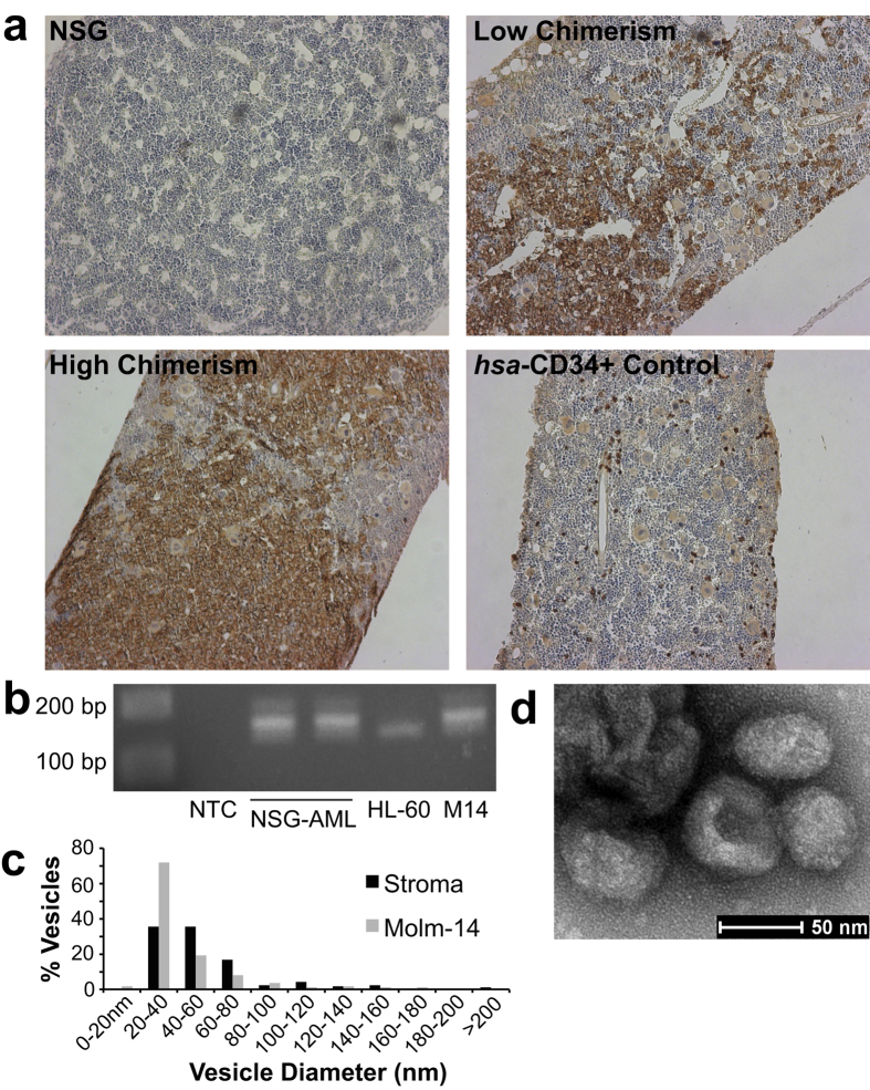
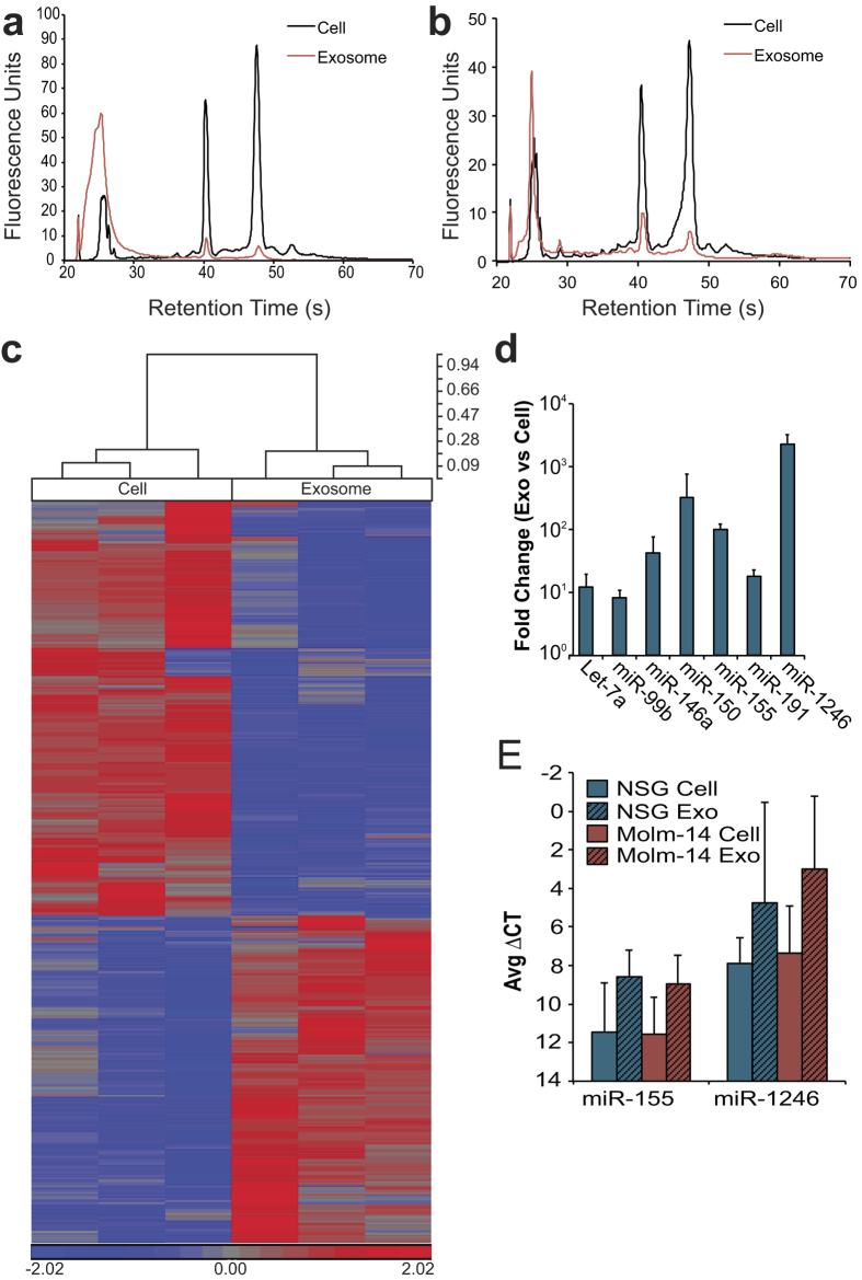
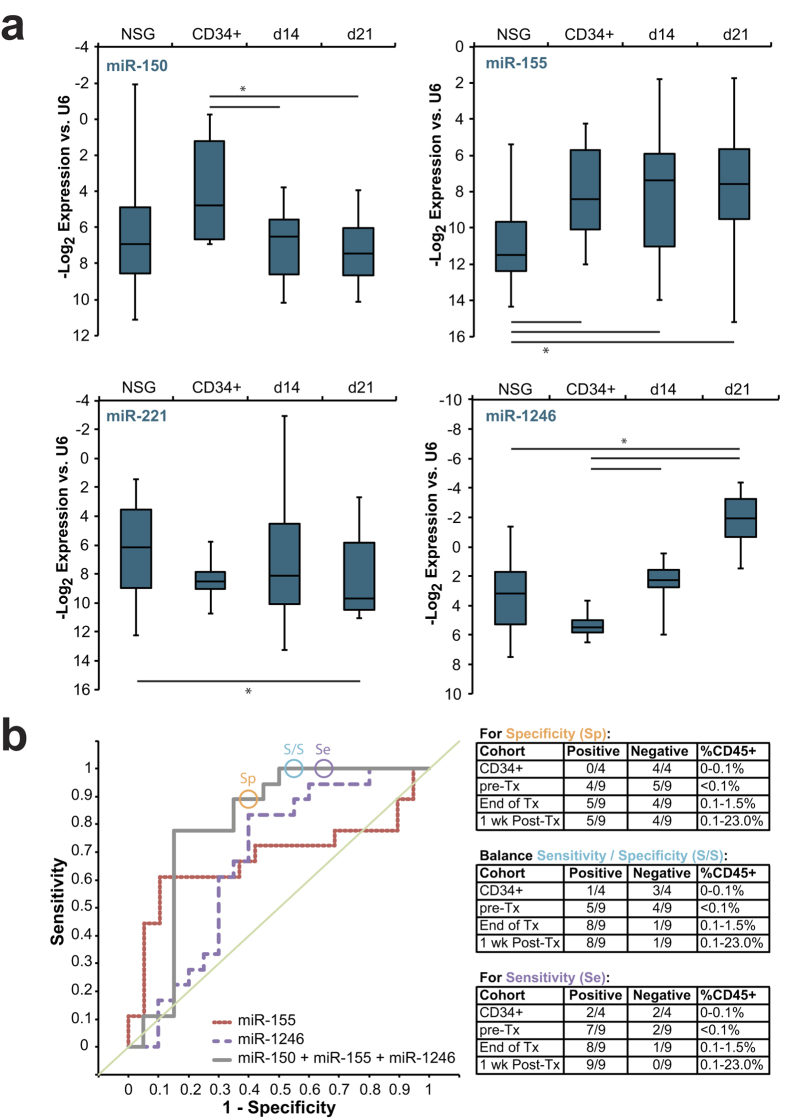
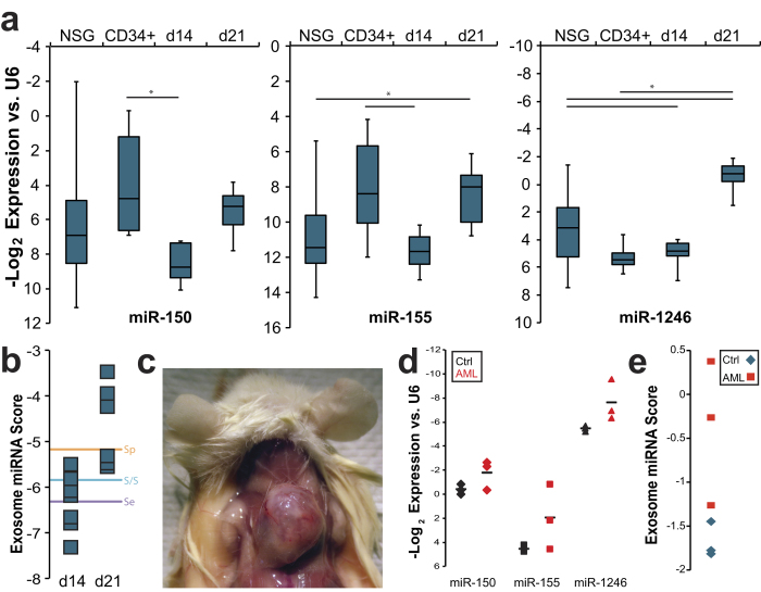
Relapse remains the major cause of mortality for patients with Acute Myeloid Leukemia (AML). Improved tracking of minimal residual disease (MRD) holds the promise of timely treatment adjustments to preempt relapse. Current surveillance techniques detect circulating blasts that coincide with advanced disease and poorly reflect MRD during early relapse. Here, we investigate exosomes as a minimally invasive platform for a microRNA (miRNA) biomarker. We identify a set of miRNA enriched in AML exosomes and track levels of circulating exosome miRNA that distinguish leukemic xenografts from both non-engrafted and human CD34+ controls. We develop biostatistical models that reveal circulating exosomal miRNA at low marrow tumor burden and before circulating blasts can be detected. Remarkably, both leukemic blasts and marrow stroma contribute to serum exosome miRNA. We propose development of serum exosome miRNA as a platform for a novel, sensitive compartment biomarker for prospective tracking and early detection of AML recurrence.


Figures

Figure 1
Figure 1. Molm-14 Engraft in NSG Mice and Contribute to Compartment Exosome Secretion
(a) Immunohistochemistry of Molm-14-engrafted NSG femurs. Femurs were collected from Molm-14-engrafted mice at 3 weeks post-engraftment, sectioned, and stained for human CD45. (b) PCR identification of NSG-engrafted AML. Molm-14 isolated from engrafted NSG mice retained their characteristic FLT3-ITD mutation, detected by 40 cycles of PCR (cropped; full gel in Figure S2). NTC: null-template control; NSG-AML: isolated Molm-14; HL-60: cell line exhibiting WT FLT3; M14: Molm-14 stock culture. (c,d) Transmission electron microscopy of exosomes. Exosomes were isolated from the culture media of Molm-14 and NSG stroma, and examined by transmission electron microscopy. Sizing of more than 100 vesicles per sample is presented in (c); representative stromal exosome image is presented in (d).
Figure 2
Figure 2. Molm-14 and Stromal Exosomes Concentrate Selected MicroRNA
(a,b) RNA size profiles of exosomes. RNA was collected from Molm-14 (a) and NSG Stromal (b) cells and their exosomes after 72 hours in culture, and miRNA was evaluated using a bioanalyzer. (c) Microarray comparison of cell, exosome miRNA. Molm-14 cell and exosome microRNA was evaluated using an Affymetrix microRNA microarray. All targets with more than 2-fold mean difference between producing cell and exosome are represented, RMA-corrected and standardized to a mean of 0 and a SD of 1. Dendrogram values are 1 - Pearson’s R. (d,e) qRT-PCR for miRNA in cells versus exosomes. Selected targets from NSG stromal (d) and Molm-14 (e) cells and exosomes were validated using Taqman qRT-PCR, normalized to U6 snRNA. Fold change was calculated by 2^-ΔΔCt.
Figure 3
Figure 3. Conventional Biomarkers Provide Limited Information; Chemotherapy is a Confounder
(a) Molm-14-engrafted NSG chimerism by flow. Flow cytometry for human CD45 was performed on the peripheral blood and bone marrow of Molm-14-engrafted NSG mice, both without and with cytarabine treatment (300 mg/kg, Q3Dx3 starting 14d post-engraftment). (b) mRNA markers of AML in serum exosomes. Exosomes isolated from the peripheral blood of Molm-14-engrafted NSG mice were tested for human FLT3 and GAPDH and murine GAPDH using 45 cycles of PCR (cropped; full gels in Figure S3). NTC: null-template control. M14: Molm-14 stock culture. PB: Peripheral blood chimerism; BM: Bone marrow chimerism by flow cytometry for human CD45. (c) CBC of Molm-engrafted NSG. Peripheral blood CBC of mice, comparing baseline NSG (n = 31), NSG engrafted with Molm-14 at 2 (n = 16) and 3 (n = 5) weeks post-engraftment, NSG engrafted with Molm-14 and treated with cytarabine (n = 8) at 3 weeks, control NSG given cytarabine (n = 4), and NSG given nonmalignant CD34+ human cells (n = 4). WBC, white blood cells; LY, lymphocytes; NE, neutrophils; Hb, hemoglobin; PLT, platelets (×1000/uL). *, p < 0.05 by Student’s t. Error bars represent SEM. (d,e) In vitro chemotherapy and exosomal miRNA. Molm-14, HL-60, and U937 cells were exposed to cytarabine (25 ng/mL, d) overnight, and Molm-14 were exposed to quizartinib (0.1 nM, e) overnight. Cellular (on the x-axis) and exosomal (on the y-axis) levels of miRNA were measured by qRT-PCR, and are presented as ddCT (a -1 ddCT represents a 2-fold increase in miRNA).
Figure 4
Figure 4. Serum Exosome miRNA Distinguishes Leukemia From Homeostatic Hematopoiesis
(a) Serum exosome miRNA levels. Exosomes were isolated from peripheral blood of control NSG mice as well as NSG mice engrafted with Molm-14 (after 14 or 21 days; d14 and d21, respectively) or human CD34+ cells. MicroRNA levels were measured by qRT-PCR. *, p < 0.05 by Student’s t. (b) Serum exosome miRNA score performance. Receiver operating characteristic (ROC) curves are presented for single miRNA and for the combination of miRNA-150, -155, and -1246. The circles represent points on the ROC curves whose alphas were chosen as cutoff values emphasizing specificity, sensitivity, or a balance of the two, respectively. These cutoff values and the coefficients generated by the regression were used to evaluate mice engrafted with Molm-14 and treated with cytarabine, alongside control mice engrafted with human CD34+ cells. Exosome miRNA panel performance for each cohort and cutoff is presented alongside peripheral blood chimerism (by flow cytometry for human CD45).
Figure 5
Figure 5. Expansion of the Scope of the Serum Exosome miRNA Biomarker
(a) Comparing serum exosome miRNA levels in HL-60. Exosomes were isolated from peripheral blood of control NSG mice as well as NSG mice engrafted with HL-60 (after 14 or 21 days; d14 and d21, respectively) or human CD34+ cells. MicroRNA levels were measured by qRT-PCR. *, p < 0.05 by Student’s t. (b) Evaluation of score performance. miRNA panel scores were calculated for HL-60-engrafted mice at 14 and 21 day time. The lines correspond to alphas chosen as described in Fig. 4b. (c) Chloromas in HL-60-engrafted NSG mice. (d) Patient serum / plasma exosome miRNA. Exosomes were isolated from serum or plasma from AML patients (red) or normal subjects (black), and miRNA levels of miR-150, -155, and -1246 were measured. Means are represented as horizontal bars. (e) Evaluation of score in human samples. miRNA panel scores were calculated for the serum miRNA levels depicted in d.

References

    1. Howlader, N. et al.. (eds). SEER Cancer Statistics Review, 1975–2008. http://seer.cancer.gov/csr/1975_2008/ (2011).
    1. Yin J. A. et al.. Minimal residual disease monitoring by quantitative RT-PCR in core binding factor AML allows risk stratification and predicts relapse: results of the United Kingdom MRC AML-15 trial. Blood 120, 2826–2835, 10.1182/blood-2012-06-435669 (2012). - DOI - PubMed
    1. Loken M. R. et al.. Residual disease detected by multidimensional flow cytometry signifies high relapse risk in patients with de novo acute myeloid leukemia: a report from Children’s Oncology Group. Blood 120, 1581–1588, 10.1182/blood-2012-02-408336 (2012). - DOI - PMC - PubMed
    1. Buonamici S. et al.. Real-time quantitation of minimal residual disease in inv(16)-positive acute myeloid leukemia may indicate risk for clinical relapse and may identify patients in a curable state. Blood 99, 443–449 (2002). - PubMed
    1. Dohner H. et al.. Diagnosis and management of acute myeloid leukemia in adults: recommendations from an international expert panel, on behalf of the European LeukemiaNet. Blood 115, 453–474, 10.1182/blood-2009-07-235358 (2010). - DOI - PubMed

Publication types

Associated data