Review
. 2015 Aug;40(8):435-45.
doi: 10.1016/j.tibs.2015.05.001. Epub 2015 Jun 9.

Peroxiredoxins: guardians against oxidative stress and modulators of peroxide signaling

Affiliations
Review

Peroxiredoxins: guardians against oxidative stress and modulators of peroxide signaling

Arden Perkins et al. Trends Biochem Sci. 2015 Aug.

Abstract

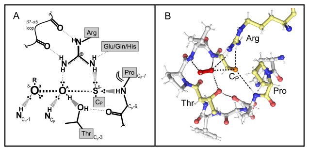
Peroxiredoxins (Prxs) are a ubiquitous family of cysteine-dependent peroxidase enzymes that play dominant roles in regulating peroxide levels within cells. These enzymes, often present at high levels and capable of rapidly clearing peroxides, display a remarkable array of variations in their oligomeric states and susceptibility to regulation by hyperoxidative inactivation and other post-translational modifications. Key conserved residues within the active site promote catalysis by stabilizing the transition state required for transferring the terminal oxygen of hydroperoxides to the active site (peroxidatic) cysteine residue. Extensive investigations continue to expand our understanding of the scope of their importance as well as the structures and forces at play within these critical defense and regulatory enzymes.

Keywords: antioxidant defense; antioxidant enzyme; peroxidase; redox signaling.


Figures

Figure I
Figure I
WebLogo plots for sequences proximal to CP and pie charts illustrating the prevalence of each CR location among all six subfamilies of Prxs. Subfamilies as defined by Nelson et al. [37] but updated from the October 2011 GenBank(nr) database were represented by the following number of members: Prx1 (1387), Prx6 (1102), AhpE (112), PrxQ (2060), Tpx (990), and Prx5 (1139). (A) The local sequences including the PxxxTxxC motif (conserved positions of the motif noted with asterisks) and 5 residues downstream of Cp were extracted from all members of each subfamily and used to create WebLogo plots to summarize sequence conservation around Cp. (B) Pie charts showing the frequency at which the CR is in a given location for each subfamily. Wedges are colored by CR position consistent with Fig. 2A: CR in the C-terminus (Ct, magenta), α2 (yellow), α3 (green), α5 (dark blue), or the N-terminus (Nt, orange). No CR (red) indicates that there is no other Cys in the protein, and uncertain (gray) indicates there are other Cys, but not in a recognized position of a CR. Hybrid proteins in the Prx5 subfamily with a C-terminally appended Grx domain, as exemplified by Haemophilus influenza Prx5-Grx, are represented in cyan; these proteins either have only one Cys (the Cp) in the Prx domain or have other Cys residues that do not align with recognized CR residues. The exact positions in canonical representatives (allowing a shift of up to 2 residues) are defined as follows: Ct aligns with Cys165 in S. typhimurium AhpC; α2 aligns with Cys50 in E. coli PrxQ (also known as BCP); α3 aligns with Cys95 in E. coli Tpx; α5 aligns with Cys151 in H. sapiens PrxV; and Nt aligns with Cys31 in S. cerevisiae Ahp1.
Figure I
Figure I
Quaternary structures of Prxs. Dimeric α2 complexes are formed using either an A-type interface, where the monomers interact near helix α3, or B-type dimers where the interaction is through the β-strands, generating an extended 10–14 strand β-sheet. Further interactions at the A-interfaces of some Prx1 and Prx6 members generate (α2)5 decamers [or in rare cases (α2)6 dodecamers]. The blue subunit is displayed at approximately the same position in each of the structures to illustrate these interaction interfaces that together build the decamer. For a number of Prx1 members, the structural change upon disulfide bond formation destabilizes the A-type dimer interface, and the decamer dissociates to B-type dimers. The structures depicted are: Aeropyrum pernix PrxQ (A-type dimer, Protein Data Bank Identifier 4GQF), and wild type S. typhimurium AhpC (B-type dimer and decamer, Protein Data Bank Identifier 4MA9).
Figure 1
Figure 1
The catalytic and regulatory cycles of 2-Cys peroxiredoxins (Prxs). Shown in brown is the normal Prx cycle with the structure of the peroxidatic Cys (CP) residue shown for each redox state (carbons are colored gray, nitrogens blue, oxygens red, and sulfurs yellow; hydrogens are not shown for simplicity). The CP thiolate (RS) in the fully folded (FF) active site is first oxidized by the peroxide to form the sulfenic acid (R-SOH) or sulfenate (R-SO) (computational approaches suggest stabilization of the CP as the sulfenate, but the true protonation state is as yet uncertain). This sulfenate, which must undergo a conformational change to become locally unfolded (LU), then forms a disulfide bond with the resolving Cys (CR) in 2-Cys Prxs. Reductive recycling by thioredoxin (Trx) or a Trx-like protein or domain (e.g., tryparedoxin or the N-terminal domain of bacterial AhpF) then restores the thiolate in the FF active site for another catalytic cycle. Shown in blue is the redox-linked regulatory cycle of predominantly eukaryotic Prxs, wherein the CP sulfenate becomes further oxidized, in the presence of high peroxide levels, to the inactive sulfinate (R-SO2). In some organisms and Prx isoforms the active enzyme is restored by the ATP-dependent activity of sulfiredoxin (Srx).
Figure 2
Figure 2
Variable locations of the resolving Cys (CR) and oxidation-driven conformational changes across Prxs with distinct CR locations. (A) Shown are the various positions of the peroxiredoxin CR (colored sidechains) in relation to the active site peroxidatic Cys (CP, circled and in red). Intramolecular CP-CR disulfides are formed for the α2 (yellow), α3 (green), and α5 (blue) types, and intermolecular disulfides are formed for the N-terminal (Nt, orange CR in the gold chain) and C-terminal (Ct, magenta CR in the black chain) types. (CR residues are mapped onto a composite structure based on S. typhimurium AhpC (StAhpC), Protein Databank Identifier 4MA9). (B) Shown are the transitions from fully folded to locally unfolded conformations for representative Prxs: Nt, Saccharomyces cerevisiae Ahp1; α2, Aeropyrum pernix PrxQ; α3, E. coli Tpx; α5, Homo sapiens PrxV; Ct, StAhpC. The fully folded conformation is shown in white and the locally unfolded conformation is shown in black, with structural changes that occur in the transition highlighted in yellow. CP is highlighted in red, and CR is highlighted in blue. For clarity, residues 169–186 of StAhpC are not shown. Note substantial movement of CR when in the C-terminus (making an intersubunit disulfide bond), and of both CP and CR when CR is in α2 [25].
Figure 3
Figure 3
The universally-conserved Prx active site. (A) Cartoon representation of the putative active-site transition state conformation. The stabilizing interactions between key atoms from the backbone and the four conserved residues, and with the ROOH substrate, are indicated. In the transition state, a bond is forming between the S atom of the CP and the OA of ROOH, and a bond is breaking between the OA and OB atoms of ROOH. The geometry of the active site appears nicely set up to stabilize the larger distance between the OA and OB atoms as the bond is broken. Based on a figure from Hall et al., 2010 [52]. (B) Shown is the Michaelis complex of a hydrogen peroxide-bound Prx (ApTpx, Protein Databank Identifier code 3A2V) with the four conserved residues of the active site motif highlighted in pale yellow and key active site hydrogen bonds noted with dashed lines. The interaction between Cp and the OA of H2O2 where the bond will form is depicted with dots.

References

    1. Segel IH. Biochemical Calculations. John Wiley & Sons, Inc; 1976.
    1. van der Kamp MW, Mulholland AJ. Combined quantum mechanics/molecular mechanics (QM/MM) methods in computational enzymology. Biochemistry. 2013;52:2708–2728. - PubMed
    1. Winther JR, Thorpe C. Quantification of thiols and disulfides. Biochim Biophys Acta. 2014;1840:838–846. - PMC - PubMed
    1. Karplus PA. A primer on peroxiredoxin biochemistry. Free Radic Biol Med. 2014 in press. - PMC - PubMed
    1. Winterbourn CC. Reconciling the chemistry and biology of reactive oxygen species. Nat Chem Biol. 2008;4:278–286. - PubMed

Publication types

LinkOut - more resources