Case Reports
. 2015 Jun 11:16:361-6.
doi: 10.12659/AJCR.892750.

Erdheim-Chester Disease: A Case Report

Affiliations
Case Reports

Erdheim-Chester Disease: A Case Report

Jean Alexiou et al. Am J Case Rep. .

Abstract

Background: The diagnosis of Erdheim-Chester disease, a rare illness, is difficult and requires increased awareness.

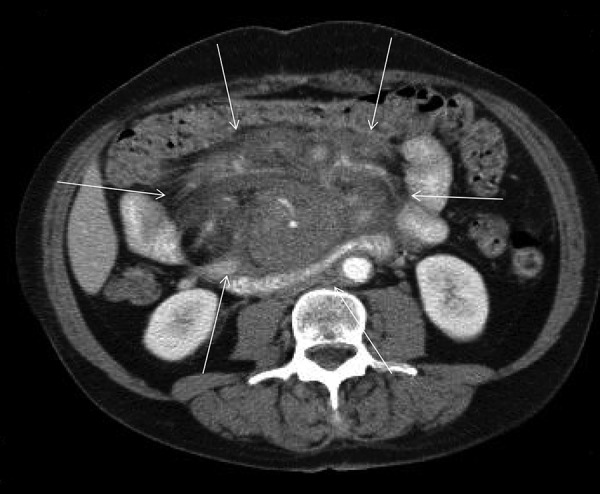
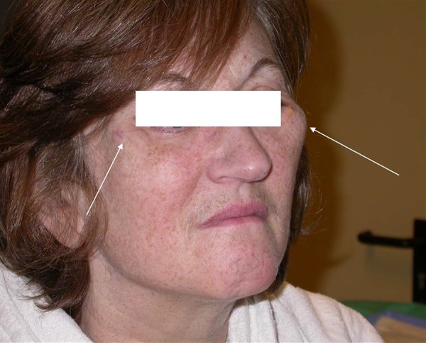
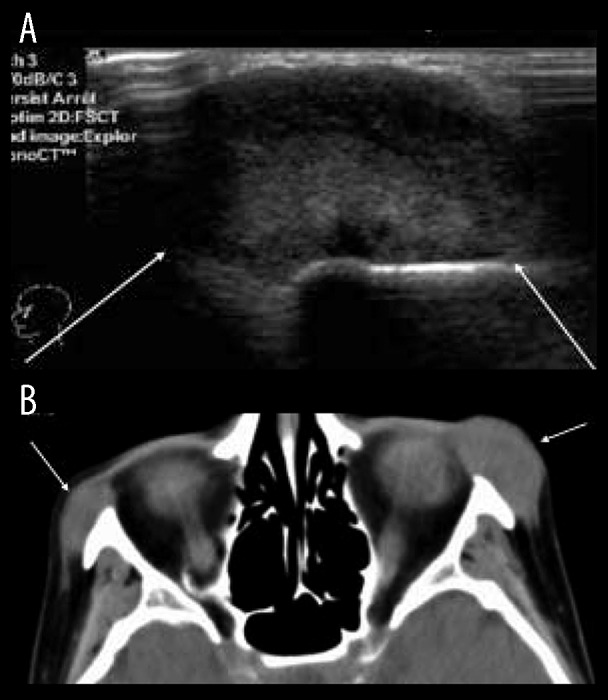
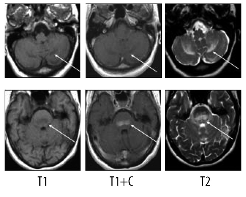
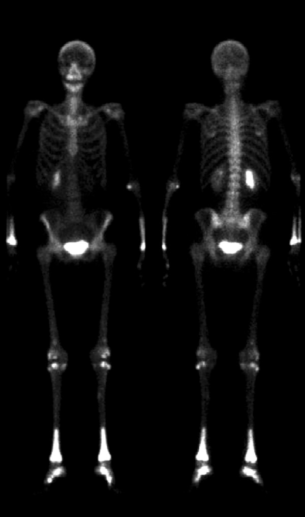
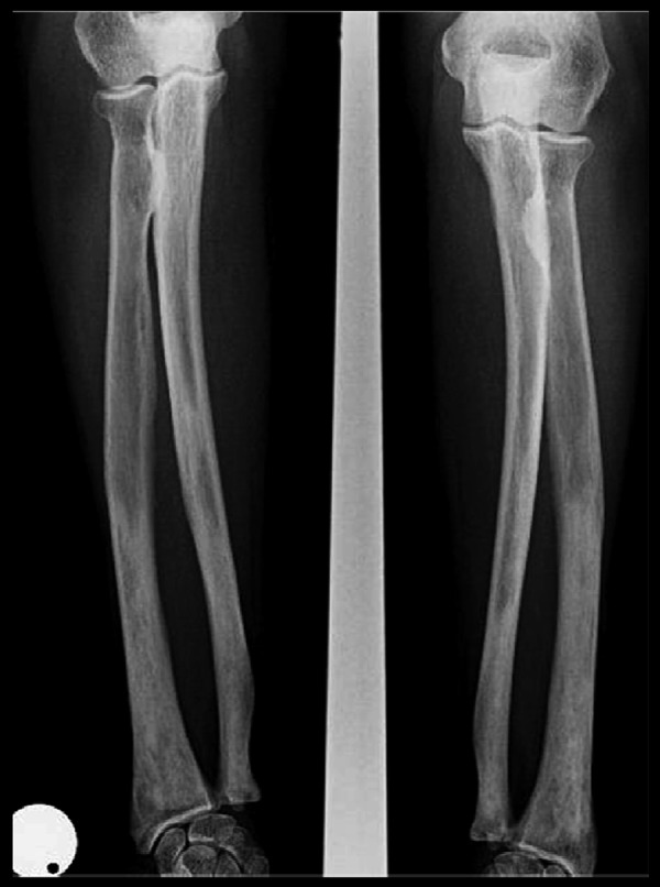
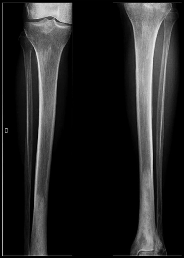
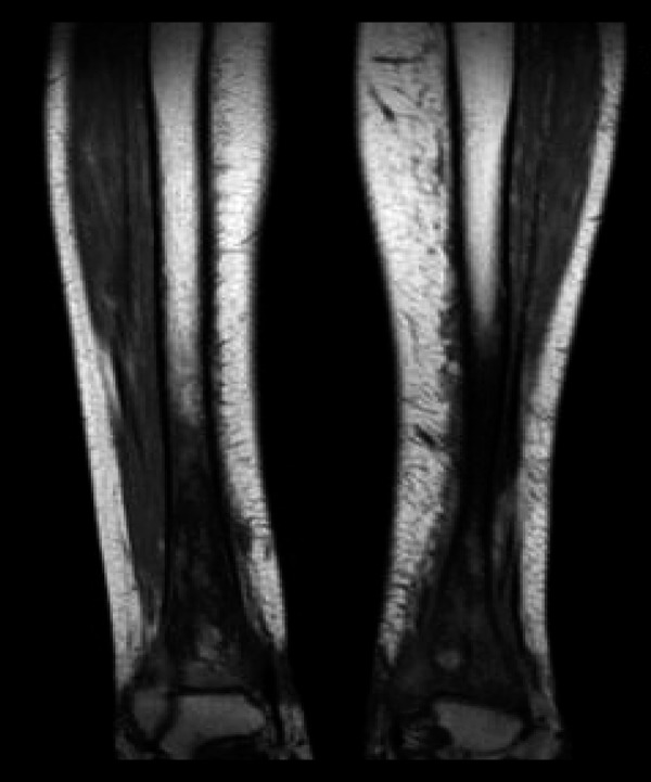
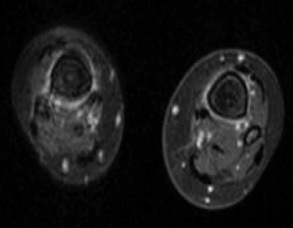
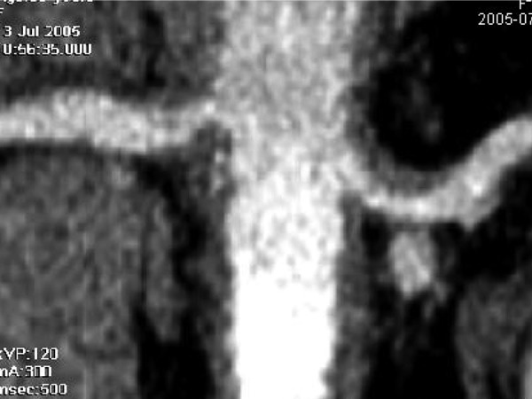
Case report: We report the case of a 56-year-old woman who initially presented with a mesenteric panniculitis and 8 years later developed neurological manifestations and bone lesions that led to a diagnosis of Erdheim-Chester disease.

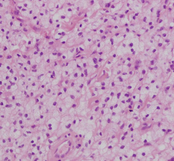
Conclusions: The rather characteristic aspect of the bone lesions as well as the presence of foamy cells in involved tissue biopsies should suggest the diagnosis. No therapy is available at present but recent biological data might suggest new approaches for the understanding and therapy of this condition.


Figures

Figure 1.
Figure 1.
Abdominal contrast enhanced computed tomography demonstrating a large infiltrating mesenteric mass (arrows).
Figure 2.
Figure 2.
Prominent zygomatic arches.
Figure 3.
Figure 3.
(A) Ultrasound of the cheekbone showing a solid hyperechoic soft tissue mass well delineated [arrows]. (B) Solid homogenous masses appearance on computed tomography. These masses append the zygomatic arches without bony erosion or periosteal reaction.
Figure 4.
Figure 4.
Histopathology: multiple foamy cells.
Figure 5.
Figure 5.
On MR, presence of lesions in the cerebellar peduncles extending to the cerebellar hemispheres.
Figure 6.
Figure 6.
99Tc bone scintgraph with increased uptake of distal long bone extremities.
Figure 7.
Figure 7.
Plain radiograph showing diffuse sclerotic changes of the distal extremity of the radius and cubitus.
Figure 8.
Figure 8.
Plain radiograph showing diffuse sclerotic changes of the distal extremity of the tibiae.
Figure 9.
Figure 9.
Diffuse hypointensity of the signal in the extremity of the tibiae, on MR T1 sequence.
Figure 10.
Figure 10.
Peripheral hyperintense signal around cortex and the level of the medullar bony infiltration, on MR T2 sequence.
Figure 11.
Figure 11.
Coronal reconstruction of a CT at the level of the renal arteries demonstrating a thickened vascular wall at the origin of these arteries.

References

    1. Veyssier-Belot C, Cacoub P, Caparros-Lefebvre D, et al. Erdheim-Chester disease: clinical and radiologic characteristics of 59 cases. Medecine. 1996;75(3):157–69. - PubMed
    1. Gomez C, Diard F, Chateil JF, Moinard M. Imagerie de la maladie d’Erdheim-Chester. J Radiol. 1996;77:1213–21. [in French] - PubMed
    1. Cattin F, Runge M, Nagy N, et al. [Case #5. Erdheim-Chester disease] J Radiol. 2005;86:527–30. [in French] - PubMed
    1. Kenn W, Stäbler A, Zachoval R, et al. Erdheim-Chester disease: a case report and literature overview. Eur Radiol. 1999;9:153–58. - PubMed
    1. Breuil V, Brocq O, Pellegrino C, et al. Erdheim-Chester Disease: typical radiological bone features for a rare xanthogranulomatosis. Ann Rheum Dis. 2002;66:199–200. - PMC - PubMed

Publication types