. 2015 Sep 15;192(6):682-94.
doi: 10.1164/rccm.201412-2278OC.

Cytokines in Chronic Rhinosinusitis. Role in Eosinophilia and Aspirin-exacerbated Respiratory Disease

Affiliations

Cytokines in Chronic Rhinosinusitis. Role in Eosinophilia and Aspirin-exacerbated Respiratory Disease

Whitney W Stevens et al. Am J Respir Crit Care Med. .

Abstract

Rationale: The mechanisms that underlie the pathogenesis of chronic rhinosinusitis without nasal polyps (CRSsNP), chronic rhinosinusitis with nasal polyps (CRSwNP), and aspirin-exacerbated respiratory disease (AERD) are not clear.

Objectives: To first evaluate the inflammatory profiles of CRSsNP and CRSwNP tissues and then to investigate whether clinical differences observed between CRSwNP and AERD are in part secondary to differences in inflammatory mediator expression within nasal polyp (NP) tissues.

Methods: Expression levels of numerous inflammatory mediators were determined by quantitative real-time polymerase chain reaction, ELISA, and multiplex immunoassay.

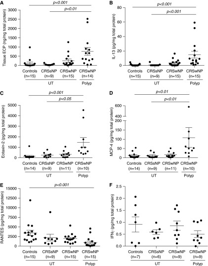
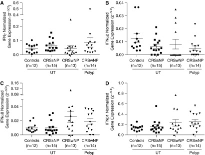
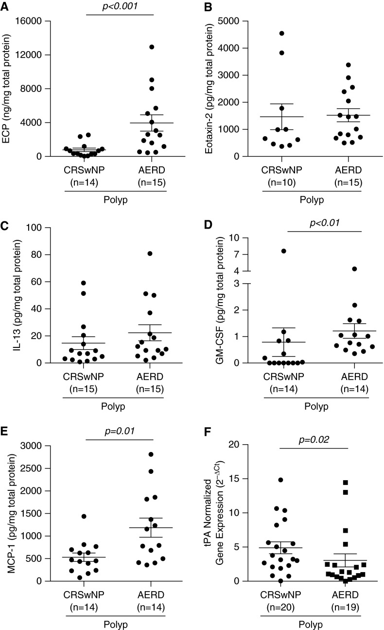
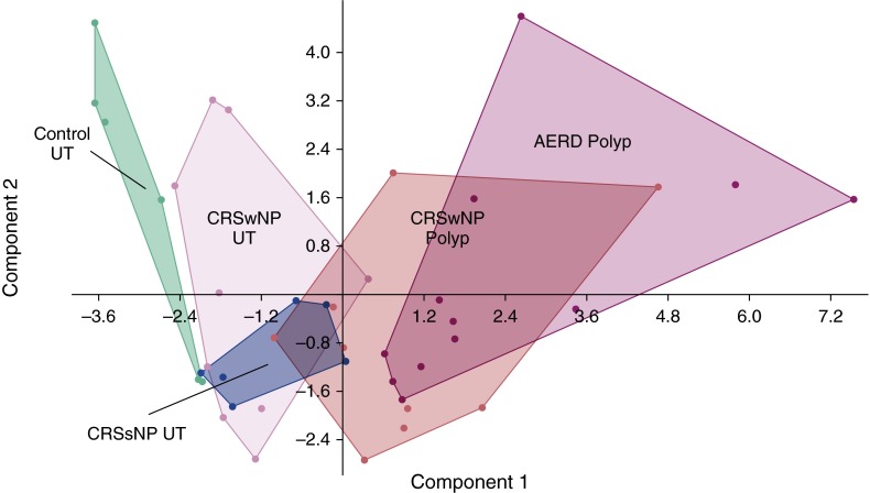
Measurements and main results: CRSwNP NP had increased levels of type 2 mediators, including IL-5 (P < 0.001), IL-13 (P < 0.001), eotaxin-2 (P < 0.001), and monocyte chemoattractant protein (MCP)-4 (P < 0.01), compared with sinonasal tissue from subjects with CRSsNP and control subjects. Expression of IFN-γ messenger RNA or protein was low and not different among the chronic rhinosinusitis subtypes examined. Compared with CRSwNP, AERD NP had elevated protein levels of eosinophil cationic protein (ECP) (P < 0.001), granulocyte-macrophage colony-stimulating factor (GM-CSF) (P < 0.01), and MCP-1 (P = 0.01), as well as decreased gene expression of tissue plasminogen activator (tPA) (P = 0.02). Despite the higher eosinophilia in AERD, there was no associated increase in type 2 mediator protein levels observed.

Conclusions: CRSwNP was characterized by a predominant type 2 inflammatory environment, whereas CRSsNP did not reflect a classic type 1 milieu, as has been suggested previously. AERD can be distinguished from CRSwNP by elevated ECP levels, but this enhanced eosinophilia is not associated with elevations in traditional type 2 inflammatory mediators associated with eosinophil proliferation and recruitment. However, other factors, including GM-CSF, MCP-1, and tPA, may be important contributors to AERD pathogenesis.

Keywords: CRSsNP; CRSwNP; eosinophil.


Figures

Figure 1.
Figure 1.
Protein expression of select inflammatory mediators in sinonasal tissues. Protein levels of (A) eosinophil cationic protein (ECP), (B) IL-13, (C) eotaxin-2, (D) monocyte chemoattractant protein (MCP)-4, (E) regulated upon activation, normal T-cell expressed and secreted protein (RANTES), and (F) IFN-γ were measured in uncinate tissue (UT) of healthy controls, UT of patients with chronic rhinosinusitis without nasal polyps (CRSsNP), and UT and nasal polyps of patients with chronic sinusitis with nasal polyps (CRSwNP). Statistical significance (P < 0.05) was determined by Kruskal-Wallis test with post hoc analysis using Dunn’s test for multiple comparison. Dot plots illustrate individual data points, and solid lines represent the mean ± SEM. Concentrations of ECP were expressed as nanograms per milligram of tissue, and all other mediators are expressed as picograms per milligram of tissue.
Figure 2.
Figure 2.
Relative gene expression of select type I interferons in sinonasal tissue. Total RNA was extracted from uncinate tissue (UT) isolated from controls, UT from patients with chronic rhinosinusitis without nasal polyps (CRSsNP) or chronic sinusitis with nasal polyps (CRSwNP), or nasal polyps from patients with CRSwNP. Expression of messenger RNA for (A) IFN-γ, (B) IFN-α2, (C) IFN-α8, and (D) IFN-β1 was measured by real-time polymerase chain reaction. Statistical significance (P < 0.05) was calculated by Kruskal-Wallis test with Dunn’s correction for multiple comparisons. Dot plots illustrate individual data points, and solid lines represent the mean ± SEM. Ct = cycle threshold.
Figure 3.
Figure 3.
Expression of select inflammatory mediators in nasal polyps of aspirin-exacerbated respiratory disease (AERD) compared with chronic sinusitis with nasal polyps (CRSwNP). Protein levels of (A) eosinophil cationic protein (ECP), (B) eotaxin-2, (C) IL-13, (D) granulocyte–macrophage colony–stimulating factor (GM-CSF), and (E) monocyte chemoattractant protein (MCP)-1 were measured in nasal polyp tissue from patients with either CRSwNP or AERD. Concentrations of ECP were measured as nanograms per milligram of tissue, and all other mediators are expressed as picograms per milligram of tissue. (F) Expression of messenger RNA for tissue plasminogen activator (tPA) was measured by real-time polymerase chain reaction. Statistical significance (P < 0.05) was determined by Mann–Whitney U test. Dot plots illustrate individual data points, and solid lines represent the mean ± SEM. Ct = cycle threshold.
Figure 4.
Figure 4.
Principal component analysis (PCA) of inflammatory mediator data. The PCA plot illustrates the first versus second principal component of inflammatory mediator levels in sinonasal tissue. Dots represent individual patients, and distinct clusters of the different disease states and/or tissue types are outlined. The colors represent the various disease states and tissue types. Green = control uncinate tissue (UT); pink = chronic sinusitis with nasal polyps (CRSwNP) UT; blue = chronic rhinosinusitis without nasal polyps (CRSsNP) UT; red = CRSwNP nasal polyp; purple = aspirin-exacerbated respiratory disease (AERD) nasal polyp.

Comment in

References

    1. Dykewicz MS, Hamilos DL. Rhinitis and sinusitis. J Allergy Clin Immunol. 2010;125(2) Suppl 2:S103–S115. - PubMed
    1. Bhattacharyya N, Orlandi RR, Grebner J, Martinson M. Cost burden of chronic rhinosinusitis: a claims-based study. Otolaryngol Head Neck Surg. 2011;144:440–445. - PubMed
    1. Fokkens WJ, Lund VJ, Mullol J, Bachert C, Alobid I, Baroody F, Cohen N, Cervin A, Douglas R, Gevaert P, et al. EPOS 2012: European position paper on rhinosinusitis and nasal polyps 2012. A summary for otorhinolaryngologists. Rhinology. 2012;50:1–12. - PubMed
    1. Meltzer EO, Hamilos DL. Rhinosinusitis diagnosis and management for the clinician: a synopsis of recent consensus guidelines. Mayo Clin Proc. 2011;86:427–443. - PMC - PubMed
    1. Tan BK, Chandra RK, Pollak J, Kato A, Conley DB, Peters AT, Grammer LC, Avila PC, Kern RC, Stewart WF, et al. Incidence and associated premorbid diagnoses of patients with chronic rhinosinusitis. J Allergy Clin Immunol. 2013;131:1350–1360. - PMC - PubMed

MeSH terms