. 2015 Aug;123(2):377-88.
doi: 10.1097/ALN.0000000000000742.

Isoflurane Ameliorates Acute Lung Injury by Preserving Epithelial Tight Junction Integrity

Affiliations

Isoflurane Ameliorates Acute Lung Injury by Preserving Epithelial Tight Junction Integrity

Joshua A Englert et al. Anesthesiology. 2015 Aug.

Abstract

Background: Isoflurane may be protective in preclinical models of lung injury, but its use in patients with lung injury remains controversial and the mechanism of its protective effects remains unclear. The authors hypothesized that this protection is mediated at the level of alveolar tight junctions and investigated the possibility in a two-hit model of lung injury that mirrors human acute respiratory distress syndrome.

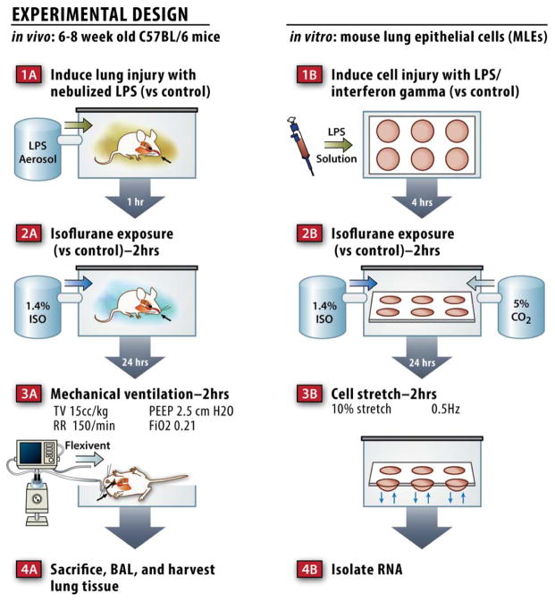
Methods: Wild-type mice were treated with isoflurane 1 h after exposure to nebulized endotoxin (n = 8) or saline control (n = 9) and then allowed to recover for 24 h before mechanical ventilation (MV; tidal volume, 15 ml/kg, 2 h) producing ventilator-induced lung injury. Mouse lung epithelial cells were similarly treated with isoflurane 1 h after exposure to lipopolysaccharide. Cells were cyclically stretched the following day to mirror the MV protocol used in vivo.

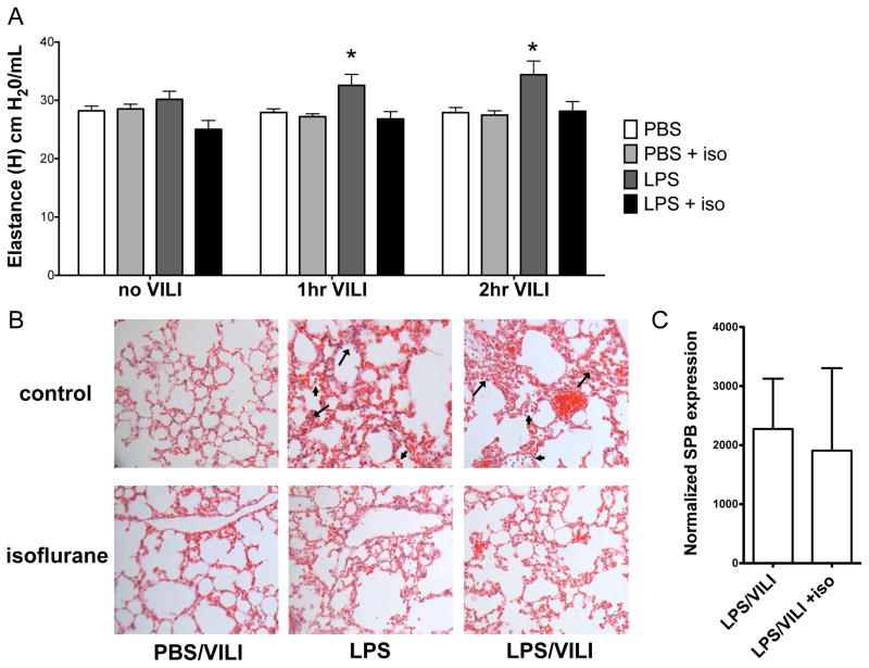
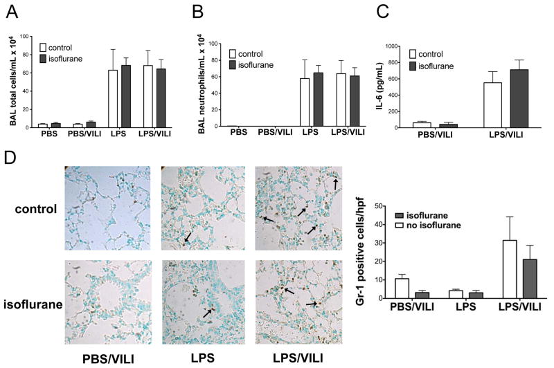
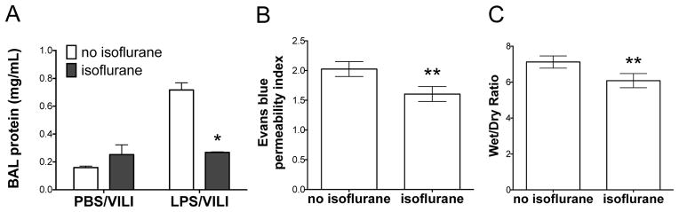
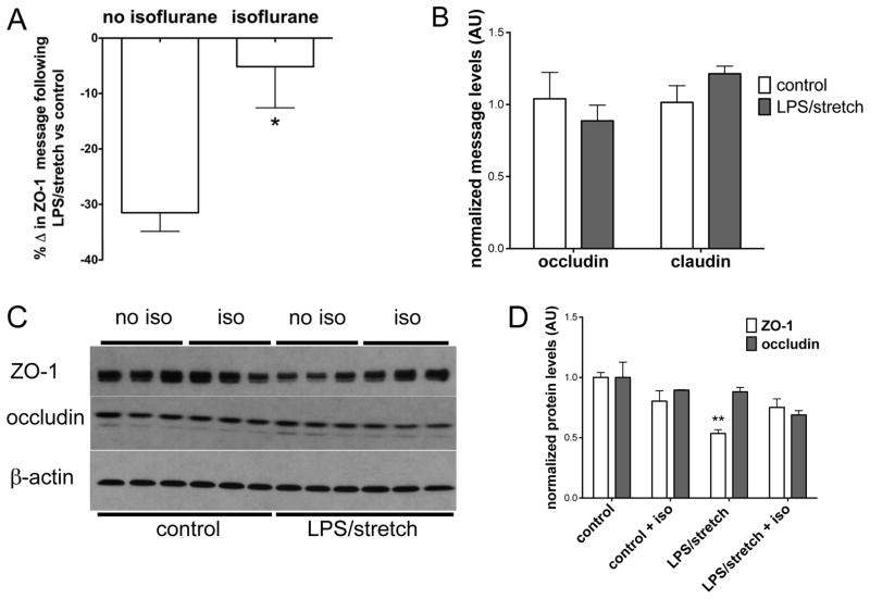
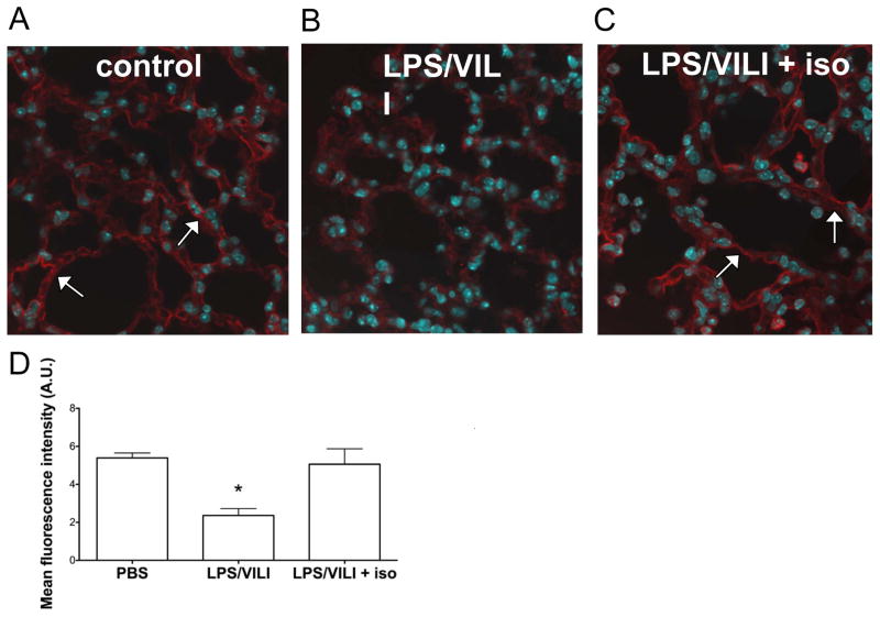
Results: Mice treated with isoflurane following exposure to inhaled endotoxin and before MV exhibited significantly less physiologic lung dysfunction. These effects appeared to be mediated by decreased vascular leak, but not altered inflammatory indices. Mouse lung epithelial cells treated with lipopolysaccharide and cyclic stretch and lungs harvested from mice after treatment with lipopolysaccharide and MV had decreased levels of a key tight junction protein (i.e., zona occludens 1) that was rescued by isoflurane treatment.

Conclusions: Isoflurane rescued lung injury induced by a two-hit model of endotoxin exposure followed by MV by maintaining the integrity of the alveolar-capillary barrier possibly by modulating the expression of a key tight junction protein.


Conflict of interest statement

Competing Interests: The authors declare no competing interests.

Figures

Figure 1
Figure 1
Schematic of the in vivo and in vitro models used.
Figure 2
Figure 2. Isoflurane attenuates lung injury in a two hit model using nebulized endotoxin and mechanical ventilation
C57BL/6 mice (n=8/group) were treated with isoflurane (or control gas) 1 hr after exposure to 10 mg nebulized lipopolysaccharide (LPS) and 24 hrs prior to mechanical ventilation (15 mL/kg, 2 hrs). A. The combination of nebulized endotoxin and mechanical ventilation to induce ventilator induced lung injury (LPS/VILI) increased lung elastance (H, stiffness) compared to mice treated with nebulized phosphate buffered saline (PBS, no VILI, n=8) or nebulized PBS followed by VILI (PBS, 2 hr VILI, n=8). Isoflurane prevented the increase in lung stiffness seen following LPS/VILI at both 1 and 2 hrs. (*p<0.05 vs LPS/VILI + isoflurane at same time point by 2-way ANOVA with Bonferroni post test) B. LPS treatment resulted in inflammatory cell infiltration (arrowhead) and interstitial thickening (arrow) consistent with edema that was more pronounced with LPS/VILI and appeared to be ameliorated by isoflurane. C. Isoflurane did not alter lung surfactant protein B (SPB) message levels following LPS/VILI.
Figure 3
Figure 3. Isoflurane does not impact lung inflammation after nebulized endotoxin, mechanical ventilation, or the combination of both injuries
Total bronchoalveolar lavage (BAL) inflammatory cells (A) and neutrophils (B) were increased following injury with lipopolysaccharide (LPS, n=3) and LPS followed by mechanical ventilation to induce ventilator induced lung injury (LPS/VILI, n=7) compared to control mice treated with nebulized phosphate buffered saline (PBS, n=2) or PBS treatment followed by mechanical ventilation (PBS/VILI, n=8), but were not affected by isoflurane treatment. Interleukin-6 (IL-6) levels in BAL fluid (C) increased following LPS/VILI (n=7) compared to PBS/VILI (n=7) but were not altered by isoflurane treatment. Staining for Gr-1 revealed increased neutrophils in animals subjected to LPS/VILI compared to either injury alone but there was no significant change in neutrophil infiltration with isoflurane (D).
Figure 4
Figure 4. Isoflurane prevents increased alveolar-capillary barrier permeability following injury with lipopolysaccharide and mechanical ventilation
C57BL/6 mice were subjected to injury with nebulized lipopolysaccharide (LPS) and mechanical ventilation to induce ventilator induced lung injury (LPS/VILI) or nebulized phosphate buffered saline followed by mechanical ventilation (PBS/VILI) prior to performing bronchoalveolar lavage (BAL). A. BAL protein increased significantly following LPS/VILI (n=3) compared to PBS/VILI (n=3). Isoflurane treatment following LPS but prior to VILI (n=4) prevented the increase in BAL protein seen with LPS/VILI without isoflurane (n=3). (*p<0.001 vs LPS/VILI no isoflurane by 2-way ANOVA with Bonferroni post-test). In a separate experiment, mice were subjected to LPS/VILI with isoflurane (n=6) or control gas (n=5). Mice treated with isoflurane had significantly less alveolar-capillary permeability as measured by Evan blue assay (B) and wet to dry ratio (C). **p<0.05 vs LPS/VILI no isoflurane by Mann-Whitney test.
Figure 5
Figure 5. Isoflurane attenuates the reduction of zona occludens 1 following lipopolysaccharide and cyclic stretch in vitro
Mouse lung epithelial cells were plated in 6-well dishes on flexible silico-elastic membranes and grown to confluence prior to treatment with lipopolysaccharide (LPS) and interferon gamma (IFN-γ). Following LPS/IFN-γ treatment, cells were exposed to isoflurane (or control gas) 1 day prior to cyclic stretch (10% stretch, 2 hrs). cDNA was synthesized, and message levels were analyzed by qPCR after normalizing for the housekeeping gene 18S. In separate experiments protein was isolated and tight junction protein levels were probed by immunoblotting. A. The combination of LPS/IFN-γ and cyclic stretch (denoted LPS/stretch, n=13 wells) decreased zona occludens 1 (ZO-1) message levels compared to control cells (no LPS/IFN-γ and no stretch, n=8 wells). The decrease in ZO-1 message was significantly attenuated by isoflurane treatment (n=15 wells, *p<0.05 vs no isoflurane by Mann-Whitney test.) B. Occludin and claudin 4 message levels did not change significantly following the combination of LPS/IFN-γ + stretch compared to control cells (no LPS/IFN-γ, no stretch) C. ZO-1 protein levels, but not occludin protein levels, decreased following LPS/IFN-γ + stretch. D. Image quantitation revealed that ZO-1 expression (panel C) decreased significantly with LPS/IFN-γ + stretch, but ZO-1 expression did not differ significantly from control (no LPS/IFN-γ, no stretch) levels when cells were treated with isoflurane. (**p<0.05 vs control/no iso by Kruskal-Wallis test with Dunn’s multiple comparisons test). All data n=3 wells/group unless otherwise noted.
Figure 6
Figure 6. Isoflurane exposure attenuates the reduction in lung epithelial zona occludens 1 after injury with lipopolysaccharide and mechanical ventilation in vivo
Wild-type C57BL/6 mice (n=3/group) were subjected to lung injury with the combination of lipopolysaccharide followed by mechanical ventilation to induce ventilator induced lung injury (LPS/VILI) with or without isoflurane treatment and compared to control mice subjected to treatment with nebulized phosphate buffered saline (PBS) without VILI. Zona occludens 1 (ZO-1) immunostaining revealed decreased ZO-1 expression in LPS/VILI (B) that was discontinuous in nature compared to control animals (A). The addition of isoflurane treatment after LPS but prior to mechanical ventilation (C) restored ZO-1 levels back to those seen in control mice, and staining was smooth and continuous in nature similar to that seen in control mice. Quantitation of ZO-1 staining (D, 3 images per mouse) from lung sections confirmed that ZO-1 levels significantly decreased with LPS/VILI and that isoflurane treatment prevented this decrease. *p<0.05 vs all other groups by 1-way ANOVA with Tukey’s multiple comparisons test.

Comment in

References

    1. ARDS Definition Task Force. Ranieri VM, Rubenfeld GD, Thompson BT, Ferguson ND, Caldwell E, Fan E, Camporota L, Slutsky AS. Acute respiratory distress syndrome: The Berlin Definition. JAMA. 2012;307:2526–33. - PubMed
    1. Rubenfeld GD, Caldwell E, Peabody E, Weaver J, Martin DP, Neff M, Stern EJ, Hudson LD. Incidence and outcomes of acute lung injury. N Engl J Med. 2005;353:1685–93. - PubMed
    1. Ware LB, Matthay MA. The acute respiratory distress syndrome. N Engl J Med. 2000;342:1334–49. - PubMed
    1. Han X, Fink MP, Uchiyama T, Yang R, Delude RL. Increased iNOS activity is essential for pulmonary epithelial tight junction dysfunction in endotoxemic mice. Am J Physiol Lung Cell Mol Physiol. 2004;286:L259–67. - PubMed
    1. Tremblay LN, Slutsky AS. Ventilator-induced lung injury: From the bench to the bedside. Intensive Care Med. 2006;32:24–33. - PubMed

Publication types