. 2015 Jun 11;6(6):e1783.
doi: 10.1038/cddis.2015.149.

Δ122p53, a mouse model of Δ133p53α, enhances the tumor-suppressor activities of an attenuated p53 mutant

Affiliations

Δ122p53, a mouse model of Δ133p53α, enhances the tumor-suppressor activities of an attenuated p53 mutant

T L Slatter et al. Cell Death Dis. .

Abstract

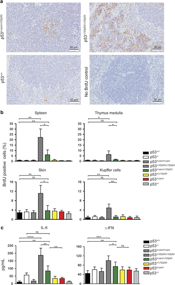
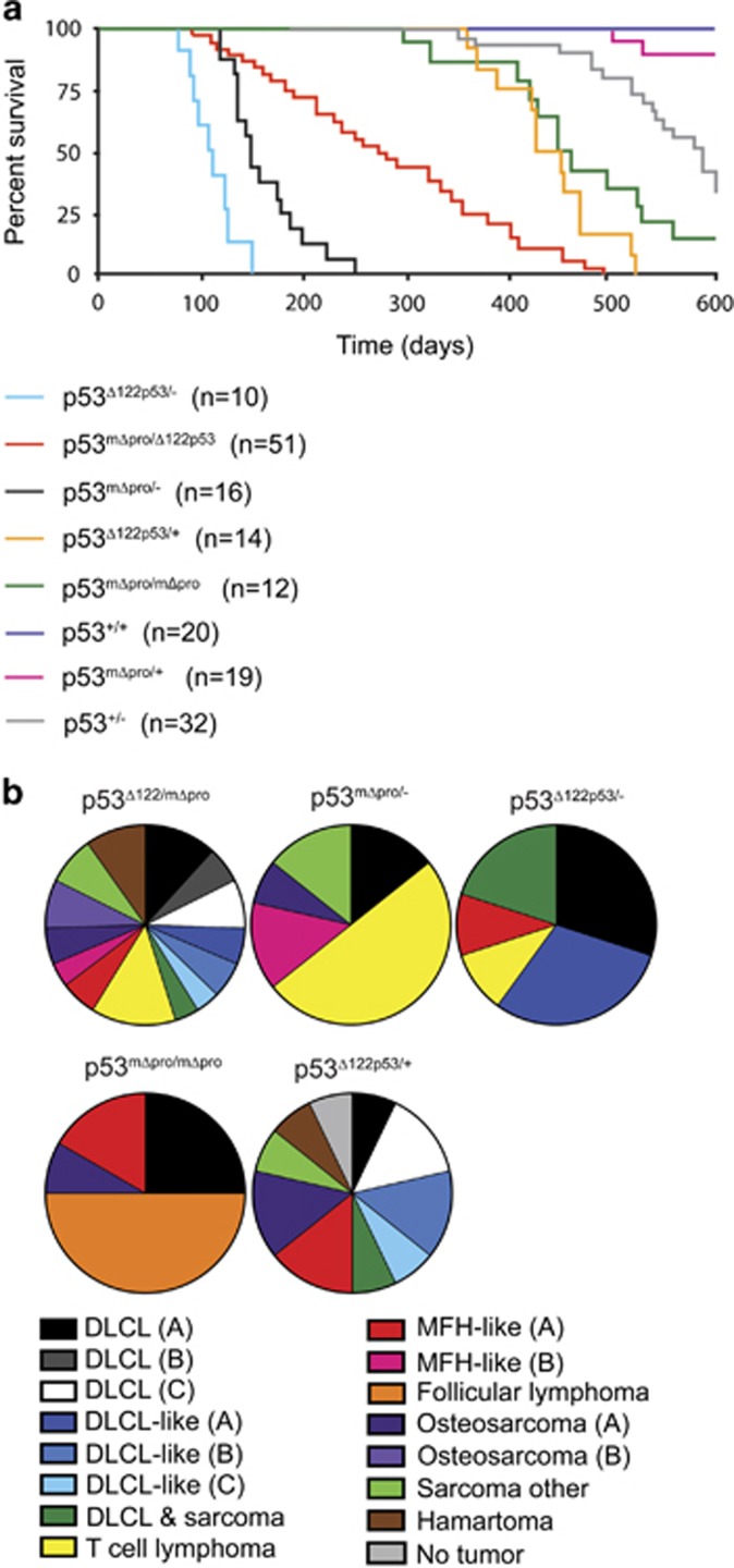
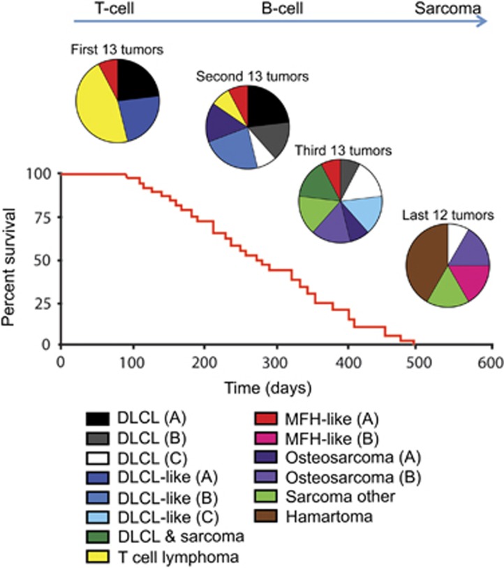
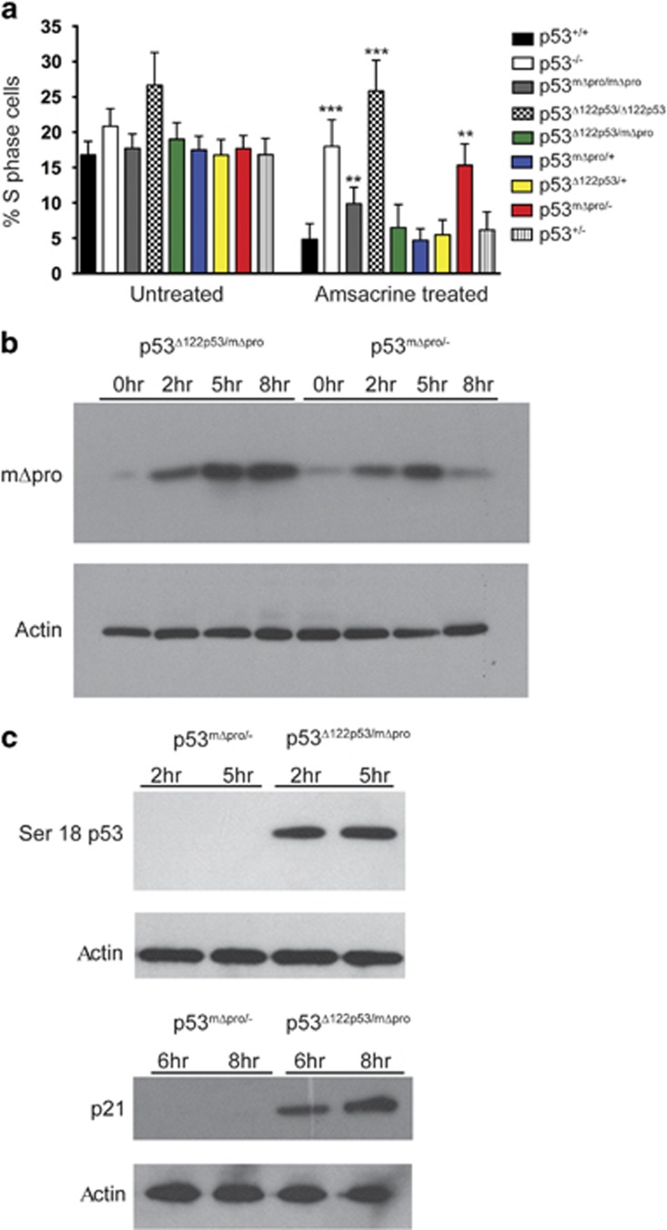
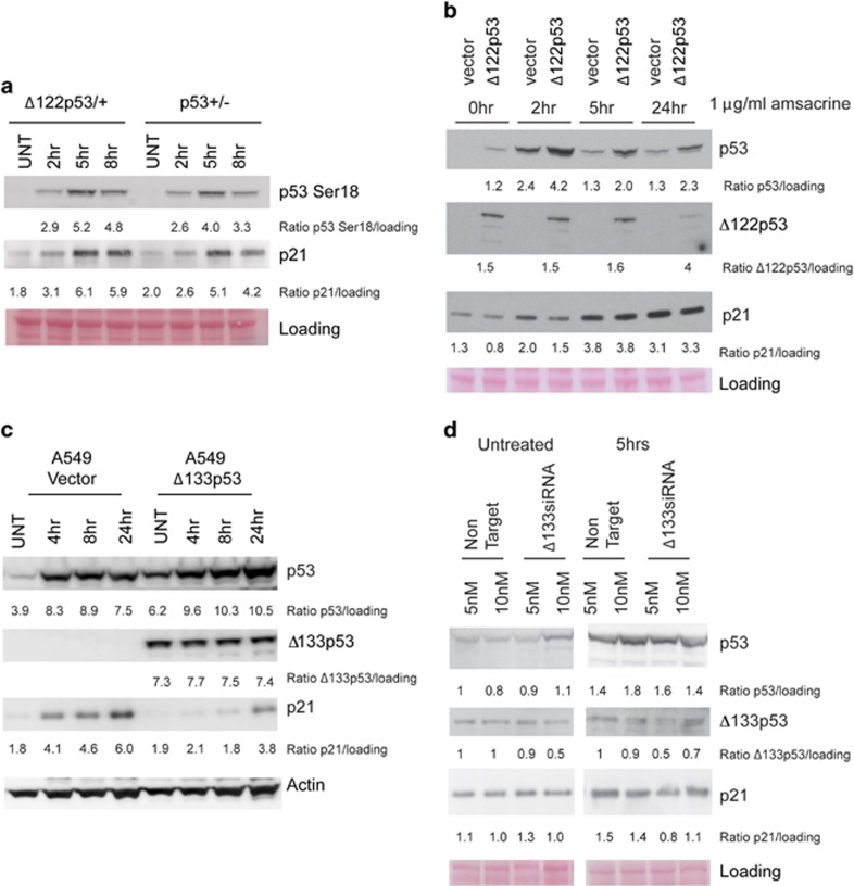
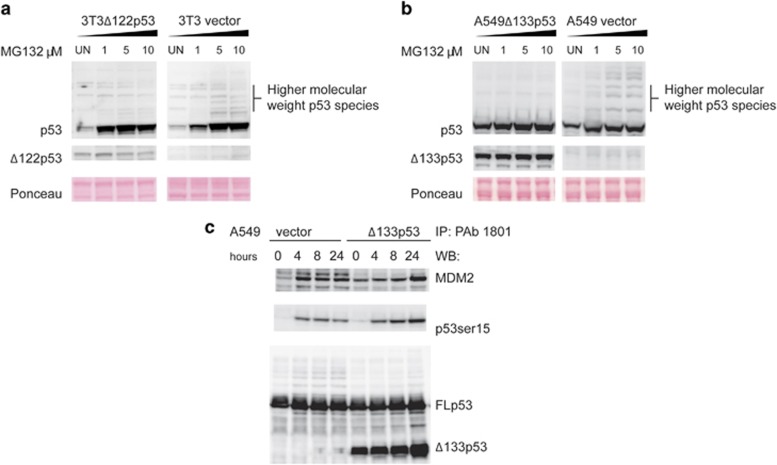
Growing evidence suggests the Δ133p53α isoform may function as an oncogene. It is overexpressed in many tumors, stimulates pathways involved in tumor progression, and inhibits some activities of wild-type p53, including transactivation and apoptosis. We hypothesized that Δ133p53α would have an even more profound effect on p53 variants with weaker tumor-suppressor capability. We tested this using a mouse model heterozygous for a Δ133p53α-like isoform (Δ122p53) and a p53 mutant with weak tumor-suppressor function (mΔpro). The Δ122p53/mΔpro mice showed a unique survival curve with a wide range of survival times (92-495 days) which was much greater than mΔpro/- mice (range 120-250 days) and mice heterozygous for the Δ122p53 and p53 null alleles (Δ122p53/-, range 78-150 days), suggesting Δ122p53 increased the tumor-suppressor activity of mΔpro. Moreover, some of the mice that survived longest only developed benign tumors. In vitro analyses to investigate why some Δ122p53/mΔpro mice were protected from aggressive tumors revealed that Δ122p53 stabilized mΔpro and prolonged the response to DNA damage. Similar effects of Δ122p53 and Δ133p53α were observed on wild-type of full-length p53, but these did not result in improved biological responses. The data suggest that Δ122p53 (and Δ133p53α) could offer some protection against tumors by enhancing the p53 response to stress.


Figures

Figure 1
Figure 1
mΔpro overrides the pro-proliferative pro-inflammatory features of Δ122p53. (a) Examples of BrdU staining on spleen tissue from p53+/+, Δ122p53/mΔpro mice, and Δ122p53 mice. Mice were pulse-labeled with BrdU for 90 min to label proliferating cells. Organs were harvested and BrdU-positive cells were detected with a horseradish peroxidase-labeled antibody and light microscopy. (b) Quantitation of BrdU-positive cells in different tissues in Δ122p53/mΔpro mice to illustrate a reduction in the percentage of proliferating cells compared with Δ122p53 homozygote mice. Mice of various p53 genotypes were pulse-labeled with BrdU and tissues collected at necropsy. BrdU-positive cells were identified using immunohistochemistry and light microscopy and the percentage of BrdU-positive cells over the total cell count calculated. Results are represented as the mean±S.D.; n=4 mice per genotype. (c) Quantitation of serum IL-6 and γ-IFN by ELISA in Δ122p53/mΔpro mice to illustrate a reduction in the pro-inflammatory phenotype compared with Δ122p53 homozygote mice. In all analyses, other genotypes with Δ122p53, mΔpro, wild-type (+) or p53-null (-) alleles were included for comparison. Results are represented as the mean±S.D.; n, at least 4 mice per genotype. *P<0.05; **P<0.01; ***P<0.001; ****P<0.0001
Figure 2
Figure 2
Broad lifespan and mixed spontaneous tumor spectrum of Δ122p53/mΔpro mice. (a) Kaplan–Meier survival curve of Δ122p53/mΔpro mice and mice with various genotype combinations (Δ122p53, mΔpro, wild-type (+), or p53-null (-) alleles and heterozygous combinations). Mice were monitored for 600 days. n=cohort size. (b) The tumor spectrum of Δ122p53/mΔpro mice in comparison with the other genotypes as identified by histo- and immuno-pathological examination. DLCL and DLCL-like tumors were further sub-grouped by cell surface markers into the following: DLCL-A, (B-cell-positive for CD34, CD10, CD45, CD45R, and CD20, and negative for CD138); DLCL-B (B-cell-positive for CD20, CD45, and CD45R, and negative for CD34, CD10, and CD138); DLCL-C (B-cell-positive for CD138, CD45, CD45R, and CD20, and negative for CD10 and CD34); DLCL-like (A), negative for all markers tested (CD3, CD10, CD20, CD45, CD45R, CD138, and cytokeratin); DLCL-like (B) lymphoma CD45-positive but negative for all other markers; DLCL-like (C), CD138- and CD45-positive, and negative for all other markers. Osteosarcoma (A) osteoblastic by morphology, (B) more differentiated by morphology; MFH-like (A) angiomatoid type, (B) non-angiomatoid type
Figure 3
Figure 3
As Δ122p53/mΔpro mice age, different tumor types become predominant. The spontaneous tumor spectrum of the Δ122p53/mΔpro mice from Figure 2 was divided into four groups based on survival time: the first 13, the second 13, the third 13, and the last 12 mice to be killed because of tumor burden, to illustrate the predominance of different tumors types at different times. The classification: DLCL, DLCL-like, osteosarcoma and MFH-tumors were subdivided based on morphological or cell surface markers using immunohistochemistry as outlined in the legend to Figure 2
Figure 4
Figure 4
Δ122p53 stabilizes mΔpro and enhances its ability to induce a cell cycle arrest after DNA damage. (a) Bone marrow from Δ122p53/mΔpro mice induced a cell cycle arrest response following DNA damage. Bone marrow was isolated from 4 to 6-week-old mice of indicated genotypes, cultured and treated with 0.2 μg/ml amsacrine. After 24 h, cells were pulse-labeled with BrdU, harvested, fixed, and stained with a fluorescent antibody to BrdU and the percentage of BrdU-positive cells was measured by flow cytometry. Bone marrow from mice with various combinations of the Δ122p53, mΔpro, wild-type (+), or p53 null (-) alleles were included for comparison. ****P<0.0001, ***P<0.001, **P<0.01, *P<0.05 in comparison with p53+/+ treated. Results are represented as the mean±S.D.; n=6 mice per genotype. (b) The presence of the Δ122p53 allele, stabilized mΔpro after DNA damage. Splenocytes from Δ122p53/mΔpro and mΔpro/- mice were cultured, exposed to 1 μg/ml amsacrine and western blots carried out with an antibody to the N terminus of p53 to detect mΔpro. (c) The presence of the Δ122p53 allele led to increased Ser18 phosphorylated mΔpro (left) and increased p21CIP1 (right) in response to DNA damage. Splenocytes from Δ122p53/mΔpro and mΔpro/- mice were cultured, exposed to 1 μg/ml amsacrine and western blots carried out with an antibody to phosphorylated Ser18 on p53 or p21CIP1. All experiments were carried out at least three times
Figure 5
Figure 5
Δ122p53 and Δ133p53α stabilize FLp53 but inhibit FLp53 activity. (a) Splenocytes from Δ122p53/mΔpro and mΔpro/- mice were cultured, exposed to 0.2 μg/ml amsacrine, and western blots carried out with an antibody to phosphorylated residue 18 of p53 (Serine 18) and an antibody to the p53 target gene, p21CIP1. Equal loading was determined by Ponceau S staining. (b) Mouse 3T3 cells were transduced with either an empty vector or a retroviral vector expressing Δ122p53. The transduced cells were then exposed to 1 μg/ml of amsacrine and western blotting carried out for p53, Δ122p53 and p21CIP1. (c) A549 cells stably transduced with either an empty vector or Δ133p53α were exposed to 1 ug/ml amsacrine and harvested at the indicated time points and protein levels determined by western blotting. (d) A549 cells were transfected with either non-targeting siRNA or siRNA targeting Δ133p53 for 48 h, treated with 1 μg/ml of amsacrine for 5 h, then protein levels determined by western blotting
Figure 6
Figure 6
Δ122p53 and Δ133p53α inhibit proteasomal degradation of FLp53. (a) Mouse 3T3 cells transduced with either an empty vector or Δ122p53 were treated with the proteasomal inhibitor MG132 at the indicated concentrations for 4.5 h. Following MG132 treatment, cells were harvested and protein levels determined by immunoblotting. (b) Experiments were repeated using human A549 cells transduced to express Δ133p53α. (c) A549 cells transduced with Δ133p53α or a vector control were treated with 1 μg/ml amsacrine for 0, 4, 8, and 24 h. Following amsacrine treatment, cells were harvested and subjected to immunoprecipitation with the p53 antibody pAb1801, followed by western blotting with a rabbit polyclonal p53 phospho-serine antibody to detect activated p53; the p53 antibody pAb240 to detect Δ133p53α; and SMP14 to detect bound MDM2

References

    1. Braithwaite AW, Del Sal G, Lu X. Some p53-binding proteins that can function as arbiters of life and death. Cell Death Differ 2006; 13: 984–993. - PubMed
    1. Braithwaite AW, Prives CL. p53: more research and more questions. Cell Death Differ 2006; 13: 877–880. - PubMed
    1. Oren M. Decision making by p53: life, death and cancer. Cell Death Differ 2003; 10: 431–442. - PubMed
    1. Speidel D. The role of DNA damage responses in p53 biology. Arch Toxicol 2015; 89: 501–517. - PubMed
    1. Bensaad K, Tsuruta A, Selak MA, Vidal MN, Nakano K, Bartrons R et al. TIGAR, a p53-inducible regulator of glycolysis and apoptosis. Cell 2006; 126: 107–120. - PubMed

Publication types

MeSH terms