. 2015 Jun 12;348(6240):1255-60.
doi: 10.1126/science.aaa0922. Epub 2015 Jun 11.

PHYSIOLOGY. Regulation of breathing by CO₂ requires the proton-activated receptor GPR4 in retrotrapezoid nucleus neurons

Affiliations

PHYSIOLOGY. Regulation of breathing by CO₂ requires the proton-activated receptor GPR4 in retrotrapezoid nucleus neurons

Natasha N Kumar et al. Science. .

Abstract

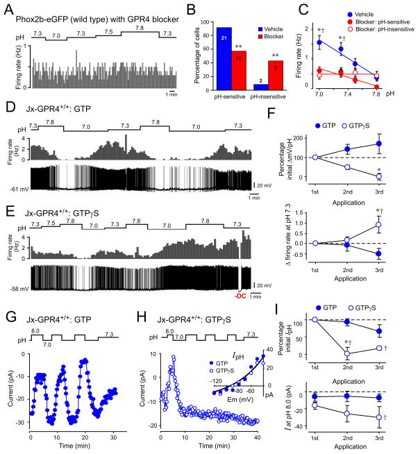
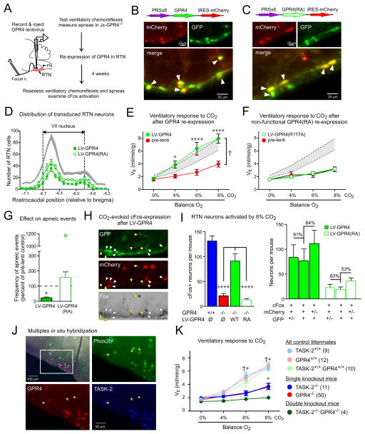
Blood gas and tissue pH regulation depend on the ability of the brain to sense CO2 and/or H(+) and alter breathing appropriately, a homeostatic process called central respiratory chemosensitivity. We show that selective expression of the proton-activated receptor GPR4 in chemosensory neurons of the mouse retrotrapezoid nucleus (RTN) is required for CO2-stimulated breathing. Genetic deletion of GPR4 disrupted acidosis-dependent activation of RTN neurons, increased apnea frequency, and blunted ventilatory responses to CO2. Reintroduction of GPR4 into RTN neurons restored CO2-dependent RTN neuronal activation and rescued the ventilatory phenotype. Additional elimination of TASK-2 (K(2P)5), a pH-sensitive K(+) channel expressed in RTN neurons, essentially abolished the ventilatory response to CO2. The data identify GPR4 and TASK-2 as distinct, parallel, and essential central mediators of respiratory chemosensitivity.


Figures

Figure 1
Figure 1. GPR4 deletion disrupts CO2-evoked ventilatory stimulation and RTN neuronal activation in vivo, and increases incidence of spontaneous apneas
(A) Left, Respiratory flow recording from Jx-GPR4+/+ and Jx-GPR4−/− mice (cross between Jx-Phox2b-eGFP and GPR4 lines) with increased CO2 concentrations in the inspired air (balance O2). Right, Minute ventilation (VE) during incremental CO2 challenge in Jx-GPR4+/+ and Jx-GPR4−/− mice (n=16 & 52; †, P<0.0001 between genotypes by 2-way repeated measures (RM)-ANOVA; ****, P<0.0001 for pairwise comparisons). (B) Jx-GPR4+/+ and Jx-GPR4−/− mice exhibited similar increases in peak VE during exposure to 10% O2 (n=16 & 52; ****, P<0.0001 hypoxia vs. normoxia, by 2-way RM-ANOVA). (C) The frequency of spontaneous apneic events during quiet hyperoxic (100% O2) breathing was greater in Jx-GPR4−/− than in Jx-GPR4+/+ mice (n=23 & 8; *, P<0.05 by unpaired t-test). (D) GPR4 expression by in situ hybridization in coronal brainstem section from an adult Jx-GPR4+/+ mouse. Inset is enlarged in a merged image; GPR4 mRNA is observed in 68.1 ± 5.1% of GFP+ neurons, n=6; counts do not exclude C1 neurons); no labeling was detected in sections from Jx-GPR4−/− mice. pyr: pyramid. Scale bar = 25 μm. (E) Jx-GPR4+/+ and Jx-GPR4−/− mice were exposed to CO2 in vivo and activated RTN (GFP+:TH-) neurons (asterisks) identified by immunoreactivity for cFos (left, black reaction product in cell nuclei denoted by arrows) and GFP (right,). Scale bars = 50 and 25 μm. (F) Upper: Parasagittal schematic of mouse brainstem depicting the RTN and surrounding neuronal populations, with areas containing Phox2b-expressing cells denoted in green. Lower: The number of RTN neurons activated by 8% CO2 challenge (cFos+:Phox2b+:TH-) was strongly reduced throughout the RTN in Jx-GPR4−/− mice, as compared to Jx-GPR4+/+ mice (n=5 & 6). (G) The total number of RTN neurons counted (every 3rd section) from Jx-GPR4+/+ and Jx-GPR4−/− mice activated in response to 0% (n=4 & 4), 6% (n=5 & 5) or 8% CO2 (n=6 & 5). (P<0.0001, between genotypes by 2-way RM-ANOVA; **, P<0.01 and ****, P<0.0001 for pairwise comparisons).
Figure 2
Figure 2. GPR4 deletion ablates pH sensitivity and a pH-sensitive background K+ current in a subset of RTN neurons in vitro
(A, B) Effects of bath pH on firing activity (cell-attached mode) in representative GFP-expressing RTN neurons. A typical pH-sensitive from a GPR4+/+ mouse is depicted (A); in GPR4−/− mice (B), RTN neurons that were either pH-sensitive (upper) or pH-insensitive (lower). (C) The percentage of pH-sensitive and pH-insensitive RTN neurons was significantly different between GPR4+/+ and GPR4−/− mice (**, P<0.0001 by χ2; n provided for each group). (D) Averaged firing rates at different bath pH for RTN neurons from GPR4+/+ mice (n=71) and for RTN neurons from GPR4−/− mice that were identified as pH-sensitive (n=45) or pH-insensitive (n=32). *, P<0.05 for GPR4+/+ vs GPR4−/−:pH-sensitive; and †, P<0.05 for GPR4+/+ vs GPR4−/−: pH-insensitive, by 2-way RM-ANOVA with Dunnett’s test. (E) Effects of bath acidification and alkalization on holding current (at −60 mV) and conductance during whole cell voltage clamp recordings in a pH-sensitive neuron from Jx-GPR4+/+ mouse (left) and a pH-insensitive neuron from a Jx-GPR4−/− mouse (right). (F) I-V relationship of pH-sensitive difference current (pH 8 minus pH 7) for RTN neurons from GPR4+/+ mice (n=16) and from GPR4−/− mice that were identified in cell-attached recordings as pH-sensitive (n=11) or pH-insensitive (n=5). Data overlaid with fits using the Goldman-Hodgkin-Katz (GHK) equation for a “leak” K+ current. **, P<0.05 vs. GPR4−/−: pH-insensitive, by 2-way RM-ANOVA with Tukey’s test.
Figure 3
Figure 3. GPR4 and G protein activation contribute to effects of pH on firing and background K+ currents in RTN neurons from wild-type mice
(A) Firing activity in a wild-type RTN neuron incubated with GPR4 antagonist (Dalton M46, 20 μM); the cell was insensitive to changes in bath pH. (B) The percentage of pH-sensitive and pH-insensitive wild-type RTN neurons was significantly different after treatment with the GPR4 blocker (**, P<0.0001 by χ2; n provided for each group). (C) Averaged firing rate at different bath pH from vehicle-treated RTN neurons (n=21) or GPR4 blocker-treated cells that were identified as pH-sensitive (n=12) or pH-insensitive (n=9). *, P<0.05 for vehicle vs blocker:pH-sensitive; and †, P<0.05 for vehicle vs blocker:pH-insensitive, by 2-way RM-ANOVA with Dunnett’s test. (D, E) Effects of repeated changes in bath pH on firing rate (upper) and membrane potential (lower) recorded from wild-type RTN neurons in the whole cell current clamp configuration with pipettes containing either GTP (D) or GTP-γ-S (E). (F) Upper: Membrane hyperpolarization (% initial) during multiple bouts of bath alkalization in RTN neurons recorded with GTP and GTP-γ-S. Lower: Change in baseline firing (at pH 7.3) following repeated bath alkalization in RTN neurons recorded with GTP and GTP-γ-S. (2-way RM-ANOVA; *, P<0.05 for GTP vs. GTP-γ-S by Bonferroni test; and †, P<0.05 vs. 1st application by Dunnett’s test; n=8 & 4). (G, H) Effects of repeated changes of bath pH on holding current in wild-type RTN neurons during whole cell voltage clamp recording with pipettes that contained GTP (G) or GTP-γ-S (H). Inset shows the pH-sensitive I-V obtained during the first sojourn from pH 8 to pH 7 for both cells, overlaid with a fit using the GHK equation. (I) Normalized pH-sensitive current (pH 8 minus pH 7 at −60 mV, upper) and holding current at pH 8 (lower) during repeated bouts of bath acidification and alkalization. (2-way RM-ANOVA; *, P<0.05 for GTP vs. GTP-γ-S by Bonferroni test; and †, P<0.05 vs. 1st application by Dunnett’s test; n=7 & 3).
Figure 4
Figure 4. Re-expression of GPR4 selectively in Phox2b-expressing neurons of the RTN restores CO2-dependent neuronal activation and breathing in Jx-GPR4−/− mice
(A) Schematic of experimental design. (B, C) White arrows denote virally-transduced (mCherry+) and Phox2b-expressing (GFP+) RTN neurons after injection of PRSx8-lentivirus driving expression of GPR4-mCherry (B) or GPR4(R117A)-mCherry (C). (D) Similar number and distribution of RTN neurons transduced with GPR4-mCherry (n=8) and GPR4(R117A)-mCherry (n=7) lentivirus. Shaded area represents 95% confidence interval for distribution of RTN (GFP+/TH-) neurons across all virus-injected animals (n=15). (E, F) Effect of CO2 on minute ventilation in Jx-GPR4−/− mice before (pre-lenti) and 4 weeks after lentiviral injection into the RTN (E: GPR4-mCherry, n=8; or F: GPR4(R117A)-mCherry, n=7). Shaded areas are 95% confidence intervals for data from Jx-GPR4+/+ mice (from Fig. 1A). (†, P<0.0001 by 2-way RM-ANOVA, comparing mice before and following lentiviral delivery; *, P<0.05, ****, P<0.0001, pairwise comparisons at each CO2 level by Bonferroni test). (G) The frequency of spontaneous apneic events was reduced following injection of GPR4-mCherry lentiviral vector into RTN (n=8; *, P<0.05 by t-test, ~22% of pre-injection control) but not GPR4(R117A)-mCherry (n=10, P>0.18 by t-test). Note the outlying data point from one mouse that received GPR4(R117A)-mCherry. (H) Many virally-transduced (mCherry+) RTN neurons (GFP+) were activated (cFos+) by 8% CO2 exposure (white arrows) after injection of PRSx8-lentivirus expressing GPR4 into RTN of Jx-GPR4−/− mice; CO2-activated GFP+ neurons that were not detectably transduced were also observed (yellow arrows). (I) Left: CO2-activated cFos expression in RTN neurons was rescued following injection of PRSx8-GPR4 but not PRSx8-GPR4(R117A) lentivirus (n=4 & 7, P<0.0001 by 1-way ANOVA, ****, P<0.0001 by pairwise comparison); data from Jx-GPR4+/+ and Jx-GPR4−/− mice are re-plotted from Fig. 1G. Right: CO2-activated, transduced RTN neurons (cFos+/mCherry+/GFP+) in comparison to all CO2-activated, transduced neurons (cFos+/mCherry+) and all CO2-activated RTN neurons (cFos+/GFP+). (J) Co-expression of Phox2b, GPR4 and TASK-2 in RTN neurons by in situ hybridization; boxed region enlarged in merged image. A few TASK-2-expressing Phox2b+ neurons without GPR4 are indicated (asterisks). Among Phox2b+ neurons that expressed GPR4 and/or TASK-2, 72.9 ± 0.8% expressed both genes, 18.1 ± 1.0% were labeled only for TASK-2 and 9.0 ± 0.2 only for GPR4 (n=3). (K) Minute ventilation during incremental CO2 challenge for the indicated genotypes. †, all controls greater than all single or double knockouts, P<0.0001; *, both single knockouts greater than double knockouts (n values provided), by 2-way RM-ANOVA with Bonferroni pairwise comparisons.

References

    1. Guyenet PG, Stornetta RL, Bayliss DA. Central respiratory chemoreception. J Comp Neurol. 2010;518:3883–3906. - PMC - PubMed
    1. Feldman JL, Mitchell GS, Nattie EE. Breathing: Rhythmicity, plasticity, chemosensitivity. Annu Rev Neurosci. 2003;26:239–266. - PMC - PubMed
    1. Mulkey DK, et al. Respiratory control by ventral surface chemoreceptor neurons in rats. Nat Neurosci. 2004;7 - PubMed
    1. Wang S, Shi Y, Shu S, Guyenet PG, Bayliss DA. Phox2b-expressing retrotrapezoid neurons are intrinsically responsive to H+ and CO2. J Neurosci. 2013;33:7756–7761. - PMC - PubMed
    1. Abbott SBG, et al. Photostimulation of retrotrapezoid nucleus Phox2b-expressing neurons in vivo produces long-lasting activation of breathing in rats. J Neurosci. 2009;29:5806–5819. - PMC - PubMed

Publication types

MeSH terms