. 2015 Aug 7;6(22):18956-65.
doi: 10.18632/oncotarget.4150.

Targeting IRAK1 in T-cell acute lymphoblastic leukemia

Affiliations

Targeting IRAK1 in T-cell acute lymphoblastic leukemia

Charles Dussiau et al. Oncotarget. .

Abstract

T-cell acute lymphoblastic leukemia (T-ALL) represents expansion of cells arrested at specific stages of thymic development with the underlying genetic abnormality often determining the stage of maturation arrest. Although their outcome has been improved with current therapy, survival rates remain only around 50% at 5 years and patients may therefore benefit from specific targeted therapy. Interleukin receptor associated kinase 1 (IRAK1) is a ubiquitously expressed serine/threonine kinase that mediates signaling downstream to Toll-like (TLR) and Interleukin-1 Receptors (IL1R). Our data demonstrated that IRAK1 is overexpressed in all subtypes of T-ALL, compared to normal human thymic subpopulations, and is functional in T-ALL cell lines. Genetic knock-down of IRAK1 led to apoptosis, cell cycle disruption, diminished proliferation and reversal of corticosteroid resistance in T-ALL cell lines. However, pharmacological inhibition of IRAK1 using a small molecule inhibitor (IRAK1/4-Inh) only partially reproduced the results of the genetic knock-down. Altogether, our data suggest that IRAK1 is a candidate therapeutic target in T-ALL and highlight the requirement of next generation IRAK1 inhibitors.

Keywords: IRAK1; T-ALL; kinases; therapeutic target.


Figures
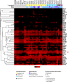
Figure 1
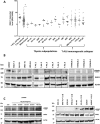
Figure 1. Kinases expression profiles of human T-ALL samples and thymic subpopulations
Transcriptional expression of major kinase receptors and receptor associated kinases in normal and malignant immature T-cells. Thymic subpopulations and T-ALL samples are displayed in a supervised classification model and ordered according to their immunogenetic status. Non-expressed (receptor)-kinases are not shown. 4ISP, CD4 immature single positive; DP TCR-, CD4/CD8 double positive surface TCR negative; DP TCR+, CD4/CD8 double positive surface TCR positive; SP4, mature CD4 single positive; SP8, mature CD8 single positive.
Figure 2
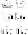
Figure 2. IRAK1 is overexpressed and functional in T-ALL
A. qRT-PCR: IRAK1 transcriptional expression is shown in T-ALL according to TCR status, and in thymic subsets. B. IRAK1 protein expression and phosphorylation were assessed by western blot on T-ALL cell lines, primary T-ALL blasts and normal thymus. C. Left panel: Activation of IRAK1 pathway at different time upon IL1β stimulation in the Jurkat cell line. Right panel: Activation of IRAK1 pathway after 45 min treatment withIL1β (10 ng/mL) in T-ALL cell lines. 4ISP, CD4 immature single positive; DP TCR-, CD4/CD8 double positive surface TCR negative; DP TCR+, CD4/CD8 double positive surface TCR positive; SP4, mature CD4 single positive; SP8, mature CD8 single positive; IM0, immature with germline TCR loci; IMB, immature with TCRβ rearrangement; Pre-ab, cTCRβ expressing T-ALL [31].
Figure 3
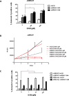
Figure 3. Genetic knock-down of IRAK1 induces apoptosis and disrupts cell cycle
A. Genetic knockdown of IRAK1 was confirmed by western blotting in Jurkat cells expressing a control or shIRAK1-expressing lentiviral vector B. Viable cell growth was assayed by trypan blue exclusion for up to 6 days. Data are represented as mean +/− SEM. C. Annexin V/IP staining was assessed in Jurkat and HPB-ALL cells by flow cytometry after transduction with shRNA-expressing lentiviral vectors. D. Cell cycle analysis of Jurkat and HPB-ALL cells after transduction was performed with Edu/7-AAD incorporation.
Figure 4
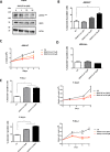
Figure 4. Genetic knockdown of IRAK1 reverses corticoresistance in Jurkat
A. AnnexinV/IP staining was assessed in Jurkat after transduction with shRNA-expressing lentiviral vector and after 72 h treatment with dexamethasone (0–100-500 μM). B. Viable cell growth was assayed by trypan blue exclusion after transduction with shRNA-expressing lentiviral vector and treatment with dexamethasone (0–100 μM) for up to 4 days. Data are represented as mean +/− SEM. C. AnnexinV/IP staining was assessed after transduction with shRNA-expressing lentiviral vector in jurkat (white) and HPB-ALL (grey) and after 72 h treatment with dexamethasone (0-1-10-100 μM) on Jurkat (light grey) and HPB-ALL (black) cell-lines. DXM, dexamethasone.
Figure 5
Figure 5. Effect of IRAK1/4-inh on T-ALL cell lines
A. IRAK1 and pIRAKT209 were evaluated by western blotting in Jurkat cells after 48 h treatment with IRAK1/4 inhibitor B–E. AnnexinV/IP staining was performed after 72 h treatment with IRAK1/4 inhibitor in Jurkat, HPB-ALL cells and in primary T-ALL blasts from 2 patients (C–E) Viable cell growth of Jurkat and in primary T-ALL blasts from 2 patients was assayed by trypan blue exclusion in the presence of IRAK1/4 inhibitor (0-10-50 μM). Data are represented as mean +/− SEM.

References

    1. Inaba H, Greaves M, Mullighan CG. Acute lymphoblastic leukaemia. Lancet. 2013;381:1943–1955. - PMC - PubMed
    1. Bassan R, Hoelzer D. Modern therapy of acute lymphoblastic leukemia. J Clin Oncol. 2011;29:532–543. - PubMed
    1. Zhang J, Ding L, Holmfeldt L, Wu G, Heatley SL, Payne-Turner D, Easton J, Chen X, Wang J, Rusch M, Lu C, Chen SC, Wei L, Collins-Underwood JR, Ma J, Roberts KG, et al. The genetic basis of early T-cell precursor acute lymphoblastic leukaemia. Nature. 2012;481:157–163. - PMC - PubMed
    1. Bandapalli OR, Schuessele S, Kunz JB, Rausch T, Stutz AM, Tal N, Geron I, Gershman N, Izraeli S, Eilers J, Vaezipour N, Kirschner-Schwabe R, Hof J, von Stackelberg A, Schrappe M, Stanulla M, et al. The activating STAT5B N642H mutation is a common abnormality in pediatric T-cell acute lymphoblastic leukemia and confers a higher risk of relapse. Haematologica. 2014;99:e188–92. - PMC - PubMed
    1. Van Vlierberghe P, Ferrando A. The molecular basis of T cell acute lymphoblastic leukemia. The Journal of clinical investigation. 2012;122:3398–3406. - PMC - PubMed

Publication types

MeSH terms

Substances