Clinical Trial
. 2015 Sep;100(9):1151-9.
doi: 10.3324/haematol.2015.124321. Epub 2015 Jun 11.

Recurrent deletions of IKZF1 in pediatric acute myeloid leukemia

Affiliations
Clinical Trial

Recurrent deletions of IKZF1 in pediatric acute myeloid leukemia

Jasmijn D E de Rooij et al. Haematologica. 2015 Sep.

Abstract

IKAROS family zinc finger 1/IKZF1 is a transcription factor important in lymphoid differentiation, and a known tumor suppressor in acute lymphoid leukemia. Recent studies suggest that IKZF1 is also involved in myeloid differentiation. To investigate whether IKZF1 deletions also play a role in pediatric acute myeloid leukemia, we screened a panel of pediatric acute myeloid leukemia samples for deletions of the IKZF1 locus using multiplex ligation-dependent probe amplification and for mutations using direct sequencing. Three patients were identified with a single amino acid variant without change of IKZF1 length. No frame-shift mutations were found. Out of 11 patients with an IKZF1 deletion, 8 samples revealed a complete loss of chromosome 7, and 3 cases a focal deletion of 0.1-0.9Mb. These deletions included the complete IKZF1 gene (n=2) or exons 1-4 (n=1), all leading to a loss of IKZF1 function. Interestingly, differentially expressed genes in monosomy 7 cases (n=8) when compared to non-deleted samples (n=247) significantly correlated with gene expression changes in focal IKZF1-deleted cases (n=3). Genes with increased expression included genes involved in myeloid cell self-renewal and cell cycle, and a significant portion of GATA target genes and GATA factors. Together, these results suggest that loss of IKZF1 is recurrent in pediatric acute myeloid leukemia and might be a determinant of oncogenesis in acute myeloid leukemia with monosomy 7.


Figures

Figure 1.
Figure 1.
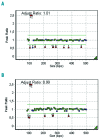
Heterozygous IKZF1 deletions in pediatric AML. (A) Multiplex ligation-dependent probe amplification results of a patient with heterozygous deletion of exons 1–8 of IKZF1 including the reference probe in POR located on 7q11.23. (B) MLPA results of a patient with heterozygous deletion of exons 1 through 4 of IKZF1. 1–8 indicates IKZF1 exons 1–8; D: reference D-fragment 96, a control probe for denaturation during the procedure; P: POR (7q11.23); X: X-chromosome specific fragment, a reference probe for gender; Y, Y-chromosome specific fragment, a reference probe for gender; Z: ZFY (Yp11.31), a reference probe for gender.
Figure 2.
Figure 2.
Array comparative genomic hybridization analysis of monosomy 7 and focal IKZF1 deletions. DNA isolated from pediatric AML samples taken at initial diagnosis and containing IKZF1 deletion based on MLPA results were analyzed using array CGH. Representative samples for monosomy 7 (patient #4, upper panel) and IKZF1 focal del7p12.2 deletions (patient #2, #3 and #10, lower panel) are shown.
Figure 3.
Figure 3.
IKZF1 haploinsufficient acute myeloid leukemia (AML) cells display an AML-specific gene expression signature. AML (n=297, red) and B-cell acute lymphoblastic leukemia (B-ALL) (n=107, blue) patients were projected onto the first two principal coordinates, obtained via multidimensional scaling of expression values of AML specific genes (A), ALL specific (B) genes and the combination of these (C) (see Thomas et al.). Focally deleted IKZF1 (dark green triangles) and monosomy 7 (light green squares) AMLs were located among other AMLs and away from B-ALLs.
Figure 4.
Figure 4.
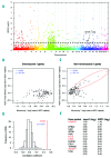
Monosomy 7 and focal IKZF1 deletion associate with gene expression profiles with significant similarity. (A) Analysis of differences in gene expression between monosomy 7 samples (n=8) and IKZF1-non-deleted samples (n=247). Genes are ranked on Bayesian false discovery rate (BFDR) (y-axis) and binned in their chromosome of origin (x-axis). Using a BFDR of 5% (dashed line), a total of 198 genes are detected differentially expressed, of which 111 are not encoded on chromosome 7. (B and C) Mean differences per gene between monosomy 7 and non-IKZF1-deleted samples versus those of IKZF1 focal deletions and non-IKZF1-deleted samples for chromosome 7 encoded genes (B) and non-chromosome 7 encoded genes (C). Pearson’s correlation coefficients (r) are indicated in the plot, together with accompanying P values. (D) Bootstrap analyses of the correlation between monosomy 7 cases (n=8) and 3 randomly picked IKZF1-non-deleted samples (n=247). The red dashed line indicates correlation coefficient from the analyses displayed in Figure 4C. (E) Top correlating genes [dashed elipse in (C)] between monosomy 7 and focal IKZF1 deletions. Highlighted in red are those reported to be positively acting on leukemic cell self-renewal or cell cycle.
Figure 5.
Figure 5.
HEMGN, FHL2, FZD6 and SETBP1 expression in IKZF1-deleted patients as compared to other cytogenetically classified AML groups. (AD) Comparison of mRNA abundances in IKZF1-deleted samples to major cytogenetic AML subgroups revealed that the median expression of HEMGN, FHL2, FZD6 and SETBP1 in the IKZF1 deleted group is clearly higher than in each of the subgroups, except for FZD6 and SETBP1 in comparison to t(7;12)/MNX1-ETV6. (E and F) When excluding the patient harboring both the IKZF1 deletion and t(7;12)/MNX1-ETV6, FZD6 and SETBP1 remain significantly higher expressed in the IKZF1 -deleted cases compared to the other pediatric AML subgroups (P=0.003 and P=0.005, respectively).
Figure 6.
Figure 6.
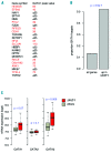
Genes commonly up-regulated in IKZF1 focally deleted and monosomy 7 cases are enriched in myeloid GATA1 targets. (A) GATA1 chromatin interaction values (GATA1 peak value) in K562 cells of commonly up-regulated genes (n=23) in monosomy 7 and focal IKZF1 deletion (ΔIKZF1). (B) Proportion of all matching genes (n=22564) or ΔIKZF1 up-regulated (n=23) displaying a GATA1 chromatin interaction in K562 cells. P value from Fisher’s exact test. (C) mRNA expression of GATA1, GATA2 and GATA3 in IKZF1 deleted samples (red) versus IKZF1 WT samples (gray). P values according to Mann-Whitney tests.

References

    1. Pui CH, Carroll WL, Meshinchi S, Arceci RJ. Biology, risk stratification, and therapy of pediatric acute leukemias: an update. J Clin Oncol. 2011;29(5):551–565. - PMC - PubMed
    1. Shih AH, Abdel-Wahab O, Patel JP, Levine RL. The role of mutations in epigenetic regulators in myeloid malignancies. Nat Rev Cancer. 2012;12(9):599–612. - PubMed
    1. Creutzig U, van den Heuvel-Eibrink MM, Gibson B, et al. Diagnosis and management of acute myeloid leukemia in children and adolescents: recommendations from an international expert panel. Blood. 2012;120(16):3187–3205. - PubMed
    1. Kaspers GJ, Zwaan CM. Pediatric acute myeloid leukemia: towards high-quality cure of all patients. Haematologica. 2007;92(11):1519–1532. - PubMed
    1. Oestreich KJ, Weinmann AS. Ikaros changes the face of NuRD remodeling. Nat Immunol. 2012;13(1):16–18. - PubMed

Publication types

Supplementary concepts